This article contains AI generated content.
· UpdatedLateral Retinacular Tightness — 5 Genes and 6 Biomarkers to Track
Introduction
If you have lateral retinacular tightness, you probably know the sensation well: a persistent pulling discomfort along the outer edge of the kneecap, pain that flares with stairs or prolonged sitting, and a frustrating pattern where standard stretching brings only partial or temporary relief. You may have been told to strengthen your VMO, stretch your IT band, or simply rest — and while that advice is not wrong, it often misses why the tissue is tight in the first place.
The lateral retinaculum is a dense fibrous sleeve on the outer side of the patella. When it becomes chronically shortened or fibrotic, it tilts and compresses the kneecap against the femur in ways that generate pain, cartilage stress, and eventually structural change. The reasons this happens vary considerably between individuals: some people have a genetic predisposition toward stiffer collagen architecture, others have silent micronutrient deficiencies that blunt their body's ability to maintain and remodel connective tissue, and still others carry inflammatory states that drive progressive fibrosis even in the absence of major injury.
Generic rehabilitation protocols do not account for these individual differences. Two people doing the same patellar mobilization protocol can have opposite outcomes — and the reason often lies beneath the surface, in blood chemistry and genetic variants that no standard orthopedic evaluation measures.
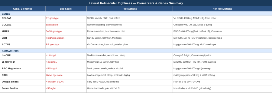
This article takes a more granular approach. The first half focuses on six specific biomarkers you can measure with standard and specialty blood tests — each one capable of revealing a distinct mechanism driving your tightness. The second half explores five genes that influence connective tissue architecture and inflammation, with practical plans for each. Together, they give you a clearer picture of what is actually happening in your tissue and what levers are actually worth pulling.
6 Biomarkers to Track for Lateral Retinacular Tightness
Biomarkers do not tell you everything, but the right ones tell you things that are otherwise invisible. For lateral retinacular tightness, the most useful markers cluster around three themes: systemic inflammation, connective tissue turnover, and micronutrient status. When these are off, rehabilitation is slower, tissue remodels poorly, and the tightness tends to return even after successful manual therapy.
Biomarker 1: High-Sensitivity C-Reactive Protein (hs-CRP)
CRP is the liver's response to circulating inflammatory signals. At elevated levels — even the so-called "low-level" elevations that sit between 1 and 3 mg/L — it reflects a systemic inflammatory state that promotes fibrosis in connective tissue. The lateral retinaculum, like all dense collagen structures, is particularly vulnerable: chronic, low-grade inflammation drives the cross-linking and thickening of the retinacular fibers in ways that make them progressively stiffer and less responsive to stretching. High hs-CRP does not cause tightness directly, but it creates the biochemical environment in which tightness becomes self-perpetuating.
How to Measure It
hs-CRP is a standard blood test ordered by most primary care physicians or available through direct-to-consumer labs. Cost typically ranges from $15 to $40. The high-sensitivity version (not standard CRP) is needed to detect subclinical inflammation. Optimal is below 0.5 mg/L; anything above 1.0 mg/L warrants attention.
If the Score Is High: The Plan Without Supplements
The most powerful non-supplement intervention for hs-CRP is dietary quality. A Mediterranean-style eating pattern — emphasizing olive oil, fatty fish, colorful vegetables, legumes, and minimal ultra-processed food — consistently reduces hs-CRP by 20 to 40% in randomized trials. Aerobic exercise (30 minutes of moderate intensity, 4 to 5 times per week) also meaningfully reduces circulating CRP, though intense training without adequate recovery can temporarily spike it. Prioritize sleep quality, as sleep deprivation of even one or two nights raises IL-6 and CRP measurably. Eliminating alcohol and reducing refined carbohydrates are the fastest dietary changes. Multiple randomized trials support these interventions.
If the Score Is High: The Plan With Supplements or Equipment
Omega-3 fatty acids (EPA + DHA): 2 to 4 grams per day with meals. Daily, long-term (no cycling needed at these doses). Side effects: fishy aftertaste, mild GI discomfort, and at doses above 4g/day, some anticoagulant effect — note if you take blood thinners. Reduces hs-CRP through prostaglandin and cytokine modulation.
Curcumin with piperine: 500 to 1000 mg of curcumin (with 5 to 10 mg piperine for absorption) daily with a fat-containing meal. Can be used for 3-month cycles with a 2-week break. Side effects: GI upset in sensitive individuals; interacts with blood thinners and some liver medications. Clinically validated for reducing CRP in joint conditions.
Infrared sauna (equipment): 15 to 20 minute sessions, 3 to 4 times per week. Some evidence for reduction in inflammatory markers with repeated use; also promotes localized circulation in the knee joint. Side effects: dehydration, dizziness if used too long — maintain hydration.
Biomarker 2: 25-OH Vitamin D
Vitamin D is not just a bone hormone. Vitamin D receptors are present in fibroblasts, the cells responsible for building and maintaining the retinaculum's collagen matrix. When vitamin D is low, collagen synthesis is impaired, inflammatory gene expression increases in connective tissue, and muscle neuromuscular control deteriorates — all factors that worsen patellar tracking and retinacular tension. Deficiency is extremely common (affecting more than 40% of adults in Western populations), which makes it a high-yield target. Peter Attia has consistently emphasized that optimal vitamin D function requires a serum level well above the conventional clinical minimum.
How to Measure It
The test is 25-hydroxyvitamin D (25-OH D), available through any standard blood panel. Cost is $25 to $60. Conventional labs flag deficiency below 20 ng/mL, but many sports medicine and longevity clinicians — including Attia — aim for 40 to 60 ng/mL for musculoskeletal function. Retest 3 months after any protocol change.
If the Score Is Low: The Plan Without Supplements
Midday sunlight exposure is the most effective natural source: 15 to 30 minutes of arms and legs exposed between 10am and 2pm, 4 to 5 times per week. This is highly latitude- and season-dependent. Dietary sources (fatty fish, egg yolks, fortified dairy) contribute but rarely correct deficiency alone. Losing excess body fat helps — vitamin D is fat-soluble and sequesters in adipose tissue, lowering circulating levels.
If the Score Is Low: The Plan With Supplements or Equipment
Vitamin D3: 2000 to 5000 IU per day depending on baseline level, always taken with a fat-containing meal. Always paired with Vitamin K2 (MK-7, 100 to 200 mcg/day) to direct calcium appropriately and prevent soft tissue calcification. Daily, no cycling required at these doses. Side effects: at doses above 10,000 IU/day, hypercalcemia risk increases; at standard doses, well tolerated. Retest levels at 12 weeks to adjust dose.
A note on RBC Magnesium
Magnesium is required for VDR (vitamin D receptor) activation. If magnesium is also low (see Biomarker 3), vitamin D supplementation may be partially ineffective — address both simultaneously.
Biomarker 3: RBC Magnesium
Standard serum magnesium is a poor indicator of cellular magnesium status — it can appear normal even when intracellular stores are significantly depleted. RBC (red blood cell) magnesium measures the concentration inside cells, giving a far more accurate picture. Magnesium deficiency contributes to lateral retinacular tightness in two key ways: it raises resting muscle tone (particularly in the vastus lateralis, whose tension directly loads the lateral retinaculum), and it impairs collagen cross-link resolution, meaning tightened tissue stays tight longer. Epidemiological data consistently show that most adults consuming a Western diet are marginally magnesium deficient. Research linking magnesium status to connective tissue and muscle function is growing.
How to Measure It
Request RBC magnesium specifically — not serum. Some labs offer it, others require a specialty request. Cost is $25 to $60. Optimal RBC magnesium is typically 5.5 to 6.5 mg/dL. Serum magnesium "normal range" (0.7 to 1.0 mmol/L) is too broad to detect functional deficiency.
If the Score Is Low: The Plan Without Supplements
Prioritize magnesium-rich whole foods: dark leafy greens (spinach, Swiss chard), pumpkin seeds, black beans, dark chocolate (85%+), and almonds. Reducing alcohol is critical — alcohol accelerates urinary magnesium excretion significantly. Stress reduction (cortisol drives renal magnesium wasting) and avoiding excessive caffeine help maintain cellular stores.
If the Score Is Low: The Plan With Supplements or Equipment
Magnesium glycinate or magnesium malate: 300 to 400 mg elemental magnesium per day, taken in the evening (also improves sleep). Daily, safe long-term with no cycling required. Side effects: loose stools or GI discomfort (far less common with glycinate than oxide or citrate forms); reduce dose if this occurs. Avoid magnesium oxide — it has poor bioavailability. Start at 200 mg and titrate up over two weeks to minimize GI adaptation.
Biomarker 4: CTX-I (C-Terminal Telopeptide of Type I Collagen)
CTX-I is a degradation product released when type I collagen is broken down by enzymes. Since the lateral retinaculum is primarily composed of type I collagen, an elevated CTX-I in blood or urine signals that collagen is being degraded faster than it is being rebuilt — a state of net connective tissue loss that produces tissue that is simultaneously weaker, less elastic, and prone to reactive fibrosis. This is a paradox of chronic collagen loss: the body tries to stabilize the tissue by laying down irregular cross-links, making it tight but structurally inferior. CTX-I is most commonly used in bone density medicine but is directly relevant to all collagenous soft tissue. Research supports CTX-I as a collagen turnover marker beyond bone.
How to Measure It
CTX-I (also called serum beta-CrossLaps) is available as a fasting morning blood test through most labs. Cost: $50 to $120. Morning fasting is important as it shows the highest reproducibility. Optimal ranges vary by age and sex; in general, lower values indicate better collagen preservation. A value above the age-appropriate upper limit of normal suggests excessive collagen breakdown.
If the Score Is High: The Plan Without Supplements
The primary non-supplement strategy is load management: reducing excessive mechanical stress on the retinaculum while maintaining circulation-promoting movement. This means modifying training volume temporarily, emphasizing low-impact cardiovascular activity (swimming, cycling at low resistance), and optimizing sleep — growth hormone secreted during deep sleep is the primary anabolic driver for collagen repair. Minimizing alcohol consumption is critical; alcohol directly impairs collagen synthesis. Adequate dietary protein (at least 1.6 g/kg body weight per day) provides the amino acid substrates for collagen synthesis.
If the Score Is High: The Plan With Supplements or Equipment
Hydrolyzed collagen peptides: 10 to 15 grams per day, taken 30 to 60 minutes before a light exercise session (timing relative to exercise appears to improve tissue uptake). Always taken with 500 mg of Vitamin C (required for prolyl hydroxylase activity, the enzyme that stabilizes collagen triple helix). Daily for 12-week cycles; reassess CTX-I at the end of each cycle. Side effects: generally very well tolerated; mild GI discomfort in some individuals.
Vitamin C: 500 to 1000 mg per day with collagen, or separately. Daily. Side effects: GI upset and loose stools at doses above 2000 mg/day; below 1000 mg, very well tolerated.
Biomarker 5: Omega-3 Index
The Omega-3 Index measures the percentage of EPA and DHA in red blood cell membranes and reflects your body's overall anti-inflammatory capacity at the cellular level. A low Omega-3 Index (below 4%) is associated with a heightened pro-inflammatory state in tissues — including connective tissue — and with slower recovery from soft tissue stress. For someone with lateral retinacular tightness, a low index means that the tissue's local inflammatory response is likely amplified and prolonged, reinforcing the fibrotic cycle that keeps the retinaculum tight. Peter Attia has specifically highlighted the Omega-3 Index as one of the most underutilized and clinically meaningful biomarkers for tissue health and longevity.
How to Measure It
The Omega-3 Index is a finger-prick or blood draw test available through specialty labs (OmegaQuant is the most validated). Cost: $50 to $90. Target range is 8 to 12% — the range associated with the lowest inflammatory burden and best cardiovascular and musculoskeletal outcomes. Most people in Western populations sit between 4 and 6%.
If the Score Is Low: The Plan Without Supplements
Eat fatty fish (wild salmon, mackerel, sardines, herring) at least 3 to 4 times per week. This realistically raises the index by 1 to 2 percentage points over 3 months. Reduce competing omega-6 intake by eliminating industrial seed oils (soybean, corn, sunflower oil) from cooking. The omega-6 to omega-3 ratio matters as much as absolute omega-3 intake.
If the Score Is Low: The Plan With Supplements or Equipment
High-quality fish oil (EPA + DHA): 2 to 4 grams per day of combined EPA and DHA (not total fish oil — read the label for the combined EPA + DHA content). Daily, with the largest meal of the day to maximize absorption. No cycling necessary at these doses. Side effects: fishy burps (minimized by enteric-coated forms or freezing capsules), mild anticoagulant effect above 3g/day, GI discomfort in some. Retest Omega-3 Index at 3 to 4 months to assess response.
Biomarker 6: Serum Ferritin
Iron is rarely discussed in the context of connective tissue health, but it serves a critical enzymatic role: prolyl hydroxylase and lysyl hydroxylase — the enzymes responsible for collagen triple-helix stabilization and cross-linking — both require iron as a cofactor. When serum ferritin is low (indicating depleted iron stores), these enzymes underperform, and the collagen laid down in the retinaculum and surrounding structures is structurally inferior. Low ferritin is especially common in premenopausal women and athletes with high training loads. The link between iron status and collagen enzyme activity is well-established in biochemical literature.
How to Measure It
Serum ferritin is a standard blood test, $20 to $50. Note that ferritin is also an acute-phase reactant — it rises during inflammation, which can mask low iron stores. If hs-CRP is elevated simultaneously, ferritin values may be falsely elevated. Optimal ferritin for connective tissue function is generally considered 50 to 100 ng/mL by functional medicine standards; conventional labs flag deficiency only below 12 to 15.
If the Score Is Low: The Plan Without Supplements
Prioritize heme iron from food: lean red meat, organ meats (liver is extremely dense), dark poultry meat, and shellfish (especially oysters). Pair iron-rich plant foods with vitamin C to enhance non-heme iron absorption. Avoid calcium-rich foods, coffee, and tea within 1 to 2 hours of iron-rich meals — these significantly reduce absorption.
If the Score Is Low: The Plan With Supplements or Equipment
Iron supplementation (only if deficient — excess iron is harmful): Work with a physician to determine appropriate dose. Typical protocols use 15 to 30 mg of elemental iron every other day (research shows alternate-day dosing improves absorption versus daily dosing by avoiding hepcidin upregulation). Pair with 500 mg Vitamin C. Side effects: constipation, GI upset, dark stools — start low and titrate. Recheck ferritin at 8 to 12 weeks. Do not supplement iron without confirmed deficiency.
The alternate-day dosing strategy is supported by published clinical research and significantly outperforms daily dosing for bioavailability in iron-deficient populations.
---
Understanding your biomarkers gives you a map of what is happening at the molecular level. The next layer — genetic variants — explains why some people are more predisposed to developing and sustaining retinacular tightness even with good habits, and what specific compensations make the most sense for each variant.
What Recent Genetics Research Reveals About Lateral Retinacular Tightness
Genetics does not determine your outcome with lateral retinacular tightness, but certain variants create meaningful differences in collagen architecture, tissue remodeling rate, and inflammatory sensitivity. Knowing which variants you carry helps you prioritize interventions — and understand why a protocol that works for someone else may not work as well for you. Consumer genetic tests (23andMe raw data analyzed through third-party tools) and medical genetic panels can identify most of these variants. Ali Torkamani's work at Scripps Research and Gary Brecka's clinical application of genetics both emphasize that variants are probabilities, not sentences — each one has compensating strategies.
Gene 1: COL5A1 — Connective Tissue Stiffness and Injury Susceptibility
COL5A1 encodes type V collagen, a regulatory component of the collagen fibril that controls the diameter and organization of type I collagen fibers — the main structural protein of the lateral retinaculum. Variants in the BstUI RFLP site (the TT genotype, sometimes called the "stiff collagen" variant) produce less compliant connective tissue, meaning the retinaculum has less natural give and is more likely to remain shortened after periods of reduced activity or increased loading. Multiple studies by Posthumus and colleagues have linked COL5A1 variants to differential flexibility, range of motion, and soft tissue injury susceptibility. Research on COL5A1 and tissue flexibility is available on PubMed.
If the Gene Is Unfavorable: The Plan Without Supplements
For people with the TT genotype, passive stretching alone is insufficient — the tissue architecture requires longer duration, lower intensity loading. Hold static stretches for 60 to 90 seconds (rather than the standard 30 seconds), repeat 3 to 5 times per session, and do this 5 to 7 days per week rather than 3. Specifically target the lateral quadriceps and iliotibial band complex using long-duration holds in positions that load the lateral retinaculum (standing quadriceps stretch with lateral hip shift). Contract-relax (PNF) stretching has stronger evidence for generating lasting change in structurally stiffer tissue — engage the target muscle at 30 to 50% contraction for 6 seconds, then stretch for 30 seconds, repeat 4 to 5 cycles. Heat before stretching (warm shower, heat pack for 10 minutes on the lateral knee) increases tissue compliance and is particularly important for the TT genotype.
If the Gene Is Unfavorable: The Plan With Supplements or Equipment
Vitamin C: 500 to 1000 mg/day — COL5A1 variants that reduce collagen quality can be partially compensated by ensuring collagen synthesis enzymes are maximally active. Daily, no cycling needed. Side effects: GI discomfort above 2000 mg/day.
MSM (Methylsulfonylmethane): 1 to 3 grams per day — sulfur donor that supports collagen cross-link quality in connective tissue. Daily for 12-week cycles; evidence for connective tissue benefit is emerging but not conclusive. Side effects: GI discomfort, headache initially (usually resolves in 1 to 2 weeks).
Equipment — foam roller with targeted lateral quad release: 2 to 3 minutes per side over the vastus lateralis and IT band, 5 to 7 days per week. Apply slowly; for stiffer tissue, sustained low-pressure holds are more effective than aggressive rolling. This is a compensating input for tissue that lacks natural compliance.
Gene 2: COL1A1 — Primary Retinacular Structure
COL1A1 encodes the alpha-1 chain of type I collagen — the dominant collagen in the lateral retinaculum itself. The Sp1 polymorphism (the "s" allele) affects the ratio of collagen alpha chain production, resulting in collagen fibrils that are structurally different in density and tensile properties. Carriers of the Ss or ss genotype may produce retinacular collagen that has a different stiffness profile and responds differently to mechanical loading and unloading cycles. This variant has been studied primarily in the context of bone mineral density, but its implications for dense fibrous tissue — including the retinaculum — are mechanistically parallel. COL1A1 Sp1 and tendon tissue studies can be found on PubMed.
If the Gene Is Unfavorable: The Plan Without Supplements
Progressive mechanical loading of the patellar and retinacular tissue stimulates healthy collagen synthesis and remodeling even in people with COL1A1 variants — the key is gradual, consistent tensile loading rather than rest. Isometric quadriceps exercises (sustained contractions at 50 to 70% of maximum) applied 5 times per day in the knee extended or slightly flexed position are a practical starting point. This loads the retinacular tissue with a stimulus that drives fibroblast activity and collagen production. Over 8 to 12 weeks, progress to slow eccentrics (Spanish squat, terminal knee extensions). Sleep quality optimization is critical as collagen deposition primarily occurs during growth hormone pulses in deep sleep.
If the Gene Is Unfavorable: The Plan With Supplements or Equipment
Hydrolyzed collagen peptides (type I): 10 to 15g/day taken 30 to 60 minutes before exercise. 12-week cycles with 4 weeks off. Side effects: minimal; minor GI discomfort. Always pair with 500 mg Vitamin C.
Silica (as orthosilicic acid): 5 to 10 mg/day — a cofactor for collagen cross-linking and fibroblast stimulation. Emerging evidence; use for 12-week cycles. Side effects: generally well tolerated at these doses; avoid high doses without supervision.
Gene 3: MMP3 — Tissue Remodeling Rate
MMP3 (stromelysin-1) is a collagenolytic enzyme that breaks down and remodels collagen in connective tissue. The MMP3 promoter 5A/6A polymorphism affects transcription rate: the 5A/5A genotype produces significantly more MMP3 enzyme, meaning connective tissue is degraded at a faster rate. In the context of lateral retinacular tightness, high MMP3 activity can actually contribute to weakened and disorganized collagen over time — the retinaculum becomes fibrotic and stiff not because collagen is accumulating well, but because it is being degraded unevenly. MMP3 polymorphism research on PubMed.
If the Gene Is Unfavorable: The Plan Without Supplements
Reducing mechanical overload is the primary behavioral intervention for high MMP3 activity — excessive stress on the retinaculum amplifies MMP3 upregulation. Avoid training through acute lateral knee pain; implement structured rest-to-load cycling. Anti-inflammatory dietary patterns (Mediterranean) also reduce MMP3 expression through NFkB pathway suppression. Avoid smoking — nicotine significantly upregulates MMP3 in connective tissue.
If the Gene Is Unfavorable: The Plan With Supplements or Equipment
EGCG (Epigallocatechin Gallate from green tea extract): 400 to 600 mg per day — EGCG is a documented MMP inhibitor across multiple published studies. Take with food to reduce GI irritation. Cycle 8 weeks on, 2 weeks off due to potential liver enzyme elevation at high doses with long-term use. Side effects: caffeine-related effects (anxiety, sleep disruption if taken late), rare hepatotoxicity at very high doses (above 800 mg/day). Monitor liver enzymes if used long-term.
Curcumin (liposomal or with piperine): 500 to 1000 mg/day — also shown to downregulate MMP activity via NFkB. Daily with cycling every 3 months. Side effects: GI discomfort, mild anticoagulant effect.
Gene 4: VDR — Vitamin D Receptor Efficiency
Even with adequate serum vitamin D levels, the VDR (vitamin D receptor) gene determines how effectively vitamin D signals are translated into cellular activity. Key polymorphisms — including Fok1, Bsm1, and Taq1 — affect receptor binding affinity and downstream gene expression in fibroblasts, immune cells, and muscle tissue. Someone with unfavorable VDR variants may need higher serum vitamin D levels to achieve the same tissue effect as someone with favorable variants. This is particularly relevant because VDR signaling directly modulates collagen gene expression and the anti-inflammatory activity that counteracts retinacular fibrosis. Gary Brecka specifically emphasizes VDR variants as a key determinant of whether standard vitamin D dosing is adequate.
If the Gene Is Unfavorable: The Plan Without Supplements
Increase dietary and environmental inputs: prioritize midday sun exposure (minimum 20 to 30 minutes of full-body exposure), fatty fish 4 to 5 times per week, and egg yolks daily. Magnesium sufficiency is particularly important for VDR function — VDR is a zinc-finger transcription factor that also requires magnesium as a cofactor. Ensure magnesium-rich foods are consistent in the diet.
If the Gene Is Unfavorable: The Plan With Supplements or Equipment
Vitamin D3: For VDR-variant individuals, target serum levels of 60 to 80 ng/mL (rather than the usual 40 to 60 ng/mL), requiring careful titration under medical supervision. May require 5000 to 10,000 IU/day — do not exceed without lab monitoring. Always with K2 (MK-7, 200 mcg/day). Retest at 12 weeks.
Magnesium glycinate: 400 mg elemental magnesium/day (VDR function requires this cofactor). Daily. Side effects: see Biomarker 3 section.
Boron: 3 to 6 mg/day — boron upregulates VDR gene expression and extends vitamin D half-life in circulation. Daily; no cycling required at these doses. Side effects: generally very well tolerated; avoid doses above 20 mg/day.
Gene 5: ACTN3 — Muscle Fiber Composition and Lateral Chain Tension
ACTN3 encodes alpha-actinin-3, a structural protein found exclusively in fast-twitch (type II) muscle fibers. The R577X polymorphism is one of the most studied in sports genetics: the RR genotype is associated with a higher proportion of type II (power/speed) muscle fibers, while the XX genotype is associated with a predominance of type I (endurance) fibers. For lateral retinacular tightness, this matters because the vastus lateralis — the primary muscle pulling the patella laterally — is more type-II dominant in people with the RR genotype. These fibers generate greater tension at rest relative to their endurance counterparts and recover more slowly from sustained loading, contributing to chronically elevated lateral tension on the patella. Yang et al. (2003) originally identified this polymorphism's functional significance in human performance.
If the Gene Is Unfavorable: The Plan Without Supplements
For RR individuals with chronically elevated vastus lateralis tension, the priority is a combination of lateral quadriceps deload and VMO strengthening to rebalance the patellar pull vector. Vastus medialis oblique (VMO) exercises — terminal knee extensions, step-ups with emphasis on the medial quad, short-arc quads — performed 3 to 4 times per week, 3 to 4 sets, build the medial counterforce. Foam rolling the lateral quadriceps and IT band is more important for RR individuals than for XX individuals — prioritize 3 to 5 minutes daily at low pressure (slow rolls, sustained holds over tight spots). Patellar mobilization (medial glide, 2 minutes 2 to 3 times per day) directly addresses the lateral pull that the genotype amplifies.
If the Gene Is Unfavorable: The Plan With Supplements or Equipment
Magnesium glycinate: 300 to 400 mg/day — directly reduces resting muscle tone and neuromuscular excitability in fast-twitch fibers. Evening dosing. Daily. Side effects: as above.
Equipment — patellar tracking brace or patellar taping (McConnell technique): For acute periods, medially directed patellar tape (McConnell taping) used during exercise can reduce lateral retinacular load and allow VMO strengthening without exacerbating tightness. Daily during activity, remove at rest. Side effects: skin irritation with extended use of tape.
Tart cherry extract: 480 mg anthocyanins per day — some evidence for reducing muscle tension and DOMS (delayed-onset muscle soreness) specifically in type-II fiber-dominant muscles. 8-week cycles. Side effects: minor GI discomfort; high sugar content in juice form (prefer extract capsules).
---
With biomarkers and genetics addressed, it is worth stepping back to look at the broader mechanical and neurological framework that governs why connective tissue tightness persists — and what a systems-level approach looks like in practice.
The Andrew Huberman Episode That Reframes How You Think About Tissue Tightness
Huberman Lab's episode on flexibility — particularly the content developed with exercise physiologist Andy Galpin — fundamentally challenges the assumption that lateral retinacular tightness is purely a structural problem. The core insight is that most tightness is not about the tissue being "short" in a mechanical sense, but about the nervous system's range of motion tolerance being set conservatively. Ten findings from this body of work that directly apply to lateral retinacular tightness:
1. Tightness Is Primarily a Neurological Setting, Not a Structural Shortening
Muscle spindles and Golgi tendon organs constantly regulate how much range of motion the nervous system allows. What we feel as "tightness" is often the nervous system refusing permission, not the tissue being physically shortened. This means training the nervous system — not just pulling on the tissue — is the primary leverage point.
2. Proprioceptive Re-education Can Be More Effective Than Passive Stretching
Exercises that teach the nervous system that the end-range position is safe — such as controlled articular rotations (CARs) or end-range isometric contractions — produce more lasting flexibility improvements than passive holds alone, because they change the neurological limit, not just the temporary elastic state of the tissue.
3. Static Stretching Gains Are Transient Without Repeated Exposure
A single stretching session produces range-of-motion gains that disappear within hours. Only high-frequency exposure (5 to 7 days per week) builds durable neurological adaptation. This explains why most home stretching programs fail — the 3-day-per-week recommendation is too infrequent.
4. Heat Before Stretching Increases Effectiveness Significantly
Raising tissue temperature by 2 to 3 degrees Celsius dramatically increases tissue compliance and viscoelasticity. For the lateral retinaculum — a deep, dense structure — this means warm bath or heat pack applied for 10 to 15 minutes before stretching is not optional but mechanistically critical.
5. Inflammatory States Block Neurological Flexibility Gains
When systemic or local inflammation is elevated, the nervous system's protective tone overrides stretching gains. This explains the biomarker connection: you cannot stretch your way out of an inflammatory state. Address the inflammation first, then the stretching will actually work.
6. PNF (Proprioceptive Neuromuscular Facilitation) Works by Resetting Stretch Reflexes
Contract-relax PNF stretching temporarily overrides muscle spindle activity through GTO engagement, allowing a deeper range. The key is not the contraction strength (30 to 50% of maximum is sufficient) but the precise timing — 6-second contraction followed immediately by 20 to 30 seconds of passive stretch, repeated 4 to 6 cycles.
7. Breathing Directly Modulates the Nervous System's Stretch Tolerance
Extended exhales (4-second inhale, 8-second exhale) during stretching activate the parasympathetic nervous system and measurably reduce muscle spindle activity, allowing deeper range without triggering the protective stretch reflex. This is one of the simplest — and most underused — technique modifications.
8. Flexibility and Mobility Are Separate Qualities Requiring Different Training
Passive flexibility (range achievable with external force) is different from active mobility (range you can control under load). Lateral retinacular tightness requires both: improving passive range of the tissue AND building active control of the VMO and hip external rotators through that range. Most programs only address one.
9. Sleep Is a Critical Flexibility Recovery Period
The nervous system resets its range-of-motion limits during sleep. Consistent sleep deprivation — even modest (6 versus 8 hours) — measurably reduces next-day range of motion and increases perceived tissue stiffness. Sleep is part of the flexibility protocol, not separate from it.
10. Long-Duration, Low-Intensity Loading (Soleus-Style Protocol) Has Unique Benefits for Dense Connective Tissue
For very dense structures like the lateral retinaculum, brief stretches are insufficient. Huberman and Galpin discuss how sustained loading at low intensity (2 to 5 minutes of continuous gentle tension) produces creep in the tissue — gradual viscoelastic deformation that passive short-duration stretches cannot match. This is a technique borrowed from physical therapy for conditions exactly like retinacular tightness.
---
The neurological and molecular approaches above create significant gains for most people. For those who want to add evidence-based complementary modalities — or who need additional tools because progress has stalled — the following approaches have meaningful clinical support.
Complementary Approaches Worth Considering
Yoga
Yoga combines the two qualities most necessary for lateral retinacular tightness: sustained passive stretching that targets the lateral quadriceps and hip complex, and neuromuscular re-education through active weight-bearing in end-range positions. The lateral retinaculum is loaded by the iliotibial band, tensor fascia latae, and vastus lateralis — structures that are directly targeted in Yin Yoga (long-duration passive holds of 3 to 5 minutes) and Hatha Yoga hip-opening sequences. Unlike passive isolated stretching, yoga postures engage the entire kinetic chain, which makes retinacular release more functional and more durable.
A 2016 randomized controlled trial published in the International Journal of Yoga found significant improvements in quadriceps flexibility and knee pain in participants practicing yoga 3 times weekly for 8 weeks. A meta-analysis on yoga and musculoskeletal pain consistently shows reduced pain and improved range of motion in knee conditions, though direct lateral retinacular evidence is limited. The most relevant postures are: reclined hero pose (Supta Virasana) for quadriceps lengthening, pigeon pose for IT band and TFL, and extended triangle for the lateral chain. All held for 2 to 5 minutes.
Apply this practically and cautiously: begin with Yin Yoga (not hot yoga or power yoga) and hold each lateral quad and hip position for 3 minutes, 3 to 4 times per week. Stop immediately if sharp pain arises at the knee — tightness sensations and loading sensations are acceptable, pain is not. Progress gradually; do not force end-range positions in the first 4 to 6 weeks.
Massage Therapy
Manual therapy applied directly to the lateral quadriceps, IT band, and lateral retinaculum itself addresses the tissue architecture in ways that stretching and exercise cannot reach independently. Skilled deep tissue massage and myofascial release can mechanically deform the fascia around the lateral retinaculum, reduce neurologically driven protective tone, and improve local circulation — accelerating the clearance of inflammatory metabolites and enhancing oxygen delivery to cells involved in collagen remodeling. A trained physical therapist or sports massage therapist can also apply direct retinacular mobilization — passive patellar glides combined with soft tissue work — which has good clinical evidence in patellofemoral pain syndrome, the condition most closely related to retinacular tightness.
A 2015 randomized trial in the Journal of Orthopaedic and Sports Physical Therapy found that patellar mobilization combined with soft tissue massage produced significantly greater short-term reductions in patellofemoral pain compared to exercise alone. Targeted lateral release massage at the IT band-retinaculum junction is a commonly applied technique with consistent clinical outcomes in sports rehabilitation settings. Clinical trials on patellar soft tissue mobilization are available on PubMed.
Realistically, 1 to 2 sessions per week with a sports-focused massage therapist or physical therapist for the first 6 to 8 weeks provides the most benefit. Between sessions, self-massage tools — a firm foam roller or a massage ball applied to the lateral quadriceps — can be used daily. Avoid direct aggressive pressure over the patella itself; target the tissue 2 to 4 centimeters above and lateral to it.
Low-Level Laser Therapy (Photobiomodulation)
Photobiomodulation (PBMT), applied using near-infrared wavelengths between 630 and 1000 nm, stimulates mitochondrial activity in fibroblasts through the cytochrome c oxidase pathway, increasing ATP production, reducing oxidative stress in the tissue, and modulating the local inflammatory response. For the lateral retinaculum, this translates into accelerated collagen remodeling, reduced fibrosis, and improved tissue healing at a cellular level — mechanisms directly relevant to the chronic tightness and poor remodeling that characterize this condition. PBMT devices are FDA-cleared for musculoskeletal conditions and have a very strong safety profile. Multiple studies on PBMT and connective tissue healing are available on PubMed.
A 2014 systematic review in Lasers in Medical Science found that PBMT significantly reduced pain and improved function in patellofemoral pain syndrome across multiple trials. A 2020 randomized trial showed that PBMT applied to the patellar retinaculum region combined with exercise produced greater functional outcomes than exercise alone. Parameters matter: most positive trials use 3 to 6 J/cm² delivered at 830 nm or 904 nm wavelength, 3 sessions per week for 6 to 8 weeks. This is increasingly available in sports medicine and physical therapy clinics; home devices (e.g., Joovv, LightStim Medical) allow daily self-application.
For practical application, target the lateral patellar region and lateral quadriceps with a near-infrared device for 10 to 20 minutes per session, 3 to 5 times per week. Maintain the device in direct contact or within 1 to 2 cm of the skin. This approach is not a standalone treatment but a meaningful adjunct that accelerates the tissue response to the mechanical and nutritional interventions described above. Evidence is moderate to strong for pain reduction and limited but promising for structural change — set realistic expectations over a 6 to 12 week timeline.
Conclusion
Lateral retinacular tightness is rarely a simple problem with a simple solution. Understanding the biomarkers driving your tissue's inflammatory and remodeling state — and the genetic variants shaping your collagen architecture and muscle tension patterns — gives you a level of precision that generic protocols cannot match. The clearest starting points are the most accessible: get an hs-CRP, vitamin D, RBC magnesium, and Omega-3 Index measured. These four markers alone will often reveal significant gaps that, once corrected, meaningfully shift the trajectory of the condition.
From there, layer in the mechanical and neurological approaches — consistent high-frequency stretching, VMO strengthening, and if available, massage or photobiomodulation. If your progress has been slower than expected, genetic testing for COL5A1 and ACTN3 is a practical next step that can explain why standard protocols are underperforming and what modifications make sense for your tissue type.
Take the next smart step: print the biomarker list from this article, discuss it with your primary care physician or sports medicine doctor, and request the tests that are not yet part of your routine panel. Track your results, track your symptoms, and give each intervention at least 8 to 12 weeks before evaluating it. Better information consistently produces better decisions — and in a condition as mechanical and biological as lateral retinacular tightness, the right information is the most important tool you have.
Musculoskeletal: Joint Conditions Tendon & Ligament Conditions Sports Injuries
Autoimmune: Inflammatory Conditions Connective Tissue Conditions