This article contains AI generated content.
Lateral Retinaculum Calcification: 5 Genes and 7 Biomarkers to Track
Introduction
If you have been told that you have calcification of the lateral retinaculum, or calcific enthesitis of the lateral patellofemoral ligament, you have probably already noticed how little practical information exists for it. Most of what comes up is generic knee-pain advice, or dense radiology language describing what a scan shows without explaining what it means for your body, your risk factors, or what to actually check next. That gap is what this article tries to close.
Generic advice about "resting the knee" or "reducing inflammation" is not wrong, but it is not built for you specifically. Soft-tissue calcification, wherever it shows up in the body, is downstream of a handful of measurable, checkable processes: how your body handles calcium and phosphate, how well it keeps calcium out of places it does not belong, how much local tissue stress and micro-injury has accumulated, and in some cases, inherited tendencies that shift the odds. Two people with the same MRI finding can have very different underlying pictures.
This article takes a more granular approach. Instead of treating "lateral retinaculum calcification" as one uniform problem, it breaks the topic into the biomarkers that are actually worth measuring, the genetic pathways with real biological plausibility, a well-known book that reframes how calcium and vitamin K interact in soft tissue, and complementary approaches with some human evidence behind them for this part of the knee.
None of this is a promise of reversal, and anyone suggesting a guaranteed fix for calcific tissue deposits is overselling. But better information changes the quality of the decisions you can make with your doctor or physical therapist. The biomarker section below is the most actionable place to start, the genetics section adds useful context for anyone with a family history of calcium-handling disorders, and the sections after that round out the picture with a notable book and a short list of complementary techniques that have actual studies behind them.
Summary
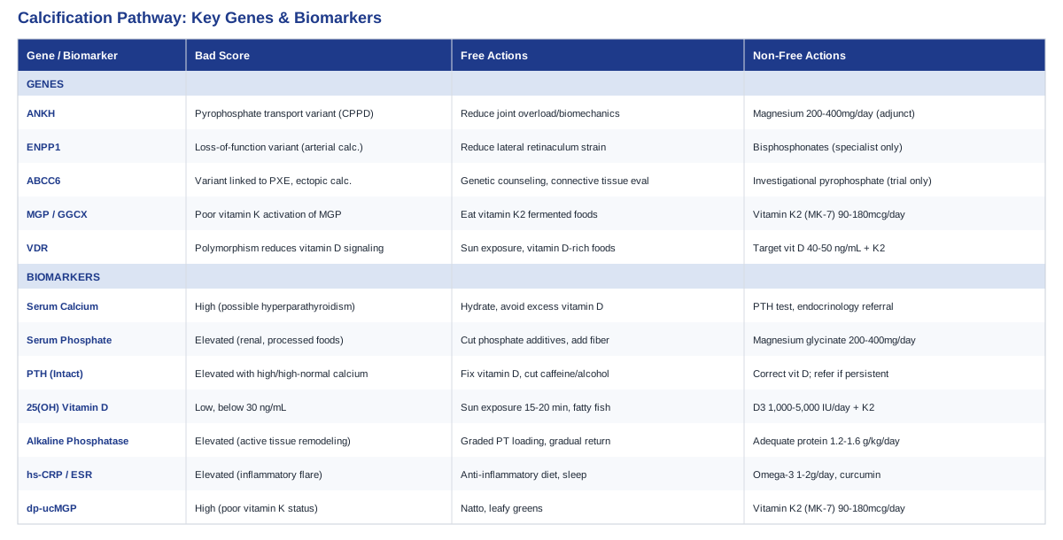
Calcific enthesitis of the lateral patellofemoral ligament is uncommon enough that most patients get a diagnosis and very little else. Underneath that one radiology finding sits a web of measurable biology: calcium and phosphate handling, parathyroid hormone, vitamin D and vitamin K status, inflammatory load, and bone turnover markers all influence whether calcium deposits stay put, grow, or slowly resorb. A smaller but real slice of cases is shaped by inherited pyrophosphate-handling genes that also drive conditions like chondrocalcinosis and pseudoxanthoma elasticum. The sections below walk through the seven biomarkers worth asking your doctor about, the five genes with genuine biological relevance, the surprising argument at the center of a well-known vitamin K book, and the hands-on techniques with the best supporting evidence for this specific part of the knee.
The 7 Biomarkers Worth Tracking for Lateral Retinaculum Calcification
Calcific enthesitis of the lateral patellofemoral ligament, first well-documented in the radiology literature as a rare but real cause of anterolateral knee pain, behaves like other forms of dystrophic soft-tissue calcification: calcium-phosphate crystals accumulate at a site of tissue stress or micro-injury, often with normal blood calcium and phosphate on standard labs (Calcific Enthesitis of Lateral Patellofemoral Ligament, soft tissue and subcutaneous calcification in connective tissue disease). That does not make lab work useless — it makes it a screening and monitoring tool rather than a single diagnostic answer. The seven biomarkers below cover the systemic factors most likely to influence whether calcium deposits form, persist, or slowly clear, ranked roughly from the most basic and affordable to the more specialized.
1. Serum Calcium (Total and Ionized)
Total and ionized calcium are the starting point for any calcification work-up because they rule out (or flag) a systemic calcium-handling problem, such as primary hyperparathyroidism, rather than a purely local, tissue-level issue. Most people with dystrophic soft-tissue calcification, including calcific enthesitis of a knee ligament, have entirely normal serum calcium — the deposit is a local phenomenon, not a sign that calcium is "too high" in the blood. Still, an elevated result changes the entire conversation and points toward a parathyroid or renal work-up instead of a purely orthopedic one.
How to Measure It
A basic metabolic panel with calcium is one of the cheapest tests in medicine, typically $10 to $30 out of pocket in the US or bundled into routine blood work covered by insurance. Ionized calcium is more accurate but requires a slightly more specialized draw and is usually $20 to $50 when ordered separately. Ask for both if a first total calcium result is borderline.
If the Score Is Off, the Plan Without Supplements
If calcium comes back high, the non-supplement plan is medical, not a lifestyle project: follow up with a PTH level and a referral to endocrinology, stay well hydrated, and avoid thiazide diuretics or excess vitamin D intake until the cause is identified. If calcium is low-normal, increasing dietary calcium through dairy, leafy greens, and fortified foods is the first-line move rather than reaching for a pill.
If the Score Is Off, the Plan With Supplements or Equipment
Supplementing calcium directly is rarely the right move for someone already forming soft-tissue calcium deposits, and doing so without a confirmed dietary deficiency can be counterproductive. If a clinician confirms a true dietary shortfall, calcium citrate 500 to 600 mg once or twice daily with food is the more bioavailable option; it should be paired with adequate vitamin K2 (see below) and reassessed with a repeat lab in 8 to 12 weeks rather than continued indefinitely. Side effects at these doses are mild (constipation, bloating) but can worsen with higher intakes.
2. Serum Phosphate
Phosphate works in tandem with calcium, and the calcium-phosphate product (the two multiplied together) is more informative for calcification risk than either value alone. Chronically elevated phosphate, often from kidney impairment or high intake of processed foods and phosphate additives, shifts the calcium-phosphate balance toward precipitation in soft tissue.
How to Measure It
Phosphate is typically included in a comprehensive metabolic panel, adding little to no extra cost ($10 to $25 standalone). It is worth requesting explicitly, since some basic panels omit it by default.
If the Score Is Off, the Plan Without Supplements
Reducing intake of phosphate-additive-heavy processed foods (packaged meats, some sodas, fast food) and increasing fiber intake to support healthy kidney and gut handling of phosphate is the first step. Regular aerobic exercise supports better renal phosphate clearance over time.
If the Score Is Off, the Plan With Supplements or Equipment
There is no supplement that "lowers" phosphate safely for someone with normal kidneys; phosphate binders are a prescription-only tool used in kidney disease under nephrology supervision, not a self-directed option. Magnesium glycinate 200 to 400 mg daily is sometimes used adjunctively because magnesium competes with calcium-phosphate crystal formation, but it is a supporting measure, not a phosphate-lowering treatment, and should be cycled off periodically (for example, five days on, two off, or reassessed every three months) to avoid GI side effects like loose stools.
3. Intact Parathyroid Hormone (PTH)
PTH is the master regulator of calcium balance, pulling calcium out of bone and increasing kidney reabsorption when levels are low, and suppressing itself when calcium is abundant. An elevated PTH alongside high or high-normal calcium points toward primary hyperparathyroidism, a condition that can independently increase soft-tissue and vascular calcification risk over years.
How to Measure It
Intact PTH is a specific blood draw, usually $50 to $150 depending on the lab and whether insurance covers it. It is best interpreted alongside same-day calcium and vitamin D, since all three interact.
If the Score Is Off, the Plan Without Supplements
Secondary hyperparathyroidism from low vitamin D is common and often corrects itself once vitamin D is optimized (see below), alongside adequate dietary calcium. Reducing excess caffeine and alcohol, both of which can modestly raise PTH, is a reasonable low-cost adjustment.
If the Score Is Off, the Plan With Supplements or Equipment
If low vitamin D is the driver, correcting it (see the vitamin D section) is the standard, evidence-based fix and usually normalizes PTH within a few months. If PTH stays elevated despite normal vitamin D and calcium, that is a referral to endocrinology for primary hyperparathyroidism work-up, not a supplement problem — self-treating a persistently high PTH with supplements is not appropriate.
4. 25-Hydroxyvitamin D
Vitamin D status affects both ends of the calcium-calcification story: too little drives secondary hyperparathyroidism and poor bone mineralization, while very high supplemental doses without adequate vitamin K2 can, in theory, push more calcium into circulation than soft tissue can safely clear. This is the biomarker most people have already had checked, but rarely with its downstream partners.
How to Measure It
25-hydroxyvitamin D is widely available, $40 to $100 out of pocket, and often covered by insurance when ordered with a relevant clinical reason. A reasonable target range discussed by longevity-focused physicians such as Peter Attia is roughly 30 to 50 ng/mL, avoiding both deficiency and unnecessarily high levels.
If the Score Is Off, the Plan Without Supplements
Regular, sensible sun exposure (roughly 15 to 20 minutes of midday skin exposure several times a week, adjusted for skin tone and climate) and dietary sources like fatty fish and fortified foods can meaningfully move levels for people who are only mildly low.
If the Score Is Off, the Plan With Supplements or Equipment
For confirmed deficiency, vitamin D3 1,000 to 2,000 IU daily is a common maintenance dose, with higher short-term repletion doses (for example 5,000 IU daily for 8 to 12 weeks) reserved for more significant deficiency and guided by repeat labs. Vitamin D3 should be paired with vitamin K2 (MK-7, 90 to 180 mcg daily) to help direct the additional calcium toward bone rather than soft tissue — this pairing is discussed at length in the book summary below. Side effects of vitamin D at appropriate doses are rare, but very high, unsupervised doses (over 10,000 IU daily for months) can raise the risk of hypercalcemia, so retesting every 3 months during correction is sensible.
5. Alkaline Phosphatase (Bone-Specific)
Alkaline phosphatase (ALP) is an enzyme central to normal mineralization, and it works in the opposite direction of the pyrophosphate pathway described in the genetics section below — where pyrophosphate inhibits calcification, ALP breaks pyrophosphate down and promotes it. Bone-specific ALP is a useful marker of local bone and connective tissue turnover, and can be modestly elevated with active healing or remodeling near a calcified enthesis.
How to Measure It
Standard ALP is included in most liver panels ($10 to $25); a bone-specific ALP isoenzyme test is more targeted and costs $60 to $120, typically ordered by a rheumatologist or endocrinologist rather than a general practitioner.
If the Score Is Off, the Plan Without Supplements
Mild elevations tied to recent injury or active tissue remodeling generally resolve as the tissue heals; graded, progressive loading of the knee under physical therapy guidance supports normal remodeling rather than either complete immobilization or continued overuse.
If the Score Is Off, the Plan With Supplements or Equipment
There is no supplement protocol to directly "lower" ALP, and attempting to suppress it is not the goal — persistently high levels warrant investigation into liver, bone, or parathyroid causes rather than self-treatment. Adequate protein intake (around 1.2 to 1.6 g per kg of body weight daily) supports the connective tissue repair process that ALP reflects.
6. High-Sensitivity CRP (or ESR)
Local calcific enthesitis is frequently accompanied by a mild inflammatory component — the perilesional edema seen on MRI in these cases reflects real tissue inflammation, even when the process is not autoimmune. Elevated hs-CRP or ESR does not diagnose the calcification itself, but it helps track whether an active inflammatory flare is present and whether it is resolving with treatment.
How to Measure It
hs-CRP costs $15 to $40 and is one of the most accessible inflammatory markers; ESR is similarly priced and often ordered alongside it. Both are widely used by physicians like Peter Attia and lipidologists such as Thomas Dayspring as general inflammatory context markers, not knee-specific tests.
If the Score Is Off, the Plan Without Supplements
An anti-inflammatory eating pattern (more oily fish, vegetables, olive oil; less ultra-processed food and added sugar), consistent sleep, and reducing training load on the affected knee during a flare are the first-line, no-cost interventions.
If the Score Is Off, the Plan With Supplements or Equipment
Omega-3 fish oil (1 to 2 g combined EPA/DHA daily) has modest, well-studied anti-inflammatory effects and is reasonable to cycle continuously with periodic reassessment every 3 months; it can mildly increase bleeding risk at high doses, so it is worth flagging before any injection or procedure. Curcumin with piperine (500 to 1,000 mg daily) is a second-line adjunct some patients use, cycling 8 weeks on and 2 weeks off, though evidence specific to calcific enthesitis is limited and it should not replace medical evaluation of a genuine flare.
7. Desphospho-Uncarboxylated Matrix Gla Protein (dp-ucMGP)
Matrix Gla protein, once activated by vitamin K, is one of the most powerful natural inhibitors of soft-tissue calcification identified so far (circulating MGP is associated with coronary calcification and vitamin K status in healthy women, inactive circulating MGP as a marker of vitamin K status and calcification risk). The inactive form, dp-ucMGP, rises when vitamin K status is poor, and functions as an indirect but meaningful marker of how well your body is protecting soft tissue, including tendons and ligaments, from unwanted calcium deposition.
How to Measure It
This is the most specialized test on this list, typically $80 to $150, offered through specialty and research-oriented labs rather than routine bloodwork, and not yet a standard clinical order in most primary care settings. It is worth requesting specifically if a physician is open to investigating vitamin K status.
If the Score Is Off, the Plan Without Supplements
Increasing dietary vitamin K2 through fermented foods (natto being the richest source, along with some aged cheeses) and vitamin K1 through leafy greens is the first, no-cost step, though K1 converts to K2 in the body relatively inefficiently.
If the Score Is Off, the Plan With Supplements or Equipment
Vitamin K2 as MK-7, 90 to 180 mcg daily, is the dose most commonly used in the studies referenced above and in the book discussed in the next section; it is generally well tolerated, with the main caution being an interaction with warfarin and other vitamin K antagonist blood thinners, which requires physician coordination before starting. This is typically taken continuously rather than cycled, with reassessment of dp-ucMGP after 3 to 6 months if the test is repeated.
Taken together, these seven markers give a far more complete picture than the MRI report alone. A calcium-phosphate-PTH-vitamin D cluster that looks completely normal shifts the likely explanation toward local, mechanical, dystrophic calcification tied to old injury or chronic friction at the retinaculum; an abnormal cluster reframes the knee finding as one visible sign of a more systemic calcium-handling issue worth investigating further.
What the Genetics Suggest, and What Can Be Done About It
Most cases of calcific enthesitis at the lateral retinaculum are not primarily genetic — they follow local tissue stress, prior injury, or age-related enthesis changes. But a real subset of soft-tissue and periarticular calcification, especially when it recurs, appears at an unusually young age, or shows up alongside chondrocalcinosis in other joints, tracks back to inherited pyrophosphate or calcification-inhibitor pathways. The evidence for each gene below ranges from well-established (in related calcification diseases) to exploratory when applied specifically to a knee ligament finding — that distinction is called out for each one.
ANKH
ANKH encodes a transmembrane channel that transports inorganic pyrophosphate out of cells; pyrophosphate is one of the body's main natural brakes on calcium crystal formation. Mutations in ANKH are a well-documented cause of familial calcium pyrophosphate deposition disease (chondrocalcinosis), where either too little or, in some mutation types, too much pyrophosphate transport disrupts the normal balance and predisposes to crystal deposition in cartilage and periarticular tissue (autosomal dominant familial CPPD is caused by mutation in ANKH, narrative review of chondrocalcinosis). This evidence is strong for chondrocalcinosis generally; its direct link to isolated lateral retinaculum calcification specifically has not been studied, so consider this plausible rather than proven for this exact presentation.
If the Gene Variant Is Unfavorable, the Plan Without Supplements
There is no lifestyle change that corrects an ANKH variant itself, but reducing joint overload (addressing biomechanics, footwear, and training volume) can lower the mechanical trigger that turns a pyrophosphate-handling tendency into an actual calcific deposit.
If the Gene Variant Is Unfavorable, the Plan With Supplements or Equipment
Magnesium (200 to 400 mg daily as glycinate or citrate) is sometimes used as a general crystal-formation inhibitor in CPPD-related conditions, cycled with periodic breaks and reassessed with a treating rheumatologist; it is a supportive measure rather than a targeted fix for the gene itself, and there is no over-the-counter supplement proven to correct ANKH-driven pyrophosphate handling.
ENPP1
ENPP1 generates extracellular pyrophosphate from ATP, working alongside ANKH in the same protective pathway. Severe loss-of-function ENPP1 mutations cause generalized arterial calcification of infancy, a rare and serious pediatric disease, underscoring just how powerful this pathway is at preventing calcification when it works correctly (Generalized Arterial Calcification of Infancy, GeneReviews). Milder, common ENPP1 variants have not been specifically studied in adult periarticular or ligamentous calcification, so this remains a mechanistic, plausible link rather than direct evidence for the knee.
If the Gene Variant Is Unfavorable, the Plan Without Supplements
Since ENPP1 function feeds the same pyrophosphate pool as ANKH, the same mechanical-load-reduction logic applies: addressing any anatomical driver of lateral retinaculum strain (patellar tracking, tight lateral structures) reduces the local trigger for calcification independent of the underlying gene.
If the Gene Variant Is Unfavorable, the Plan With Supplements or Equipment
No supplement restores ENPP1 enzyme activity. Bisphosphonates, which mimic pyrophosphate's calcification-inhibiting structure, are used medically for severe ENPP1-related disease but are prescription drugs used under specialist care for serious systemic calcification, not an appropriate self-directed option for an isolated knee finding.
ABCC6
ABCC6 is expressed mainly in the liver and helps export ATP that is subsequently converted into pyrophosphate, again feeding the same protective system. Mutations cause pseudoxanthoma elasticum, a connective tissue disease marked by ectopic calcification of skin, eyes, and blood vessels (ABCC6, pyrophosphate, and ectopic calcification: therapeutic solutions). People with pseudoxanthoma elasticum can develop periarticular calcification, which makes ABCC6 biologically relevant to this topic, but again there is no dedicated study connecting common ABCC6 variants to isolated lateral retinaculum calcification.
If the Gene Variant Is Unfavorable, the Plan Without Supplements
For anyone with a personal or family history of pseudoxanthoma elasticum-type skin or eye findings alongside knee calcification, the useful non-supplement step is a referral for genetic counseling and a broader connective tissue evaluation, not a home remedy.
If the Gene Variant Is Unfavorable, the Plan With Supplements or Equipment
Research into oral pyrophosphate and etidronate supplementation for ABCC6-related disease is ongoing but investigational; this is not an established or self-directed supplement strategy and should only be pursued through a clinical trial or specialist, not general over-the-counter use.
MGP / GGCX (Matrix Gla Protein Pathway)
The MGP gene itself is rarely mutated, but the vitamin K-dependent activation of the MGP protein (carried out by the enzyme GGCX) is the most directly modifiable part of this entire pathway, and it is the mechanism explored in depth in the next section. Even with normal MGP genetics, poor vitamin K status leaves MGP inactive and unable to do its job of keeping calcium out of soft tissue.
If the Score Is Bad, the Plan Without Supplements
Regular intake of vitamin K2-rich fermented foods, discussed above, is the direct lever here regardless of genetic status.
If the Score Is Bad, the Plan With Supplements or Equipment
Vitamin K2 (MK-7) 90 to 180 mcg daily, taken continuously with meals containing some fat for absorption, is the standard approach; anyone on warfarin must coordinate this with their prescriber first, since it directly opposes how that medication works.
VDR (Vitamin D Receptor)
Common VDR polymorphisms (BsmI, ApaI, TaqI, FokI) modestly affect how efficiently vitamin D signaling occurs at the tissue level, and have been studied in relation to osteoarthritis and connective tissue metabolism (association of vitamin D receptor, COL1A1, and estrogen receptor genes with osteoarthritis). This is early-stage, population-level evidence rather than a strong individual predictive tool, but it offers one more reason someone might need higher vitamin D intake to reach the same functional effect as someone without the variant.
If the Score Is Bad, the Plan Without Supplements
Consistent, moderate sun exposure and vitamin D-rich foods remain useful regardless of receptor efficiency, since more circulating substrate partially compensates for a less efficient receptor.
If the Score Is Bad, the Plan With Supplements or Equipment
For someone with a known VDR variant and low-normal vitamin D levels, targeting the upper-middle of the normal range (40 to 50 ng/mL, per the earlier biomarker section) rather than the bare minimum is a reasonable, low-risk adjustment, again paired with vitamin K2 and rechecked periodically rather than escalated indefinitely.
The Book That Reframes How Most People Think About Calcium
Vitamin K2 and the Calcium Paradox, by Dr. Kate Rheaume-Bleue, is the most widely read popular explanation of exactly the mechanism running through the biomarker and gene sections above: that calcium supplementation without adequate vitamin K2 can push calcium toward the wrong tissues. It draws on the same MGP and vitamin K research already cited here, and its central argument directly challenges a habit most doctors still recommend by default — take calcium and vitamin D for bone health, without asking where in the body that calcium actually ends up. Below are the ten most useful points from the book for anyone dealing with soft-tissue or ligamentous calcification.
1. Calcium Needs a Traffic Director
The book's central claim is that calcium alone does not know where to go in the body; it is vitamin K2, through activation of proteins like MGP and osteocalcin, that directs calcium into bone and away from arteries, cartilage, and ligaments. Without that direction, calcium supplementation can theoretically make soft-tissue calcification worse rather than better.
2. Osteoporosis and Soft-Tissue Calcification Can Coexist
Rheaume-Bleue describes the "calcium paradox" of the book's title: the same person can have weak, undermineralized bones and calcium deposits building up in soft tissue simultaneously, because the problem is not too little or too much calcium overall, it is misallocation.
3. Vitamin K1 and K2 Are Not Interchangeable
K1, abundant in leafy greens, is mostly directed toward liver-based clotting functions, while K2 is far more effective at reaching bone, blood vessels, and connective tissue. This is the practical reason the supplement recommendations throughout this article specify K2, not K1.4. MK-7 Has the Longest Half-Life of the K2 Forms
Among K2 subtypes, MK-7 (found in natto and available as a supplement) stays active in the bloodstream far longer than MK-4, making once-daily dosing realistic and giving it the strongest research base for once-daily supplementation protocols like the ones described above.
5. Uncarboxylated MGP Is a Measurable Warning Sign
The book popularized the idea, now supported in the peer-reviewed literature cited earlier in this article, that inactive (uncarboxylated) MGP circulating in blood is a usable, testable proxy for whether soft tissue is protected from calcification — directly matching biomarker seven above.
6. Standard Vitamin D Advice Is Incomplete Without K2
One of the book's more provocative arguments is that increasing vitamin D intake without also ensuring adequate K2 may increase calcium absorption faster than the body can safely direct it, reinforcing why this article pairs the two rather than treating vitamin D as a stand-alone fix.
7. Fermented Foods Are an Underused K2 Source
Natto, certain aged cheeses (particularly Gouda and Brie), and some fermented foods carry meaningful K2 content that most Western diets simply do not include, which the book argues explains part of the population-level gap in K2 status.
8. Antibiotics Can Disrupt K2-Producing Gut Bacteria
Some gut bacteria contribute to endogenous K2 production, and the book raises the point that repeated courses of antibiotics may reduce this contribution over time, which is a reasonable, if harder to quantify, additional factor in overall K2 status.
9. The Effect Is Slow, Not Immediate
The book is careful to frame vitamin K2's role as a years-long influence on where calcium accumulates, not a fast-acting treatment — a useful, honest caveat that applies directly to expectations around any existing calcific deposit at the retinaculum.
10. This Is a Prevention-Oriented Framework, Not a Cure Claim
Even within the book, the argument is about shifting long-term risk and supporting the body's own calcium-directing systems, not reversing an already-formed calcific deposit — a distinction worth holding onto given how this general area of health information tends to get oversold online.
Complementary Approaches Worth Considering
Alongside lab work and any medical treatment plan, a small number of complementary approaches have genuine, condition-relevant human evidence, either specifically at the lateral retinaculum or in closely related knee conditions. These are adjuncts to standard care, not substitutes for it, and the evidence quality varies by technique.
Deep Friction Massage of the Lateral Retinaculum
Deep transverse friction massage is a manual therapy technique aimed directly at fibrous, thickened, or calcified soft tissue, applied across the fibers rather than along them, with the goal of improving local tissue mobility and reducing pain at an enthesis or ligament. It is directly relevant here because it targets the exact structure discussed throughout this article rather than a nearby or generic knee region.
A randomized controlled trial on patellofemoral pain syndrome specifically included deep friction massage of the lateral retinaculum, combined with iliotibial band release and patellar mobilization, as part of a manual therapy protocol added to standard hip and knee exercises, and found added benefit over exercise alone (efficacy of adding manual therapy to hip and knee exercises in patellofemoral pain syndrome). A separate review of deep transverse friction massage for tendinitis more broadly found modest supportive evidence, with the caveat that trial quality across this technique is mixed (deep transverse friction massage for treating tendinitis).
In practice, this technique should be performed by a licensed physical therapist familiar with the knee, typically for one to two minutes per session directly over the affected retinaculum, two to three times weekly for four to six weeks, with a break if pain increases rather than decreases after the first few sessions; it is not appropriate as a self-administered technique on an actively inflamed or acutely painful area without professional guidance first.
EMG Biofeedback for Quadriceps Retraining
Electromyographic (EMG) biofeedback uses surface sensors to show real-time muscle activity on a screen, most often used at the knee to retrain the balance between the vastus medialis oblique and the lateral quadriceps and iliotibial structures that place ongoing tension on the lateral retinaculum. Since abnormal lateral pull on the patella is a plausible mechanical contributor to chronic strain and calcification at the lateral retinaculum, correcting that muscular imbalance is a reasonable, targeted goal.
A randomized controlled trial in young male athletes with patellofemoral pain syndrome combined patellar taping with EMG-biofeedback-guided isometric quadriceps training at multiple knee angles and found measurable improvements in quadriceps strength and function compared with sham taping alone (efficacy of patellar taping and EMG biofeedback training in patellofemoral pain syndrome).
A realistic protocol involves two to three sessions weekly for six weeks with a physical therapist or athletic trainer using a portable EMG unit, focusing on isometric quadriceps activation at 30, 60, and 90 degrees of knee flexion; equipment costs for a home EMG biofeedback unit range from roughly $100 to $400, and side effects are essentially limited to mild, transient muscle soreness from the exercise component itself.
Low-Level Laser Therapy (Photobiomodulation)
Low-level laser therapy (LLLT), also called photobiomodulation, uses specific wavelengths of light to stimulate cellular activity in soft tissue, and has been studied specifically in calcific tendinopathy, a closely related process to the calcific enthesitis discussed throughout this article, even though the affected structure in most trials is the rotator cuff rather than the knee.
A 2024 prospective randomized controlled trial in patients with confirmed calcific rotator cuff tendinopathy found that adding LLLT to a home exercise program, delivered five sessions weekly for three weeks, outperformed the home exercise program alone for pain, disability, and even reduced the size and severity of the calcific deposits on imaging (effects of low-level laser therapy on symptomatic calcific rotator cuff tendinopathy). Broader systematic reviews of LLLT in shoulder tendinopathy show more mixed results, so this should be read as promising rather than settled (efficacy of low-level laser therapy for shoulder tendinopathy: a systematic review and meta-analysis).
Direct evidence for LLLT at the lateral retinaculum specifically does not yet exist, so applying it here is an extrapolation from a mechanistically similar calcific tendon condition, best done under a physical therapist's guidance using a clinical-grade device, typically three sessions weekly for three to four weeks; side effects are minimal, mainly mild transient warmth at the treatment site, though clinical-grade sessions typically cost $30 to $75 each if not covered by insurance.
Tai Chi for Overall Knee Load Management
Tai chi is a low-impact, weight-bearing movement practice that combines slow, controlled joint loading with balance and proprioceptive training, both of which are relevant to a knee condition where uneven mechanical load on the lateral structures is a plausible contributing factor. It is not a targeted treatment for a calcified ligament, but it is one of the better-studied movement-based interventions for chronic knee conditions generally.
A large randomized comparative-effectiveness trial found that a 12-week tai chi program produced knee osteoarthritis symptom improvements equivalent to standard physical therapy at 52 weeks (comparative effectiveness of tai chi versus physical therapy for knee osteoarthritis). While this trial was conducted in osteoarthritis rather than calcific enthesitis specifically, the shared mechanism, gradual, controlled loading that improves knee mechanics and reduces uneven strain, makes it a reasonable general adjunct.
A practical starting point is a beginner tai chi class twice weekly for 12 weeks, ideally supervised initially to ensure knee alignment is appropriate for an irritated lateral structure; there is essentially no equipment cost, and the main caution is stopping or modifying any movement that sharply increases lateral knee pain rather than pushing through it.
Conclusion
Lateral retinaculum calcification is usually a local, mechanical finding rather than a sign of a body-wide calcium problem, but it sits at the intersection of measurable biology worth checking: calcium, phosphate, PTH, vitamin D, alkaline phosphatase, inflammatory markers, and vitamin K status through MGP all shape whether calcium deposits form, stay put, or slowly resolve. A smaller number of cases trace back to inherited pyrophosphate-handling genes like ANKH, ENPP1, and ABCC6, which is worth knowing if there is a personal or family history of chondrocalcinosis or connective tissue calcification elsewhere in the body. None of this replaces a proper orthopedic or rheumatologic evaluation, but it gives you a much clearer set of questions to bring to that appointment.
The most useful next step is practical rather than dramatic: get the core biomarker panel checked, ask specifically about vitamin D and vitamin K2 status if you have not already, and discuss with a physical therapist whether targeted manual therapy or muscle retraining around the knee is appropriate for your specific case. Tracking these numbers over time, rather than checking them once and moving on, is what turns this information into an actual decision-making tool.
Musculoskeletal: Bone Conditions Tendon & Ligament Conditions