This article contains AI generated content.
Lateral Retinaculum Tear: 4 Genes and 7 Biomarkers to Track
Introduction
A lateral retinaculum tear rarely shows up alone. It usually follows a patellar dislocation, a hard pivot on a loaded knee, or a lateral release procedure that went further than the tissue could tolerate — and once it happens, the advice you get tends to be the same three sentences everyone gets: rest, ice, physical therapy, give it time. That advice isn't wrong, but it treats every knee as if it were built from identical tissue, healing at an identical rate, with an identical capacity to rebuild collagen under load.
It isn't. Two people with the same MRI report can heal at very different speeds, and a meaningful part of that difference comes down to things a standard orthopedic visit never measures: how efficiently your body cross-links collagen, how much background inflammation is competing with repair, whether your vitamin D and copper status are supporting or slowing the rebuilding process, and whether your genetic makeup nudges your connective tissue toward being more elastic — or more injury-prone — in the first place.
This article goes past the generic recovery script. It looks at the biomarkers that reflect how well your body is currently equipped to repair fibrous connective tissue like the lateral retinaculum, and at the genetic variants that researchers in sports genomics — the kind of work popularized by geneticists like Ali Torkamani and health-optimization figures like Gary Brecka — have linked to soft tissue injury risk. Neither list is a diagnosis, and neither replaces an orthopedic surgeon or a physical therapist who has actually examined your knee.
What better information does give you is leverage. Knowing which biomarkers are working against your healing lets you correct what's correctable, set a realistic recovery timeline, and stop guessing. This article walks through the most useful biomarkers to track first, then a shorter look at the genetic side of the story, a set of takeaways from a book that reframes how injury prevention should actually be trained, and a review of complementary approaches with real evidence behind them for knee soft tissue problems.
Summary
Before diving in, here's what this article actually covers and why it's worth the full read. The core idea is simple: a lateral retinaculum tear heals inside a biochemical environment, and that environment is measurable. Seven blood biomarkers — vitamin D, hs-CRP, vitamin C, homocysteine, HbA1c/fasting insulin, copper/ceruloplasmin, and estrogen/relaxin — each tell a different part of the story of whether your body is currently primed to rebuild collagen efficiently or working against itself.
Beyond the blood work, four genes (COL5A1, COL1A1, GDF5, and MTHFR) shape how resilient your connective tissue is at baseline, and while you can't edit your genes, you can build a plan around what they predict. Further down, you'll find ten practical takeaways from Peter Attia's Outlive on why stability training — not just strength — is the piece most rehab plans skip, plus a review of which complementary therapies (biofeedback, massage, laser therapy, tai chi) actually have human trial data behind them for knee soft tissue issues, and which don't. If you've been told to "just wait it out," this is the more precise version of what to actually do while you wait.
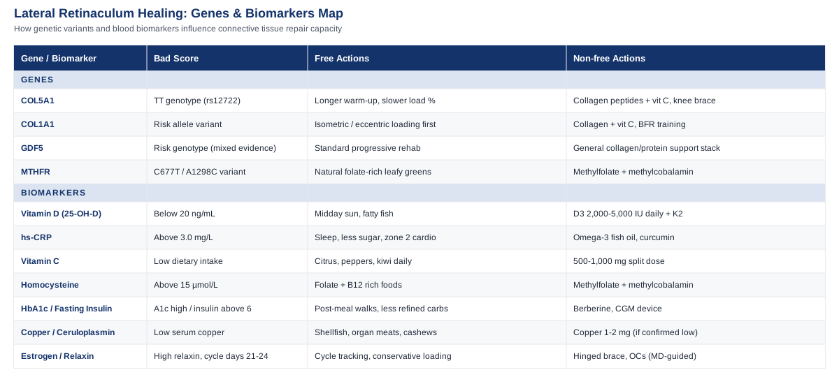
Biomarkers That Reveal Your Healing Capacity
The lateral retinaculum is dense fibrous connective tissue, structurally closer to a ligament than to muscle. Its repair depends almost entirely on collagen synthesis, cross-linking, and remodeling — processes that are directly sensitive to nutrient status, inflammation, and hormonal signaling. That's why the biomarker approach tends to be the more actionable of the two strategies in this article: unlike a gene variant, most of these numbers can move within weeks to months, and the interventions are well studied outside of this specific injury, even where retinaculum-specific trials don't exist.
The seven biomarkers below aren't an exhaustive lab panel — they're the ones with the clearest mechanistic link to fibrous tissue repair, prioritized the way clinicians like Peter Attia, Thomas Dayspring, and Allan Sniderman prioritize markers generally: start with what's cheap, common, and actionable, and only escalate to specialized testing when the basic picture doesn't explain your symptoms.
1. 25-Hydroxyvitamin D
Vitamin D status is one of the strongest predictors of postoperative and post-injury tendon and ligament outcomes currently in the literature. A scoping review of vitamin D's role in postoperative tendon healing found that deficiency was consistently associated with delayed healing, higher retear rates, and worse functional recovery, largely in rotator cuff and other soft tissue repairs (The Role of Vitamin D in Postoperative Tendon Healing: A Scoping Review). Vitamin D also modulates the local immune response during soft tissue repair, which matters directly for a retinaculum tear healing in the joint capsule's inflammatory environment (Vitamin D and the immunomodulation of rotator cuff injury).
How to measure it
A basic serum 25-OH-D blood draw, available through any primary care visit or direct-to-consumer lab, costing roughly $40 to $90 without insurance. Target is generally considered 30–50 ng/mL for musculoskeletal health; below 20 ng/mL is deficient.If the score is bad, the plan without supplements
Fifteen to twenty minutes of midday sun exposure on bare arms and legs, three to five times per week, is the most reliable non-supplement lever, though it's weather- and latitude-dependent and slower to correct a significant deficiency. Fatty fish (salmon, sardines, mackerel) two to three times weekly adds a modest amount.If the score is bad, the plan with supplements or equipment
Vitamin D3 at 2,000–5,000 IU daily with a fat-containing meal is the standard correction dose for mild-to-moderate deficiency, re-tested at 8–12 weeks. Severe deficiency (under 12 ng/mL) is sometimes corrected with a supervised loading protocol (e.g., 50,000 IU weekly for 6–8 weeks) under a physician's guidance rather than self-directed. Pair with 1–2 mg of vitamin K2 daily, since D increases calcium absorption and K2 helps direct that calcium appropriately. Side effects are rare at these doses but include hypercalcemia at chronic high intake (above 10,000 IU/day sustained) — this is why re-testing rather than indefinite high dosing matters.2. High-Sensitivity C-Reactive Protein (hs-CRP)
hs-CRP is a general marker of systemic inflammation, and while acute local inflammation is a normal and necessary part of the first days of retinaculum healing, chronically elevated systemic hs-CRP suggests your body's inflammatory resources are already committed elsewhere — often to metabolic dysfunction, poor sleep, or an unrelated low-grade issue — which can blunt the resolution phase of tissue repair. Emerging research is reframing CRP as more than a passive bystander in tissue injury, showing it actively participates in the repair cascade itself (Redefining CRP in tissue injury and repair: more than an acute pro-inflammatory mediator).
How to measure it
A standard hs-CRP blood test, $15–$40, often bundled into routine metabolic panels. Optimal is under 1.0 mg/L; above 3.0 mg/L suggests meaningful background inflammation worth investigating.If the score is bad, the plan without supplements
Prioritize sleep consistency (7–9 hours, stable wake time) for 4–6 weeks before re-testing — sleep debt is one of the most reliable ways to raise CRP independent of injury. Reduce ultra-processed food and added sugar intake, and add two to three weekly sessions of zone 2 cardio (an easy pace where you can still hold a conversation), which lowers inflammatory markers over 8–12 weeks in most people.If the score is bad, the plan with supplements or equipment
Omega-3 fish oil (2–3 g combined EPA/DHA daily, taken with food to reduce GI upset) is the best-studied supplement for lowering CRP, typically re-tested at 12 weeks. Curcumin (500–1000 mg with piperine, cycled 8 weeks on, 2 weeks off) has modest supporting evidence but interacts with blood thinners and can cause GI discomfort at higher doses — avoid combining with anticoagulants without medical guidance.3. Plasma Vitamin C
Vitamin C is a required cofactor for prolyl and lysyl hydroxylase, the two enzymes that stabilize collagen's triple helix. Without adequate vitamin C, the body still produces collagen, but it's structurally weaker and less tensile — this is the same biochemical pathway that fails catastrophically in scurvy (Biochemistry, Collagen Synthesis — StatPearls). For a tear that depends entirely on rebuilding collagen fiber strength, this is a foundational marker, not an optional one.
How to measure it
Plasma ascorbate testing isn't part of routine panels and usually requires a specialty or functional-medicine lab, running $50–$100. Because it's less accessible, many people substitute a dietary-intake review with their clinician rather than testing directly.If the score is bad, the plan without supplements
One cup of red bell pepper, kiwi, or citrus fruit provides well over the daily requirement; two to three servings of vitamin C-rich produce daily is generally enough to correct a mild insufficiency within 2–3 weeks.If the score is bad, the plan with supplements or equipment
500–1,000 mg vitamin C daily, split into two doses to improve absorption, taken during the active healing window (typically the first 6–8 weeks post-injury) rather than indefinitely. Doses above 2,000 mg/day can cause GI upset and diarrhea and offer no added collagen benefit — this is not a "more is better" nutrient.4. Homocysteine
Homocysteine is a byproduct of methionine metabolism, and chronically elevated levels are associated with impaired collagen cross-linking and reduced connective tissue integrity, largely because the same B-vitamin pathways (B6, B12, folate) that clear homocysteine also support healthy collagen maturation. It's also one of the more actionable markers here, since elevated homocysteine is often a straightforward reflection of B-vitamin insufficiency.
How to measure it
A fasting blood draw, $40–$80. Optimal is generally under 8–9 µmol/L; above 15 µmol/L is considered elevated and worth addressing regardless of injury status.If the score is bad, the plan without supplements
Increase dietary folate (leafy greens, lentils) and B12 (eggs, fish, poultry) for 8–12 weeks before re-testing; this alone corrects mild elevations in people without an absorption or genetic issue.If the score is bad, the plan with supplements or equipment
A B-complex with active forms — methylfolate (400–800 mcg) and methylcobalamin (500–1000 mcg) rather than folic acid and cyanocobalamin — daily for 8–12 weeks, then re-test. This is particularly relevant if genetic testing (see the MTHFR section below) shows reduced methylation efficiency. Side effects are minimal, though very high B6 (above 100 mg/day, sustained for months) has been linked to peripheral neuropathy, so stick to standard complex doses rather than megadosing.5. HbA1c and Fasting Insulin
Chronically elevated blood glucose accelerates the formation of advanced glycation end-products (AGEs) — sugar molecules that bind to collagen and stiffen it through non-enzymatic cross-linking. Multiple biomechanical studies show AGE accumulation reduces collagen fiber sliding and impairs the tissue's ability to absorb load and remodel properly during repair (Advanced glycation end-products diminish tendon collagen fiber sliding). This matters for anyone with prediabetes or insulin resistance recovering from a retinaculum tear — the tissue you're trying to rebuild is being glycated in the background.
How to measure it
HbA1c ($15–$30) and fasting insulin ($30–$50) are both standard lab tests. Target HbA1c under 5.5%; fasting insulin ideally under 6 µIU/mL.If the score is bad, the plan without supplements
A 10–15 minute walk after meals reliably blunts post-meal glucose spikes and, done consistently for 8–12 weeks, measurably improves HbA1c in people with mild elevations. Reducing refined carbohydrate intake and prioritizing protein and fiber at the start of meals has a similar effect.If the score is bad, the plan with supplements or equipment
Berberine (500 mg two to three times daily with meals, cycled 3 months on, 1 month off) has trial support comparable to metformin for improving insulin sensitivity, but can cause GI upset and should not be combined with other glucose-lowering medication without medical supervision. A continuous glucose monitor (roughly $50–$100/month) is the equipment option — useful for identifying which specific foods spike your glucose rather than guessing.6. Copper and Ceruloplasmin
Copper is the required cofactor for lysyl oxidase, the enzyme that cross-links collagen and elastin fibers into mature, load-bearing tissue. This step happens after the vitamin-C-dependent hydroxylation step above — without adequate copper, newly synthesized collagen fails to mature into strong fibers, regardless of how much raw collagen the body is producing.
How to measure it
Serum copper and ceruloplasmin, typically ordered together, $60–$100. True dietary copper deficiency is uncommon in people eating a varied diet, but it's worth checking in anyone with a restrictive diet, prior bariatric surgery, or high zinc supplement intake (zinc competes with copper for absorption).If the score is bad, the plan without supplements
Shellfish, organ meats, cashews, and dark chocolate are the most concentrated dietary sources; adding two to three servings weekly typically corrects a mild deficiency within 6–8 weeks.If the score is bad, the plan with supplements or equipment
Copper supplementation (1–2 mg daily) only if a deficiency is confirmed by blood work — copper toxicity from unsupervised supplementation is a real risk and can cause liver damage, so this is not a marker to self-treat without a test confirming low levels first. If you're taking zinc for another reason, keep the zinc:copper ratio in mind and check with your clinician about co-supplementing a small amount of copper.7. Estrogen and Relaxin (for female athletes and active women)
This one is specific but important: estrogen and the hormone relaxin both act on knee ligament and retinaculum tissue, increasing laxity and reducing collagen and type I/III expression during specific windows of the menstrual cycle. Relaxin receptors are present on female connective tissue but not male, and higher serum relaxin concentrations have been associated with more than a fourfold increase in ACL injury risk, with peak risk occurring around cycle days 21–24 (Menstrual Cycle Hormone Relaxin and ACL Injuries in Female Athletes: A Systematic Review). While this evidence base centers on the ACL, the same hormonal mechanism plausibly affects other knee-stabilizing connective tissue, including the retinaculum, given the shared collagen biology.
How to measure it
A hormone panel including estradiol and, where available, relaxin, $80–$150, ideally timed to a known point in the menstrual cycle for interpretability.If the score is bad, the plan without supplements
This marker isn't about "fixing" a hormone level — it's about timing. Tracking your cycle and being more conservative with high-load, high-pivot activity during the late follicular/ovulatory window (roughly days 10–16) and again around days 21–24 reduces exposure during the highest-laxity windows.If the score is bad, the plan with supplements or equipment
There's no supplement that safely lowers relaxin. Oral contraceptives have been shown to reduce serum relaxin concentration and may offer a protective effect, but that's a decision to make with a physician based on broader health context, not an injury-recovery supplement choice. The more realistic equipment-based option is a hinged knee brace during the highest-risk cycle windows for anyone returning to pivoting sports post-injury.The thread connecting all seven of these markers is that connective tissue repair is not a single pathway — it's a chain, and a weak link anywhere along vitamin D, inflammation, vitamin C, homocysteine, glycemic control, copper, or hormonal timing can slow the whole process down. Testing narrows down which link is actually weak in your case instead of guessing.
What Your Genes May Be Telling You About Connective Tissue Resilience
Biomarkers describe your current internal environment; genetics describes the hardware that environment operates on. Sports genomics research — the kind of population-level analysis Ali Torkamani has written about extensively, and the kind of practical gene-panel interpretation Gary Brecka has popularized — has identified a handful of variants tied to connective tissue strength and soft tissue injury risk. The evidence here is generally weaker than the biomarker section above: most studies are modest in size, population-specific, and describe relative risk shifts rather than deterministic outcomes. Still, for a tear involving fibrous connective tissue like the lateral retinaculum, the genes below are worth knowing.
COL5A1
COL5A1 encodes a component of type V collagen, which regulates the diameter and structure of type I collagen fibrils — the main structural collagen in ligaments, tendons, and retinacular tissue. A meta-analysis across multiple studies found the rs12722 and rs13946 variants of COL5A1 are associated with altered risk of tendon and ligament injury, with certain genotypes linked to reduced risk and others, including the TT genotype at rs12722, linked to higher susceptibility to Achilles tendon and ACL injuries (Association of COL5A1 gene polymorphisms and risk of tendon-ligament injuries among Caucasians: a meta-analysis). A separate study in skiers found specific COL5A1 variants were associated with a reduced risk of ACL injury (The Variants Within the COL5A1 Gene are Associated with Reduced Risk of Anterior Cruciate Ligament Injury in Skiers), suggesting the effect runs in both directions depending on which allele you carry.
If the gene is bad, the plan without supplements
Since a higher-risk COL5A1 genotype is associated with stiffer, less compliant connective tissue, the most useful non-supplement response is extending your warm-up and load-progression timelines rather than following generic rehab week counts. Add 5–10 minutes of dynamic mobility work before loaded knee activity, and increase training load in smaller weekly increments (aim for under 10% week-over-week) rather than the more aggressive progressions that lower-risk genotypes might tolerate.If the score is bad, the plan with supplements or equipment
Collagen peptides (10–15 g daily, taken with 50 mg vitamin C about 30–60 minutes before loading exercise) have modest RCT support for improving tendon and ligament collagen synthesis markers, best cycled during active rehab phases rather than used indefinitely. A hinged knee brace during return-to-pivot-sport activity is a reasonable equipment-based hedge for a known higher-risk genotype. There are no significant side effects to collagen peptide supplementation beyond mild GI upset in some people at higher doses.COL1A1
COL1A1 encodes the major chain of type I collagen, the dominant structural protein in retinacular and ligamentous tissue. Variants in this gene have been studied across multiple sports-injury contexts, including ACL rupture and stress fracture risk, though the associations are less consistently replicated than COL5A1's (The Role of COL1A1, COL5A1, ACTN3, MMP3, and GDF5 Gene Variants in Common Sports Injuries).
If the gene is bad, the plan without supplements
Focus rehab on progressive isometric and eccentric loading rather than rushing to dynamic, high-impact drills — eccentric loading protocols are the best-supported non-supplement way to stimulate type I collagen remodeling in fibrous connective tissue generally.If the score is bad, the plan with supplements or equipment
The same collagen-peptide-plus-vitamin-C protocol described above applies here, since both genes converge on type I collagen quality. Blood flow restriction (BFR) training equipment, used under supervision during early-stage rehab, allows lower-load strengthening that's gentler on healing collagen while still stimulating adaptation — a reasonable option specifically for higher-genetic-risk individuals who want to load tissue earlier without overloading it.GDF5
GDF5 encodes a growth factor in the TGF-beta family involved in tendon and ligament development and repair signaling. The evidence for GDF5's role in ACL and connective tissue injury risk is genuinely mixed: one study in a Chinese population linked a specific GDF5 genotype to increased ACL rupture risk, theorized to work through reduced GDF5 protein levels disrupting ligament homeostasis, while a separate case-control study of 126 ACL-injured patients versus 214 controls found the commonly studied rs143383 variant showed no association with ACL rupture risk at all (The GDF5 gene and anterior cruciate ligament rupture). This is a case where it's worth being explicit: the human evidence here is preliminary and inconsistent, and no strong practical conclusion should be drawn from a GDF5 result alone.
If the gene is bad, the plan without supplements
Given the inconsistent evidence, the most defensible response to a higher-risk GDF5 result is simply not over-indexing on it — keep to general sound rehab principles (progressive loading, adequate rest between sessions) rather than building a specific protocol around this one gene.If the score is bad, the plan with supplements or equipment
There's no supplement shown to specifically compensate for a GDF5 variant. If you want to hedge anyway, the general tissue-support stack (vitamin C, collagen peptides, adequate protein intake of roughly 1.6 g/kg bodyweight daily) is reasonable, since it supports connective tissue repair broadly regardless of which specific gene is driving your risk profile.MTHFR
MTHFR encodes the enzyme responsible for converting folate into its active, usable form, and reduced-function variants (commonly C677T and A1298C) are a major driver of elevated homocysteine — the same biomarker discussed above that impairs collagen cross-linking. This is one of the genes Gary Brecka discusses most often in his functional-genomics work, precisely because it's both common (up to 40% of some populations carry at least one reduced-function allele) and directly actionable through the biomarker it produces.
If the gene is bad, the plan without supplements
Increase intake of naturally folate-rich foods (leafy greens, lentils, asparagus) rather than relying on folic-acid-fortified processed foods, since reduced-function MTHFR variants convert folic acid less efficiently than natural folate.If the score is bad, the plan with supplements or equipment
Methylfolate and methylcobalamin, as described in the homocysteine biomarker section, are the direct correction — this is the clearest case in this article where a gene result and a blood marker point to the exact same fix. Re-test homocysteine at 8–12 weeks rather than assuming the supplement is working; standard doses have essentially no side effect profile, but there's no benefit to exceeding them either.Genetics sets the baseline; it doesn't set the ceiling. A higher-risk COL5A1 or COL1A1 result is a reason to be more patient and more deliberate with rehab pacing, not a reason to expect a worse outcome — the biomarker work above tends to matter more day-to-day than any single gene result.
What Outlive Gets Right About Preventing the Next Injury
Peter Attia's book Outlive: The Science and Art of Longevity isn't written about knee injuries specifically, but its framework for musculoskeletal durability is directly relevant to anyone recovering from a lateral retinaculum tear and trying to avoid a repeat. Attia's central argument challenges a habit most rehab programs default to: chasing strength and range of motion first, while treating stability as an afterthought squeezed into the last few minutes of a session. Here are the ten most useful ideas from that framework, translated for retinaculum and knee soft-tissue recovery specifically.
1. Stability is the foundation the other pillars sit on
Attia argues that strength, aerobic capacity, and stability form a hierarchy, not a checklist — and stability has to come first, because building strength or speed on top of poor joint stability is what produces injuries like retinaculum tears in the first place.2. Most people under-train stability by years, not weeks
He suggests genuinely unstable joints may need six months of dedicated stability work before heavy loading resumes — a timeline far longer than the two-to-six-week "core and balance" phase most standard rehab protocols allocate.3. Ten to twenty minutes of daily stability work, done as a warm-up, changes outcomes
Rather than a separate long session, Attia recommends folding short stability blocks into every training day as a non-negotiable warm-up, which is realistic to sustain long after formal physical therapy ends.4. Foot and ankle control drives knee stability more than people expect
Single-leg balance work and toe control exercises address a link in the kinetic chain that's easy to ignore after a knee injury but directly affects how load transfers through the knee, including the lateral structures.5. Hip strength and mobility protect the knee from the top down
Weak or stiff hips force the knee to absorb rotational forces it isn't built for — a mechanism directly relevant to lateral retinaculum stress during pivoting movements.6. Neuromuscular control training has some of the strongest injury-prevention data in sports medicine
This is the point in Attia's framework with the most direct trial support outside the book itself: a meta-analysis of neuromuscular training programs found they reduced overall knee injury risk by roughly 22% and ACL injury risk by around 50% in female athletes, with the effect driven largely by proximal (hip and core) control exercises and program compliance (Neuromuscular training for preventing knee injuries in female team athletes: a meta-analysis).7. Compliance matters more than program sophistication
The same body of research shows adherence above 75% is the single strongest predictor of whether a prevention program actually works — a simple program done consistently outperforms an elaborate one done sporadically.8. Spine and postural control under load is a frequently missed piece
Attia's use of Dynamic Neuromuscular Stabilization (DNS)-based breathing and core exercises reflects the idea that poor trunk control changes how force travels down to the knee during everyday movement, not just during sport.9. "Rehab" should not have a fixed end date
Attia frames stability and neuromuscular work as a permanent part of training, not a phase you graduate from once symptoms resolve — relevant for anyone tempted to drop their exercises the moment the knee "feels normal."10. Training age matters more than calendar age for injury resilience
The book's broader point — that consistent, patient, multi-year investment in movement quality outperforms short bursts of intense effort — applies directly to someone rebuilding confidence and load tolerance in a knee that's already failed once.The common denominator across all ten points is patience with the process most people rush: rebuilding control before rebuilding load. That same principle carries directly into the complementary approaches below, several of which work specifically because they retrain neuromuscular control rather than just addressing pain.
Complementary Approaches Worth Considering
None of the following replace physical therapy or surgical consultation when either is warranted, but each has genuine human evidence relevant to knee soft-tissue recovery and lateral knee structures specifically — not just general wellness claims borrowed from unrelated conditions.
Biofeedback
Electromyographic (EMG) biofeedback gives real-time visual or audio feedback on muscle activation, most commonly used around the knee to retrain the vastus medialis relative to the vastus lateralis — a balance that directly affects lateral patellar tracking and the load placed on lateral structures like the retinaculum. For a tear tied to poor patellar tracking or a lateral release history, this addresses a mechanical driver, not just a symptom.
A systematic review of 12 randomized controlled trials involving 513 patients with patellofemoral pain found biofeedback combined with exercise may improve pain, function, and vastus medialis/lateralis activation balance over 6–8 weeks, particularly with EMG-based feedback, though the authors were careful to note the evidence isn't yet definitive enough to draw firm conclusions (The Role of Biofeedback in Patellofemoral Pain Conservative Treatment: A Systematic Review). A separate randomized trial combining patellar taping with EMG biofeedback training at varying knee angles found measurable improvements in quadriceps strength and functional performance in young athletes with patellofemoral pain (Efficacy of Patellar Taping and Electromyographic Biofeedback Training at Various Knee Angles on Quadriceps Strength and Functional Performance).
Realistically, this means working with a physical therapist who has EMG biofeedback equipment for 6–8 weeks during the mid-rehab phase, once acute pain has settled but before returning to full activity — the review's own authors note that low-cost alternatives like mirror or verbal feedback are reasonable substitutes if dedicated EMG equipment isn't accessible.
Massage Therapy
Manual therapy targeting the iliotibial band and lateral knee structures is relevant here because the retinaculum shares fascial continuity with the IT band — chronic tightness in that band increases lateral tension on the retinaculum and can both contribute to injury and slow healing afterward.
A study on percussive massage therapy found a majority of field athletes had a positive response in pain and range of motion related to iliotibial band tightness immediately after treatment (The Immediate Effect of Percussive Massage Therapy on Iliotibial Band Tightness in Field Athletes). Deep transverse friction massage, a more targeted manual technique, has also been studied specifically for lateral knee tendinitis, though the review found evidence quality overall to be limited and mixed rather than strongly conclusive (Deep transverse friction massage for treating lateral elbow or lateral knee tendinitis).
In practice, this looks like 10–15 minutes of IT band and lateral thigh massage or foam rolling, two to three times weekly, ideally after the acute inflammatory phase has passed (typically past week 2–3 post-injury) rather than directly over an acutely inflamed retinaculum. It's a reasonable adjunct, not a primary treatment, and shouldn't replace targeted strengthening.
Low-Level Laser Therapy (Photobiomodulation)
Photobiomodulation uses low-intensity light to stimulate mitochondrial activity in cells, theoretically accelerating fibroblast proliferation, collagen production, and angiogenesis during tissue repair — directly relevant to a collagen-dependent healing process like a retinaculum tear.
A systematic review of LLLT's histological, physiological, and biomechanical effects on tendon healing across animal and human studies found generally promising but inconsistent results depending on dosing parameters and tissue type (Histological, Physiological and Biomechanical Effects of Low-Level Laser Therapy on Tendon Healing in Animals and Humans: A Systematic Review). A randomized trial in flexor tendon injuries found early LLLT treatment significantly improved passive range of motion and reduced pain compared to controls (Early Low-Level Laser Therapy Improves the Passive Range of Motion and Decreases Pain in Patients with Flexor Tendon Injury), though a separate double-blind trial on extensor tendon injuries found no significant difference from placebo — a reminder that results vary by tissue type and injury context, and retinaculum-specific data doesn't exist yet.
If pursued, this typically means clinic-based sessions two to three times weekly for 4–6 weeks during active healing, done through a physical therapist or sports medicine clinic with proper dosing equipment rather than a consumer-grade device, given how dose-dependent the results appear to be.
Tai Chi
Tai chi's slow, controlled, weight-shifting movements directly train knee joint proprioception and balance — the same neuromuscular control system emphasized in the Outlive section above, and one that's often compromised after any knee injury involving pain-driven movement compensation.
A pilot randomized controlled trial in knee osteoarthritis patients found a 12-week tai chi program significantly improved knee proprioception at certain test angles compared to a stretching and education control group, though the improvement didn't clearly persist at 24 and 48 weeks without continued practice (The Influence of Tai Chi Exercise on Proprioception in Patients with Knee Osteoarthritis: Results from a Pilot Randomized Controlled Trial). While this trial was conducted in osteoarthritis rather than retinaculum tears specifically, the underlying proprioceptive mechanism is directly transferable to any knee recovering from a soft-tissue injury.
A realistic approach is two 60-minute sessions weekly for at least 12 weeks, ideally continued indefinitely given the evidence that benefits fade without ongoing practice — consistent with Attia's broader point that this kind of work isn't a phase you graduate from.
Conclusion
A lateral retinaculum tear heals inside a specific biochemical and mechanical environment, and that environment is far more knowable than the standard "give it time" advice suggests. The seven biomarkers in this article — vitamin D, hs-CRP, vitamin C, homocysteine, HbA1c/fasting insulin, copper, and estrogen/relaxin timing — give you a concrete, testable picture of whether your body is currently set up to rebuild collagen efficiently, and each has a clear, proportionate correction path. The genetic variants add useful context about your baseline connective tissue resilience, though the evidence there is generally earlier-stage and should inform patience rather than panic. Layered on top, retraining neuromuscular stability and control — the piece both Attia's framework and the biofeedback and tai chi research point to — addresses the mechanical side that blood work alone can't capture.
None of this replaces a proper diagnosis or a rehab plan built with a physical therapist or orthopedic specialist who has actually seen your knee. What it does is turn a vague recovery timeline into a specific set of numbers you can track, retest, and act on. A reasonable next step: ask your physician for the basic panel — vitamin D, hs-CRP, HbA1c, and homocysteine are the four most accessible and highest-yield — and bring the results into your next physical therapy conversation rather than treating recovery as something that simply happens to you on a schedule.
Women's Health Endocrine & Metabolic
Autoimmune: Inflammatory Conditions