This article contains AI generated content.
· UpdatedLeg Length Discrepancy: 5 Genes And 6 Biomarkers To Track
Introduction
Living with leg length discrepancy means navigating a condition that most clinicians treat as a purely mechanical problem. You get a heel lift, maybe an orthotic, perhaps a referral to physiotherapy. The structural intervention is real and often helpful. But the question of why one leg grew differently, why the gap may progress or worsen with age, and how your biology is currently supporting or undermining your skeletal system almost never gets asked.
That gap matters. Whether your LLD is idiopathic, post-traumatic, or linked to a growth disorder, the underlying biology is doing something specific — and some of that biology is measurable. Certain biomarkers reveal how your bones are currently remodeling, how your inflammatory environment looks, and whether your growth hormone axis is functioning well. Certain gene variants make you statistically more prone to asymmetric skeletal development, cartilage degradation, or poor bone quality. Neither set of information replaces imaging or a specialist assessment, but together they give you a much sharper picture than symptoms alone.
Generic advice — "eat more calcium, exercise more" — is not wrong, but it is too broad to be useful when applied to a structural condition with real biological drivers. The evidence increasingly supports a more targeted approach: identify what is actually off in your biology, then address those specific gaps. Several of the most informative biomarkers cost under $50 and are available through standard labs.
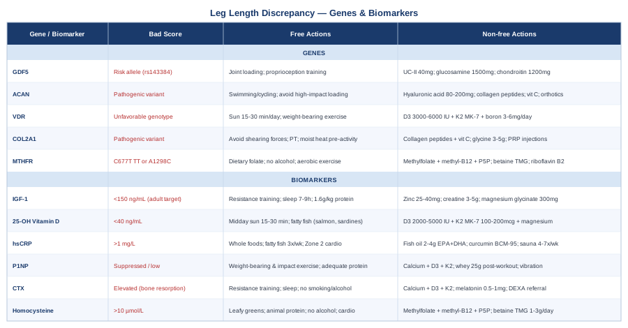
This article covers two angles. The main section examines six concrete biomarkers that are particularly relevant to bone remodeling, cartilage health, and inflammation — what they measure, how to test them affordably, and what to do when results come back unfavorable. A second section examines five gene variants that influence bone and joint development. Supplementing both is a synthesis of Peter Attia's bone health framework and four complementary physical modalities with meaningful clinical evidence for LLD-related complications. Better information leads to better decisions.
6 Biomarkers to Track When You Have Leg Length Discrepancy
Biomarker testing is most useful when treated as a baseline, not a one-time check. For LLD, the relevant biomarkers cluster around three themes: bone formation and resorption, systemic inflammation, and the hormonal and nutritional environment that drives skeletal maintenance. Understanding where you stand on each axis can meaningfully inform whether conservative management, nutritional correction, or specialist referral is the right next step.
1. IGF-1 (Insulin-Like Growth Factor 1)
Why it matters: IGF-1 is the primary downstream mediator of growth hormone signaling. During development it drives longitudinal bone growth through the growth plates. In adults it continues to regulate bone remodeling, muscle-bone interaction, and skeletal mineral density. In LLD patients, sub-optimal IGF-1 may contribute to inadequate bone response to mechanical loading and reduced chondrocyte activity — directly relevant to growth plate function. Research consistently links low IGF-1 to reduced bone mineral density and increased fracture risk.
How to Measure It
A standard serum IGF-1 test is available through most commercial labs for $40–$90 without insurance. The optimal adult range is age-specific; practitioners like Peter Attia who track longevity biomarkers typically target values in the 150–300 ng/mL window for adults in their 30s and 40s. Request the test fasted, in the morning.
If the Score Is Low: Plan Without Supplements
The most powerful non-pharmacological IGF-1 lever is resistance training. Heavy compound movements — squats, deadlifts, rows — performed 3–4 times per week consistently elevate IGF-1 in both young and older adults. Sleep quality is the second major driver: IGF-1 is predominantly released during slow-wave sleep, making 7.5–9 hours of uninterrupted sleep non-negotiable. Dietary protein adequacy is foundational — aim for 1.6–2.2 g/kg/day distributed across meals. Prolonged caloric restriction suppresses the GH-IGF-1 axis and should be avoided.
If the Score Is Low: Plan With Supplements or Equipment
Zinc (25–40 mg/day with food, cycled with copper at 2 mg/day to prevent depletion) supports growth hormone secretion. Magnesium glycinate at 300–400 mg taken at night improves sleep architecture and indirectly supports IGF-1 pulsatility. Creatine monohydrate (3–5 g/day, no cycling needed) amplifies the anabolic response to resistance training and augments the IGF-1 response. Colostrum (2–4 g/day in 8–12 week cycles) contains bioactive IGF-1 precursors, though human evidence remains early. If IGF-1 stays very low despite lifestyle changes, evaluation for adult growth hormone deficiency by an endocrinologist is warranted.
2. 25-OH Vitamin D
Why it matters: Vitamin D is essential for calcium absorption and, at the growth plate level, regulates chondrocyte differentiation and bone mineralization. In LLD, impaired mineralization can worsen structural asymmetries over time. Vitamin D deficiency is common even in populations without obvious risk factors — estimates suggest 40–50% of adults in northern countries have suboptimal levels. In patients who have undergone limb lengthening surgery, vitamin D optimization is explicitly part of the rehabilitation protocol in many centers.
How to Measure It
A 25-OH vitamin D test (the correct form to measure status, not 1,25-dihydroxy) costs $20–$60 at most labs. Peter Attia targets 40–60 ng/mL (100–150 nmol/L). Many standard labs flag anything above 20 ng/mL as "normal," but this threshold is now considered insufficient by most precision medicine practitioners for bone and immune benefit.
If the Score Is Low: Plan Without Supplements
Direct midday sun exposure — 15–30 minutes on arms and legs, 3–4 days per week — is the primary non-supplemental source. This works best at latitudes below 35°N and during spring and summer months. Dietary sources are limited but include fatty fish (salmon, sardines, mackerel), egg yolks, and liver. UVB lamps (Sperti-type) can replicate sunlight exposure during winter months in higher latitudes.
If the Score Is Low: Plan With Supplements or Equipment
Vitamin D3 (cholecalciferol) at 2,000–5,000 IU/day is the standard starting dose, always paired with vitamin K2 (MK-7 form, 100–200 mcg/day) to direct calcium to bone rather than soft tissue. Magnesium is required to convert vitamin D to its active form — many people supplementing D3 without magnesium see limited improvement. Retest at 90 days and adjust dose. Avoid D2 (ergocalciferol), which is less bioavailable. For severe deficiency below 15 ng/mL, a physician may prescribe short-term high-dose D3 followed by maintenance dosing.
3. High-Sensitivity CRP (hsCRP)
Why it matters: Chronic low-grade inflammation quietly disrupts bone remodeling, accelerates cartilage degradation, and impairs the tissue environment around joints — all directly relevant to LLD management. Elevated hsCRP is associated with reduced bone mineral density and faster joint space narrowing in observational data. For anyone with LLD managing secondary hip or knee loading abnormalities, knowing their inflammatory status provides an actionable target.
How to Measure It
The hsCRP blood test costs $10–$30. Optimal is under 1 mg/L; values of 1–3 mg/L indicate moderate risk; above 3 mg/L is high. Test outside of acute illness or injury, as any acute stress will temporarily elevate CRP independent of chronic status.
If the Score Is High: Plan Without Supplements
The strongest lifestyle levers are: eliminating ultra-processed foods and refined seed oils; increasing dietary omega-3 through fatty fish (3+ servings/week); moderate-intensity aerobic exercise (150+ minutes/week); and improving sleep. Poor sleep is a powerful CRP driver in its own right. Weight reduction in those with excess adiposity has one of the largest documented CRP-lowering effects in the clinical literature.
If the Score Is High: Plan With Supplements or Equipment
Fish oil at 2–4 g of combined EPA+DHA per day has robust randomized trial evidence for CRP reduction. Curcumin (BCM-95 or Meriva formulations, 500–1000 mg/day with food) shows meaningful anti-inflammatory effects in short-term trials. Magnesium (350–400 mg/night) modestly reduces CRP. Sauna use — 4–7 sessions per week of 15–20 minutes at 80°C — has been associated with reduced inflammatory markers in Finnish population data. If hsCRP remains above 5 mg/L without obvious cause, evaluate for occult infection or autoimmune condition before attributing it to lifestyle alone.
4. P1NP (Procollagen Type I N-Terminal Propeptide)
Why it matters: P1NP is the gold-standard bone formation marker, recommended by the International Osteoporosis Foundation as one of two reference bone turnover markers for clinical use. It reflects the rate at which new bone matrix is being laid down. For LLD patients, understanding whether bone formation is active or suppressed helps explain why some structural asymmetries worsen with age while others stabilize. Thomas Dayspring and other preventive medicine practitioners increasingly include bone turnover markers in broad metabolic panels, as they provide insight into skeletal metabolism that DEXA alone cannot capture in the short term.
How to Measure It
P1NP is measured via a blood test, typically $60–$120 outside of insurance. Collect the sample fasted, before 9 AM. Age- and sex-specific reference ranges apply. Significantly suppressed P1NP in a young adult suggests inadequate bone formation that warrants investigation.
If the Score Is Low: Plan Without Supplements
Weight-bearing exercise — particularly impact activity including jumping, brisk walking, and resistance training — is the most potent stimulus for P1NP elevation. Even brief bouts of jumping (10–20 repetitions/day) show measurable effects on bone formation markers in controlled trials. Progressive resistance training 3x/week is foundational. Adequate protein and caloric intake are preconditions; bone will not form efficiently in a catabolic environment.
If the Score Is Low: Plan With Supplements or Equipment
Calcium from food (targeting 1000–1200 mg/day total) combined with optimized vitamin D3 and K2 provides the raw material for bone matrix synthesis. Orthosilicic acid as ch-OSA (5–10 mg/day in liquid form) has shown modest evidence for increasing P1NP in a randomized trial. Whey protein (25–30 g post-workout) acutely stimulates bone formation markers. Whole body vibration platforms (15–20 minutes/day at 30–50 Hz) have shown P1NP increases in postmenopausal women in several trials, though LLD-specific data is limited.
5. CTX (C-Terminal Telopeptide of Type I Collagen)
Why it matters: CTX is the reference bone resorption marker — the complement to P1NP. High CTX indicates rapid bone breakdown, potentially outpacing formation. In LLD, the longer leg and its associated hip bear asymmetric mechanical stress. In individuals with low estrogen or testosterone, this accelerated resorption in overloaded areas may compound the structural burden over years. The P1NP-to-CTX ratio — formation versus resorption balance — is more informative than either marker alone.
How to Measure It
CTX is extremely time-sensitive: levels are highest fasted in the early morning and drop 30–50% after eating and with physical activity. Always draw fasted before 9 AM. Cost: $50–$100. Significantly elevated CTX in a premenopausal woman or young man warrants further investigation.
If the Score Is High: Plan Without Supplements
Resistance training combined with adequate protein and caloric intake suppresses CTX within weeks in most individuals. High-impact exercise performed consistently also shifts the remodeling balance toward formation. Eliminating smoking (a potent bone resorption driver) and reducing alcohol are significant. Chronic psychological stress elevates cortisol, which directly drives bone resorption — stress management and sleep prioritization have measurable effects on CTX.
If the Score Is High: Plan With Supplements or Equipment
Optimizing calcium, D3, and K2 intake as described provides the biological substrate that reduces the drive for resorption. Melatonin at low physiological doses (0.5–1 mg at bedtime) has antiresorptive properties documented in several small trials. Ipriflavone (600 mg/day in cycles) has shown modest CTX reduction in postmenopausal women. If CTX is very high, DEXA and endocrinology referral are warranted before pursuing unguided supplementation — high CTX can reflect an underlying hormonal or metabolic condition.
6. Homocysteine
Why it matters: Elevated homocysteine impairs the cross-linking of collagen molecules in bone matrix, directly reducing structural integrity. Studies consistently link elevated homocysteine to increased fracture risk, reduced bone mineral density, and impaired cartilage health. For LLD patients managing secondary complications — hip degeneration, spinal compensation patterns, stress fractures in overloaded limbs — homocysteine is an underappreciated lever. It is largely driven by B-vitamin status (B6, B12, folate) and is highly responsive to nutritional intervention.
How to Measure It
Serum homocysteine is a standard test costing $15–$45. Thomas Dayspring consistently flags homocysteine as underordered in standard panels. Optimal is below 8 µmol/L; values above 10 µmol/L are concerning; above 15 µmol/L is a significant signal for bone and cardiovascular risk.
If the Score Is High: Plan Without Supplements
Dietary optimization first: dark leafy greens (folate), animal protein sources (B12), poultry and fish (B6), eggs (full B-vitamin spectrum). Eliminating heavy alcohol consumption is important — alcohol depletes B vitamins and raises homocysteine significantly. Regular aerobic exercise modestly lowers homocysteine independent of diet. Very high caffeine intake (above 4 cups/day) raises homocysteine slightly, though moderate coffee does not.
If the Score Is High: Plan With Supplements or Equipment
The methylated B-vitamin combination is the backbone: methylfolate (400–800 mcg/day) + methylcobalamin or adenosylcobalamin (500–1000 mcg/day) + pyridoxal-5-phosphate (25–50 mg/day). This combination consistently lowers homocysteine across dozens of clinical trials. For those with the MTHFR C677T variant — discussed in the next section — active methylated forms are essential, as regular folic acid may be ineffective or counterproductive. Betaine (TMG, 1–3 g/day) provides an alternative methylation pathway and is additive to B vitamins. Retest at 8–12 weeks to confirm response.
These six biomarkers give a working snapshot of bone formation activity, inflammatory burden, and nutritional adequacy — the three domains most relevant to the biological management of LLD. None replaces imaging or clinical evaluation, but together they create the kind of actionable baseline that makes future data interpretable.
The Genetic Picture: 5 Key Variants That Influence Skeletal Development
Genetics in the context of LLD is not deterministic. A variant that predisposes to slower cartilage repair or weaker bone density does not guarantee worse outcomes — but it raises the probability that certain interventions matter more than average for that individual. The five genes below have the strongest human evidence base relevant to skeletal development, bone quality, and joint health in the context of LLD.
Gene 1: GDF5 (Growth Differentiation Factor 5)
What it does: GDF5 encodes a signaling protein critical to limb development, joint formation, and cartilage maintenance. During fetal development it is one of the master regulators of chondrogenesis — the process that shapes growth plates. Variants in GDF5 have been reproducibly associated with asymmetric joint development, shorter stature, and susceptibility to osteoarthritis.
Key variant: The rs143384 single nucleotide polymorphism (C>T change in the 5' UTR) reduces GDF5 expression in joint tissues. It is one of the most replicated genetic risk factors for osteoarthritis identified in genome-wide association studies, validated across European and Asian populations, and directly relevant to LLD-associated joint degeneration.
If the Gene Is Unfavorable: Plan Without Supplements
GDF5 expression is modulated by mechanical loading — consistent evidence from animal models and in vitro studies shows that cyclic mechanical strain upregulates GDF5 in chondrocytes. Structured progressive joint loading (resistance training, controlled walking, low-impact activity) is not merely beneficial but mechanistically appropriate for this gene. Avoid prolonged sedentary behavior, which suppresses mechanotransduction signals. Proprioception training — balance boards, single-leg exercises — supports joint tissue health independent of GDF5 status.
If the Gene Is Unfavorable: Plan With Supplements or Equipment
Undenatured type II collagen (UC-II, 40 mg/day on an empty stomach) has clinical trial evidence for joint cartilage support through a tolerogenic mechanism that may be relevant when GDF5-related cartilage quality is compromised. Glucosamine sulfate (1500 mg/day) and chondroitin sulfate (1200 mg/day) have mixed evidence but a reasonable safety profile and provide precursor molecules for cartilage matrix synthesis. Avoid chronic NSAID use — it suppresses proteoglycan synthesis and may worsen cartilage quality over time.
Gene 2: ACAN (Aggrecan)
What it does: Aggrecan is the primary structural proteoglycan of cartilage. It gives cartilage its compressive strength by trapping water within the extracellular matrix. ACAN variants are associated with short stature, premature growth plate fusion, intervertebral disc disease, and — critically for LLD — asymmetric skeletal development. Heterozygous loss-of-function variants in ACAN are now recognized as a cause of idiopathic short stature with advanced bone age in children, sometimes manifesting as LLD.
Key variants: Multiple pathogenic variants across ACAN have been documented through whole-exome sequencing studies. Children with ACAN haploinsufficiency show accelerated skeletal maturation with premature growth plate closure, sometimes affecting one limb more than another — a mechanism directly relevant to idiopathic LLD.
If the Gene Is Unfavorable: Plan Without Supplements
In adults with residual LLD from ACAN-related growth asymmetry, protecting cartilage integrity through controlled loading is the practical priority — not avoidance of exercise, but avoidance of excessive high-impact loading on already-compromised joints. Swimming and cycling offer cardiovascular fitness with minimal cartilage compression. Custom orthotics reduce asymmetric joint loading directly, lessening the mechanical burden placed on ACAN-compromised cartilage.
If the Gene Is Unfavorable: Plan With Supplements or Equipment
High-molecular-weight oral hyaluronic acid (80–200 mg/day) supports the proteoglycan environment that ACAN maintains. Collagen peptides (10–15 g/day of type II-specific or mixed collagen) provide amino acid precursors for matrix repair. Vitamin C (500–1000 mg/day) is necessary for collagen hydroxylation and aggrecan biosynthesis. Well-fitted shoe lifts and custom orthotics reduce the asymmetric joint loading that accelerates cartilage breakdown when ACAN function is compromised — arguably the most impactful equipment intervention for this gene.
Gene 3: VDR (Vitamin D Receptor)
What it does: The vitamin D receptor mediates all genomic effects of active vitamin D, including calcium absorption, bone mineralization, and regulation of osteoblast and osteoclast activity. VDR polymorphisms alter receptor affinity and downstream signaling efficiency, meaning two individuals with identical serum vitamin D levels can have substantially different bone responses. This was one of the first gene-nutrient interactions systematically studied in human bone biology, documented in a landmark Nature paper in 1994.
Key variants: The BsmI, FokI, ApaI, and TaqI polymorphisms have been studied in relation to bone mineral density. The FokI ff genotype produces a longer receptor isoform with reduced transcriptional efficiency. Meta-analyses suggest these variants explain a modest but real proportion of population variance in bone density — enough to matter when LLD has already placed asymmetric demands on skeletal structure.
If the Gene Is Unfavorable: Plan Without Supplements
High sun exposure and weight-bearing exercise are doubly important for individuals with reduced VDR efficiency. Resistance training upregulates VDR expression in muscle and bone tissue — the same mechanical stimulus that builds bone strength also compensates for receptor inefficiency. Dietary calcium optimization (dairy, sardines, tofu, cooked kale) helps because the system can absorb more even when the receptor is less efficient, if substrate intake is sufficient.
If the Gene Is Unfavorable: Plan With Supplements or Equipment
Higher vitamin D3 supplementation targets (3,000–6,000 IU/day rather than 1,000–2,000 IU/day) may be warranted to compensate for reduced receptor efficiency — always guided by 25-OH vitamin D blood levels to achieve the 40–60 ng/mL target without overshooting. Vitamin K2 (MK-7, 150–200 mcg/day) becomes even more critical to direct calcium to bone. Boron (3–6 mg/day from food or supplementation) supports VDR signaling and prolongs the half-life of active vitamin D. Practitioners with access to genetic panels can use VDR genotype to calibrate D3 dosing more precisely.
Gene 4: COL2A1 (Collagen Type II Alpha 1)
What it does: COL2A1 encodes the primary structural protein of cartilage collagen. Pathogenic mutations cause a spectrum of skeletal dysplasias, from lethal achondrogenesis at one extreme to mild late-onset spondyloepiphyseal dysplasia at the other. In LLD, COL2A1 variants that reduce collagen quality or quantity directly compromise the growth plate scaffold, potentially driving asymmetric longitudinal bone growth and accelerating cartilage degeneration in loaded joints.
Key variants: Common polymorphisms in COL2A1 have been associated with intervertebral disc degeneration and hip dysplasia risk in large cohort studies. Pathogenic variants are identified through clinical genetic testing — whole-exome sequencing or targeted skeletal dysplasia panels.
If the Gene Is Unfavorable: Plan Without Supplements
Avoiding joint positions that place shearing force on cartilage (deep heavy squatting, high-impact jumping on hard surfaces) preserves collagen-compromised cartilage longer. Physical therapy focused on muscle strengthening distributes load away from cartilage and onto well-functioning musculature — the core conservative strategy. Moist heat before movement (warm bath or heating pad for 10–15 minutes) reduces stiffness and facilitates safer tissue loading.
If the Gene Is Unfavorable: Plan With Supplements or Equipment
Collagen peptides (10–15 g/day with vitamin C, taken 30–60 minutes before exercise) show emerging evidence for stimulating cartilage matrix synthesis. Glycine (3–5 g/day) is the rate-limiting amino acid in collagen synthesis — inexpensive and often overlooked. Adequate iron status is required for prolyl hydroxylase activity, the enzyme that stabilizes collagen triple helices. For those with documented COL2A1 variants and significant cartilage involvement, platelet-rich plasma (PRP) injections into affected joints have clinical trial support, though outcomes are operator-dependent and coverage varies.
Gene 5: MTHFR (Methylenetetrahydrofolate Reductase)
What it does: While not a bone gene per se, MTHFR is included here because of its direct impact on homocysteine metabolism and its consequent relevance to bone collagen integrity. MTHFR converts folate to its active methylated form, which is required for the methylation cycle. Individuals with the C677T or A1298C variants have reduced enzyme efficiency, higher homocysteine, and — as multiple studies confirm — lower bone mineral density and higher fracture risk, particularly in the homozygous TT genotype at C677T.
Key variants: MTHFR C677T (rs1801133) and A1298C (rs1801131) are among the most commonly tested variants in direct-to-consumer genetics panels. Homozygous TT at C677T shows roughly 30–70% reduced enzyme activity. Compound heterozygotes show intermediate reduction.
If the Gene Is Unfavorable: Plan Without Supplements
Emphasize natural dietary folate (leafy greens, legumes, liver) over synthetic folic acid. Those with MTHFR inefficiency metabolize natural folate differently than synthetic folic acid, and excess synthetic folic acid can block the pathway. Eliminating smoking and heavy alcohol consumption is doubly important, as both independently impair methylation. Regular moderate-intensity cardio modestly improves methylation efficiency through epigenetic mechanisms.
If the Gene Is Unfavorable: Plan With Supplements or Equipment
This is the clearest supplement protocol in this genetics section: methylfolate (400–1000 mcg/day) + methylcobalamin (500–1000 mcg/day) + pyridoxal-5-phosphate (25–50 mg/day). Do not use regular folic acid for C677T carriers. Betaine (TMG, 1–3 g/day) provides an alternative methylation pathway and is additive to B vitamins. Riboflavin (vitamin B2, 10–15 mg/day) specifically stabilizes the MTHFR enzyme for the TT genotype — an underappreciated and inexpensive intervention with modest trial evidence. Retest homocysteine at 8 weeks to confirm response.
The genetic landscape for LLD is cumulative. Most individuals carry variants across several of these loci simultaneously, and the net biological effect is shaped by lifestyle, nutrition, and the epigenome. What is consistent across all five genes: mechanical loading and adequate micronutrient status are first-order interventions regardless of genotype.
What Peter Attia's Bone Health Framework Means for LLD
Peter Attia's Outlive: The Science and Art of Longevity (2023) does not address LLD directly, but his precision medicine framework for skeletal health translates cleanly and usefully to the condition. These are the ten most impactful ideas from his approach applied to LLD.
1. Bone Density Is a Long-Game Investment
Attia frames bone density as something built in the 20s and 30s that is spent in the 60s, 70s, and 80s. For LLD, this framing has a specific implication: asymmetric loading on the longer leg and its associated hip depletes structural reserves faster than the population average. Starting bone density optimization in middle age is late but still meaningful. Starting it earlier is always better. Establishing a DEXA baseline in the 30s or 40s costs little and provides a longitudinal reference that makes future data interpretable.
2. DEXA Scanning Is Non-Negotiable
Attia recommends baseline DEXA scanning well before age 50, particularly for women but increasingly for men. He specifically tracks not just global bone density but regional values — spine and hip separately. For LLD patients, requesting separate hip values for each side can reveal lateralized differences in bone quality that standard DEXA reports summarize away.
3. Zone 2 Cardio Protects Bone Indirectly
Steady-state aerobic exercise at a conversational pace (Zone 2, 180+ minutes/week) preserves the metabolic environment that supports bone formation. Attia links mitochondrial efficiency and chronic inflammation through a common pathway — and elevated chronic inflammation directly suppresses bone formation. This is why the hsCRP biomarker connects so directly to this framework: Zone 2 exercise is one of the most robust CRP-lowering interventions available without a prescription.
4. Resistance Training Specificity Matters for Asymmetric Loading
Bone responds specifically to the forces applied to it. Attia's training philosophy for bone involves heavy compound loading — deadlifts, farmers carries, sled work — combined with impact. For LLD patients, symmetrical loading protocols must be modified: an appropriately placed heel lift during bilateral lifts prevents the compensatory pelvic tilt that shifts mechanical loading away from the shorter limb's femur. Working with a physical therapist who understands LLD biomechanics during this phase is genuinely worth the investment.
5. The Estrogen and Testosterone Tipping Point
Attia discusses the bone-protective roles of both estrogen and testosterone extensively, noting that their decline is the primary driver of post-midlife bone loss. For LLD, the implication is specific: if baseline bone quality is already challenged by asymmetric loading and potentially unfavorable genetics, the hormonal decline of perimenopause or andropause will accelerate divergence between skeletal sites. Early tracking of estradiol (women) and total/free testosterone (men) adds useful early warning to the biomarker panel above.
6. Protein Is the Underappreciated Bone Nutrient
Calcium and vitamin D absorb most of the nutritional attention in bone health, but Attia emphasizes protein as equally foundational. Bone matrix is 30% organic material — predominantly collagen — and collagen synthesis requires amino acid supply. The RDA of 0.8 g/kg/day is almost certainly insufficient for bone maintenance in anyone doing resistance training. Attia targets 1.6–2.2 g/kg/day of high-quality protein. For LLD, adequate protein is particularly important during any rehabilitative phase involving tissue remodeling or post-surgical recovery.
7. Tracking the P1NP:CTX Ratio Longitudinally
Attia and other precision medicine practitioners track bone turnover markers longitudinally rather than as a single snapshot. The ratio of P1NP to CTX reveals whether the skeleton is in net positive balance (building) or negative balance (losing tissue). For LLD patients with asymmetric loading, this ratio provides meaningful data that DEXA alone cannot capture in the short term — bone turnover markers change within weeks, while DEXA-detectable density changes take 1–2 years to accumulate.
8. Sleep Optimization Is a Bone Health Intervention
Attia frames sleep as the single most upstream health intervention. For bone specifically: growth hormone and IGF-1 are released predominantly during slow-wave sleep; cortisol — the main bone-resorbing stress hormone — is dysregulated by poor sleep; and the circadian control of bone turnover is disrupted by irregular sleep schedules. Seven to nine hours of quality sleep is not optional for anyone taking skeletal health seriously.
9. Metabolic Health Is Upstream of Bone Health
Attia's framework emphasizes that bone health is downstream of metabolic health. Insulin resistance and chronically elevated insulin create a pro-inflammatory, pro-resorptive bone environment. Individuals with LLD who are metabolically compromised — elevated fasting insulin, elevated triglycerides, low HDL — are operating with a biological headwind that accelerates bone and joint degradation. Fasting insulin below 8 µIU/mL is Attia's functional target and worth adding to any metabolic panel.
10. Define What You're Training For: The Centenarian Decathlon
Perhaps the most useful conceptual contribution: Attia asks patients to define what they want to be able to do at age 90 and work backward from those physical requirements. For LLD, this reframes the goal. The target is not "fixing" the discrepancy — it often cannot be changed without surgery. The target is maintaining the biomechanical and structural reserves necessary to walk, carry groceries, climb stairs, and live independently into old age, despite the asymmetry. Every biomarker and genetic intervention in this article ultimately serves that specific, tangible goal.
Complementary Approaches With Meaningful Evidence for LLD
The biological interventions above are most effective when combined with physical and structural management. The following modalities have clinical evidence relevant to LLD-related compensations — primarily spinal alignment, pelvic asymmetry, hip loading, and chronic musculoskeletal pain.
Spinal Manipulation
Leg length discrepancy consistently produces secondary spinal adaptations: pelvic obliquity, lumbar scoliosis, and compensatory thoracic curves. These secondary changes generate their own pain and functional limitation, often more debilitating than the discrepancy itself. Spinal manipulation targets the restricted spinal segments and hypertonic paraspinal muscles that develop as consequences of LLD.
Clinical guidelines from the American College of Physicians (2017) support spinal manipulation as a first-line approach for non-specific chronic low back pain — the most common secondary complaint in LLD. Research published in Spine has also specifically found that combined manipulation plus orthotics produced superior outcomes to orthotics alone for LLD-related low back pain. Side effects are generally mild (temporary local soreness); serious adverse events are rare when appropriate screening excludes contraindications including osteoporosis and disc herniation with neurological signs.
Practically: 6–12 sessions over 6–8 weeks provides a reasonable trial period. A chiropractor, osteopath, or physiotherapist experienced in LLD should assess pelvic level before and after heel lift fitting — manipulation improves the response to orthotics by releasing compensatory spinal fixations that otherwise resist correction. Some patients benefit from monthly maintenance; others achieve lasting improvement within the initial course.
Yoga
Yoga's relevance to LLD lies primarily in its ability to address the muscular imbalances created by living with an asymmetric pelvis over years: the quadratus lumborum, piriformis, psoas major, and hip flexors develop systematic shortening and lengthening patterns. Iyengar-style alignment-based practice provides targeted, controllable stretching and strengthening of these structures with a level of precision not available in generic exercise.
A randomized noninferiority trial by Saper et al. published in Annals of Internal Medicine (2017) found that yoga was non-inferior to physical therapy for chronic low back pain and superior to educational materials at 12 weeks and 52 weeks of follow-up. For LLD, standing balance poses (tree pose, warrior III) retrain proprioception in the shorter limb while stretching compensatory hip structures.
Practically: 2–3 classes per week focused on hip and spinal alignment, with disclosure to the instructor about LLD. Props — blocks, bolsters, blankets — allow modification for height differences. Avoid advanced inversions and deep twists until spinal adaptations have been professionally evaluated. Progress toward yin or restorative yoga for deep fascial release once acute compensations are managed.
Massage Therapy
The muscular consequences of LLD — chronic hypertension in the quadratus lumborum on the high-pelvis side, piriformis overload on the long-leg side, Achilles and plantar fascia strain on the short-leg side — respond well to targeted soft-tissue work. Massage therapy addresses these secondary muscular consequences, reducing pain and improving the mechanical environment for bone and cartilage interventions.
A Cochrane review on massage therapy for low back pain found moderate-quality evidence for pain reduction and improved function when combined with active exercise. For LLD specifically, myofascial release targeting the iliotibial band, thoracolumbar fascia, and hip capsule has mechanistic plausibility supported by several small randomized trials on biomechanical asymmetry management.
Practically: 60-minute sessions focused on hip rotators, lumbar paraspinals, and calf/Achilles on the short-leg side provide the most targeted benefit. Monthly maintenance is reasonable for most individuals; weekly sessions during symptomatic flares. Communicate clearly to the therapist that you have documented LLD — pressure and positioning need to be adapted accordingly, and a therapist unfamiliar with the condition may inadvertently reinforce asymmetric patterns.
Biofeedback for Gait Retraining
Gait abnormalities in LLD — asymmetric step length, trunk lean, hip drop — are not purely structural; they involve learned neuromuscular patterns that persist even after structural correction with heel lifts or orthotics. Biofeedback retrains these patterns by providing real-time sensory information that enables conscious correction of habitual compensations.
Wearable EMG-based and force plate-based biofeedback devices have shown effectiveness for gait retraining in populations with lower limb asymmetry. A 2019 randomized controlled trial on real-time gait feedback demonstrated meaningful reduction in asymmetric joint loading at the hip and knee in participants with lower extremity asymmetry. Force plate-based biofeedback with more granular data is available in clinical physiotherapy settings.
Practically: formal biofeedback sessions with a physiotherapist — 6–10 sessions targeting one or two key gait parameters — produce more durable changes than unsupported home use. Smartphone-based gait analysis apps (Plantiga, Dorsavi) provide an accessible starting point for identifying dominant asymmetric patterns between clinic visits. Walking barefoot on varied terrain also provides natural proprioceptive feedback that complements formal biofeedback training.
Low-Level Laser Therapy (Photobiomodulation)
Low-level laser therapy uses non-thermal photonic energy (600–1000 nm wavelength) to stimulate mitochondrial cytochrome c oxidase in target tissues, promoting cellular energy production, reducing oxidative stress, and modulating inflammatory mediators. In bone tissue, LLLT has been shown to accelerate healing, stimulate osteoblast activity, and reduce periarticular pain in several clinical contexts relevant to LLD.
A systematic review in Photobiomodulation, Photomedicine, and Laser Surgery (2021) found that LLLT accelerated bone healing in clinical trials and consistently stimulated bone formation markers in cell culture and animal studies. Human RCTs in fracture healing and orthodontic bone remodeling show measurable effects. LLD-specific trials are not yet available, but the most practical application is periarticular treatment of overloaded joints — hip, knee, lumbar facets — which bear excess burden secondary to the discrepancy.
Practically: clinical LLLT at a physiotherapy practice (6–12 sessions for musculoskeletal conditions) is the most evidence-anchored approach. Consumer-grade red-light therapy panels (630–850 nm) are increasingly affordable ($200–$500) for home use, with protocols of 10–20 minutes/day to affected joints. Target a total fluence of 4–10 J/cm² per treatment site — too low a dose has no effect; too high may be inhibitory. Evidence is strongest for pain and healing; direct limb-length modification in humans remains unproven, but the anti-inflammatory and bone-stimulatory mechanisms are biologically plausible.
Conclusion
Leg length discrepancy is a structural condition, but the biology surrounding it responds meaningfully to targeted intervention. The six biomarkers covered here — IGF-1, 25-OH vitamin D, hsCRP, P1NP, CTX, and homocysteine — give you a working map of your bone formation activity, inflammatory burden, and nutritional adequacy. The five gene variants — GDF5, ACAN, VDR, COL2A1, and MTHFR — reveal predispositions that make certain interventions more or less important for your specific biology. Most of these biomarkers are available through standard commercial labs for under $300 total, and direct-to-consumer genetics panels cover several of the variants discussed.
The next smart step is not to optimize everything at once. Pick the one or two areas where your data suggests the greatest gap, address them systematically over 8–12 weeks, and retest. Arriving at an appointment with a physician, rheumatologist, or sports medicine specialist armed with biomarker data and genetic context means you are no longer relying solely on symptoms — and that changes the quality of the conversation significantly.
Musculoskeletal: Bone Conditions Joint Conditions Spine Conditions
Autoimmune: Inflammatory Conditions Connective Tissue Conditions