This article contains AI generated content.
· UpdatedLeprosy Arthritis – 8 Genes And 6 Biomarkers To Track
Introduction
Leprosy arthritis sits at an unusual crossroads of infectious disease and chronic joint inflammation. Caused by Mycobacterium leprae, leprosy can trigger several distinct patterns of joint involvement — from acute reactive arthritis during erythema nodosum leprosum reactions to chronic deforming polyarthritis that can look remarkably like rheumatoid disease. For people navigating this, the frustrating part is often how little precision is built into standard care: you're monitored for nerve damage and bacterial load, but the inflammatory cascades attacking your joints are frequently left to run unchecked in the background.
Generic anti-inflammatory approaches may blunt symptoms without addressing the underlying drivers. What actually determines who develops severe joint involvement, how fast it progresses, and how well someone responds to treatment is shaped in large part by individual biology — specifically, the immune genes they carry and how their inflammatory systems are calibrated. Two people on the same treatment protocol can have dramatically different joint outcomes, and genetics and measurable biomarkers explain a significant share of that gap.
This article takes a more targeted approach. Rather than general advice about rest and NSAIDs, it focuses on the biological variables that are actually worth tracking and understanding. The biomarker section gives you a practical monitoring framework — six markers that collectively reveal infection activity, inflammation severity, immune system status, and complication risk. The genetics section then digs into eight genes that influence susceptibility and severity, with specific compensatory strategies for each.
Better information does not guarantee a cure, but it consistently leads to better decisions. Knowing your inflammatory baseline, your vitamin D status, and whether you carry immune variants that make you prone to overreaction changes how you discuss treatment with your physician, what you prioritize, and where you focus your energy.
6 Key Biomarkers to Monitor in Leprosy Arthritis
Tracking inflammation from a single angle gives an incomplete picture. The six biomarkers below cover disease activity, acute-phase response, cytokine-driven joint damage, immune competence, and long-term complication risk. Together, they give a much more useful view of what is actually happening in your joints and immune system.
1. Anti-PGL-I Antibodies (Phenolic Glycolipid-I)
Why it matters: Anti-PGL-I IgM antibodies are the most specific serological marker for M. leprae infection available. Phenolic glycolipid-I is a unique antigen found only in this bacterium. In leprosy arthritis, elevated anti-PGL-I levels reflect ongoing bacterial antigenic load — the principal trigger driving immune-mediated joint inflammation. In lepromatous forms with arthritis, this marker stays elevated and correlates with disease activity, relapse risk, and the intensity of type 2 lepra reactions that are the most joint-damaging episodes in the disease course.
How to measure it: ELISA-based serology, available at reference infectious disease laboratories or specialized public health labs. Cost ranges from $50 to $150 in the US and significantly less in endemic regions. Results are expressed as titers or optical density ratios relative to controls. Paucibacillary patients often test negative despite active disease, so clinical context matters.
If the result is abnormal, the plan without supplements: Elevated anti-PGL-I despite completing treatment is a red flag for bacterial persistence or relapse. The priority is a formal reassessment with an infectious disease specialist. Skin smear bacterial index (BI), slit-skin smear microscopy, and if available PCR for M. leprae DNA should follow. Joint symptoms combined with a rising anti-PGL-I titer justify re-evaluation for retreatment under WHO MDT (multidrug therapy) guidelines. Cold applications and joint rest reduce acute synovitis during reactional episodes; thalidomide or corticosteroids may be prescribed medically for type 2 reactions. Tracking this marker every 3 to 6 months during treatment and for two years post-treatment is reasonable clinical practice.
If the result is abnormal, the plan with supplements or equipment: Directly, no supplement reverses elevated anti-PGL-I — that requires proper antibiotic treatment. However, supporting bacterial clearance through immune optimization is valid adjunctive strategy. Vitamin D3 supplementation (2,000–5,000 IU daily, adjusted to blood levels) has been shown to enhance macrophage-driven clearance of mycobacteria by upregulating cathelicidin. Zinc at 15–30 mg/day supports T-cell mediated immunity, which is characteristically suppressed in lepromatous forms. Both should be cycled with regular blood testing; excess zinc depletes copper (monitor copper and ceruloplasmin every 6 months). No known severe interactions at these doses when used alongside MDT.
2. High-Sensitivity C-Reactive Protein (hs-CRP)
Why it matters: CRP is an acute-phase protein produced by the liver in response to IL-6, IL-1β, and TNF-α — the same cytokines that drive synovitis. In leprosy arthritis, hs-CRP spikes dramatically during lepra reactions (both type 1 reversal reactions and type 2 erythema nodosum leprosum), and its trajectory correlates directly with joint inflammation severity. Chronically elevated hs-CRP between reactions suggests persistent low-grade inflammation and predicts accelerated joint damage and cartilage degradation over time. For ongoing monitoring between clinic visits, hs-CRP is among the most accessible and informative tools available.
How to measure it: Standard blood panel, ordered as hs-CRP specifically (conventional CRP lacks sensitivity below 5 mg/L). Cost is $15–$40 in the US. Target: below 1 mg/L for low cardiovascular and inflammatory risk; 1–3 mg/L is intermediate; above 3 mg/L persistently suggests active systemic inflammation warranting investigation. In active lepra reactions, values above 30–100 mg/L are common and expected.
If the score is elevated, the plan without supplements: First, rule out infection exacerbation, reaction episodes, or concurrent illness. If elevated between reactions, a structured anti-inflammatory lifestyle becomes the foundation: eliminate ultra-processed foods and refined oils high in omega-6; prioritize cold-water fish, leafy greens, and low-glycemic carbohydrates. Regular moderate-intensity aerobic exercise (30 minutes, 5 days/week) consistently reduces hs-CRP by 15–25% in clinical studies of chronic inflammatory disease. Adequate sleep (7–9 hours) and stress reduction lower cortisol, which if chronically elevated drives upward CRP drift. Smoking cessation is mandatory — smoking raises basal CRP by roughly 40%.
If the score is elevated, the plan with supplements or equipment: Omega-3 fatty acids (EPA+DHA combined, 2–4 g/day) have strong meta-analytic support for reducing hs-CRP in inflammatory conditions. Take with meals; common side effect is fishy reflux, minimized with enteric-coated forms. Cycle is continuous at therapeutic dose; check hs-CRP after 12 weeks. Curcumin with piperine (500–1,000 mg curcuminoid with 5–10 mg piperine, twice daily) reduced hs-CRP significantly in arthritis trials. Avoid high-dose piperine if on MDT drugs as it can affect drug metabolism. Infrared sauna sessions (3–4 per week, 20 minutes at 55–65°C) have been reported to lower systemic inflammatory markers in chronic arthritis contexts, though direct leprosy arthritis data is absent.
3. Erythrocyte Sedimentation Rate (ESR)
Why it matters: ESR is an older but still clinically meaningful marker of systemic inflammation and plasma protein changes. In leprosy, especially multi-bacillary and borderline forms, ESR rises with bacterial load, immunological reactions, and secondary amyloidosis — a serious long-term complication in poorly controlled lepromatous disease that can cause destructive joint and organ damage. ESR's slower response time compared to CRP makes it useful for tracking sustained inflammatory burden rather than acute spikes. When both ESR and CRP are elevated together, the combination strongly suggests significant active systemic inflammation rather than localized joint issues.
How to measure it: Westergren method, routinely included in most inflammatory panels. Cost is $10–$25. Normal is under 20 mm/hr in men and under 30 mm/hr in women (age-adjusted). In leprosy arthritis with active reactions, values of 60–120 mm/hr are common. A falling ESR over months confirms treatment response.
If the score is elevated, the plan without supplements: Persistently elevated ESR warrants evaluation for secondary amyloidosis (serum amyloid A measurement, urinary protein, renal function) and for treatment adequacy. Beyond treating underlying disease, dietary strategies that directly lower fibrinogen and immunoglobulin concentrations — the main ESR drivers — overlap with anti-inflammatory nutrition. Hydration is often overlooked; chronic mild dehydration elevates ESR. Structured low-intensity daily movement (walking 45–60 minutes) improves vascular and inflammatory markers more sustainably than sporadic intense exercise in this population.
If the score is elevated, the plan with supplements or equipment: Serrapeptase (10 mg–60 mg, between meals, away from food) has modest evidence for reducing circulating fibrinogen and inflammatory debris. Side effects include GI upset at higher doses; cycle 5 days on, 2 days off. Nattokinase has similar mechanisms but limited direct ESR data. For patients with secondary amyloid concern, taurine (2–3 g/day) has some evidence for amyloid protein modulation in animal models — human evidence remains preliminary. Consult with your physician before adding enzymatic supplements if on anticoagulants.
4. Interleukin-6 (IL-6)
Why it matters: IL-6 is the central cytokine coordinator of the acute-phase response and a key driver of synovitis in inflammatory arthritis. In leprosy, IL-6 is markedly elevated during type 2 lepra reactions and correlates with the severity of joint swelling, pain, and soft tissue destruction. Elevated baseline IL-6 between reactive episodes predicts who will develop more severe chronic joint involvement. Because IL-6 signals through STAT3 to upregulate pannus formation and cartilage-degrading enzymes, persistent elevation acts as a slow, erosive force on joint structures even when symptoms seem manageable.
How to measure it: Serum IL-6 ELISA or multiplex cytokine panel; available at reference labs and increasingly at academic medical centers. Cost ranges from $60 to $200 depending on panel configuration. Normal fasting values are below 7 pg/mL in most adults; active inflammatory disease pushes values to 10–100 pg/mL or higher during reactions. Testing during a stable interval gives the most interpretable baseline.
If the score is elevated, the plan without supplements: Reducing IL-6 without pharmacology centers on reducing its main upstream drivers: excess adipose tissue (particularly visceral fat), chronic sleep deprivation, and dysbiotic gut microbiome. Each kilogram of visceral fat reduction corresponds to a measurable IL-6 decrease. Time-restricted eating (10–12 hour feeding window) combined with reduced refined carbohydrate intake is among the most evidence-supported lifestyle approaches for lowering IL-6 in inflammatory conditions. Cold exposure (cold showers or cold immersion at 10–15°C for 2–5 minutes) transiently suppresses pro-inflammatory cytokines including IL-6, with regular practice producing longer-term modulation, though this is contraindicated if you have active neuropathic involvement or poor temperature sensation.
If the score is elevated, the plan with supplements or equipment: Melatonin (0.5–3 mg at bedtime) has demonstrated IL-6 lowering effects via its anti-inflammatory antioxidant properties in autoimmune and chronic inflammatory conditions; use at the lower end of the dose range first. Boswellia serrata extract (400–500 mg standardized to 65% boswellic acids, twice daily) inhibits leukotriene synthesis and reduces synovial IL-6 in arthritis models. Allow 4–6 weeks before reassessing. Photobiomodulation (LLLT, 630–810 nm wavelengths) applied over affected joints inhibits NF-κB and downstream IL-6 production at the synovial level; sessions of 10–15 minutes, 3 times weekly, have clinical support in rheumatoid and reactive arthritis contexts.
5. 25-Hydroxyvitamin D
Why it matters: Vitamin D is not simply a bone mineral. Its active form, 1,25-dihydroxyvitamin D3, acts as a steroid hormone with profound effects on macrophage function, T-regulatory cell differentiation, and the antimicrobial peptide cathelicidin — which directly kills M. leprae inside macrophages. Vitamin D deficiency is disproportionately common in people with leprosy and correlates with both higher bacillary loads and more severe immunological reactions. Low 25-OH-D is independently associated with greater joint inflammation, worse reactional episodes, and impaired nerve recovery. The VDR gene (discussed in the genetics section) determines how efficiently the body responds to vitamin D, making both the level and the genetic context important.
How to measure it: Serum 25-hydroxyvitamin D by immunoassay; standard panel at any lab. Cost is $30–$60. Optimal range for immune function is 40–60 ng/mL (100–150 nmol/L) according to researchers like Rhonda Patrick and broader immunology literature. Below 30 ng/mL is deficiency in most clinical frameworks; below 20 ng/mL is severe deficiency. Test in the morning, fasted.
If the score is low, the plan without supplements: Solar UVB exposure (10–30 minutes of midday sun on face, arms, and legs, depending on skin tone and latitude) is the most natural route to vitamin D synthesis. In endemic leprosy regions closer to the equator, sun exposure is often adequate in theory but skin pigmentation, clothing, and indoor work patterns create real deficits. Foods rich in vitamin D — wild salmon, mackerel, sardines, egg yolks, beef liver — contribute modestly. Magnesium sufficiency is required for vitamin D metabolism; low magnesium blunts the effect of any vitamin D intervention.
If the score is low, the plan with supplements or equipment: Vitamin D3 (cholecalciferol, 2,000–5,000 IU/day) is the standard supplementation approach for deficiency in the immune-relevant range. Take with the largest meal of the day for best absorption. Always co-supplement with vitamin K2 (MK-7 form, 100–200 mcg/day) to direct calcium appropriately and prevent soft-tissue calcification at higher vitamin D doses. Retest 25-OH-D every 3 months during correction. Upper tolerable limit is 10,000 IU/day for most adults, but toxicity (hypercalcemia) can occur at sustained high doses without monitoring. Magnesium glycinate or malate (300–400 mg/day) as cofactor. This combination is the single most impactful supplement intervention for immune function in leprosy-related inflammatory disease.
6. Serum Amyloid A (SAA)
Why it matters: Serum amyloid A is an acute-phase reactant that rises faster and higher than CRP during inflammatory episodes, making it one of the most sensitive markers of active systemic inflammation. More critically for leprosy arthritis, sustained elevation of SAA over months to years is the precursor to AA amyloidosis — the most serious long-term complication of chronic multi-bacillary leprosy. Amyloid deposits in the kidneys, joints, and liver cause progressive organ dysfunction that is largely irreversible once established. SAA monitoring is therefore both a disease activity marker and a secondary complication surveillance tool that is underused in routine leprosy care.
How to measure it: Serum immunoassay, available at reference and academic labs. Cost ranges from $70 to $180, and it is not always part of routine panels — you may need to request it specifically. Normal baseline is below 10 mg/L; acute phase reactions push values above 200 mg/L. Chronically elevated values between 10–100 mg/L outside of acute reactions are the warning zone for amyloid risk.
If the score is elevated, the plan without supplements: SAA elevation outside of acute reactions demands an aggressive anti-inflammatory lifestyle review and an honest conversation with your physician about whether disease is truly controlled. Renal function tests (creatinine, cystatin C, urine albumin:creatinine ratio) should be performed at least annually in patients with persistently elevated SAA. Mediterranean-style eating patterns (olive oil, legumes, leafy vegetables, cold-water fish, minimal processed meat) reduce circulating SAA in chronic inflammatory disease across multiple dietary intervention studies. Elimination of sugar-sweetened beverages and refined starches is among the most powerful dietary levers for SAA reduction.
If the score is elevated, the plan with supplements or equipment: Colchicine at low doses (0.5–1 mg/day, prescribed) significantly reduces SAA in AA amyloidosis-prone inflammatory conditions including familial Mediterranean fever; this is a medical intervention requiring a physician. From the non-prescription side, EGCG from green tea extract (400–800 mg standardized extract, with meals) has evidence for disrupting amyloid fibril formation in vitro; human data is early but side-effect profile is low at moderate doses. Avoid on empty stomach. Taurine (2–3 g/day) has preliminary evidence for amyloid protein modulation and broadly supports hepatic and renal detoxification. Check liver enzymes at 3 months if using higher-dose EGCG long-term.
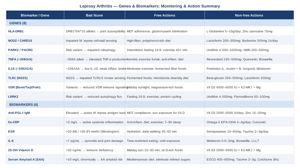
Tracking even three or four of these markers — particularly anti-PGL-I, hs-CRP, and vitamin D — gives a workable monitoring framework that most standard leprosy follow-up protocols do not include. The genetics context below explains why those same numbers may look different from one person to the next.
8 Genes That Shape Your Risk and Response in Leprosy Arthritis
Susceptibility to leprosy, severity of joint involvement, and risk of reactional episodes all carry significant genetic components. The eight genes below represent the most replicated and clinically meaningful variants identified across GWAS studies and candidate gene analyses. Understanding which variants you carry does not change the diagnosis, but it does sharpen the strategy.
HLA-DRB1
What this gene does: HLA-DRB1 encodes a component of the MHC class II molecule, which presents bacterial antigens to CD4+ T-helper cells. Specific alleles — particularly HLA-DRB1*04 and *10 — have been associated with lepromatous leprosy susceptibility and with the development of arthritis. The HLA-DRB1 shared epitope, well-known from rheumatoid arthritis research, appears in some leprosy arthritis patients, contributing to molecular mimicry-driven autoimmune joint destruction where immune responses against M. leprae proteins cross-react with joint tissue.
If the gene is unfavorable, the plan without supplements: Since HLA-DRB1 shapes antigen presentation efficiency, the practical goal is reducing antigenic provocation from non-bacterial sources — primarily gut barrier dysfunction, which allows microbial fragments into systemic circulation and amplifies inflammatory priming. Elimination of gluten and casein for a trial period of 8–12 weeks is supported by the autoimmune protocol literature for HLA-associated inflammatory conditions. Consistent MDT compliance remains the single most important modifier — reducing bacterial antigen load directly reduces the trigger for HLA-mediated joint responses.
If the gene is unfavorable, the plan with supplements or equipment: L-glutamine (5–10 g/day) and zinc carnosine (75 mg twice daily) support intestinal epithelial integrity, reducing bacterial translocation that amplifies HLA-mediated responses. Cycle L-glutamine for 8 weeks with 4-week breaks. Digestive enzymes with meals may reduce the inflammatory antigen load from incompletely digested proteins that can engage HLA antigen presentation pathways.
NOD2 / CARD15
What this gene does: NOD2 (also known as CARD15) encodes a cytosolic pattern recognition receptor that detects muramyl dipeptide, a component of bacterial cell walls including M. leprae. Variants in NOD2 that impair innate immune recognition of mycobacteria compromise the first line of bacterial containment, leading to higher bacterial survival in macrophages and — downstream — a more intense and prolonged adaptive immune response that drives reactional episodes and joint inflammation. Published work in PubMed has associated NOD2 variants with increased susceptibility to multibacillary leprosy.
If the gene is unfavorable, the plan without supplements: NOD2 dysfunction impairs the gut mucosal immune barrier as well as systemic mycobacterial recognition. A high-fiber, polyphenol-rich diet supports butyrate-producing bacteria that independently activate mucosal immunity via parallel pathways, partially compensating for NOD2 insufficiency. Reducing ultra-processed food intake and increasing dietary diversity improves microbial signals that feed into NOD2-independent innate pathways like TLR signaling.
If the gene is unfavorable, the plan with supplements or equipment: Lactoferrin (200–300 mg/day) enhances macrophage activation and mycobacterial containment via pathways partially distinct from NOD2. Berberine (500 mg twice daily, with meals) modulates innate immune signaling and NF-κB activation downstream of NOD2 and has direct antimicrobial activity. Cycle berberine for 8–12 weeks; avoid concurrent use with prescription immunomodulatory agents without medical supervision.
PARK2 / PACRG
What this gene does: The PARK2 and PACRG genes share a regulatory region whose variants were among the first GWAS-confirmed leprosy susceptibility loci, described in the landmark Mira et al. 2004 study. PARK2 encodes parkin, a ubiquitin E3 ligase important for mitophagy — the clearance of damaged mitochondria. PACRG is co-regulated. Risk variants in this locus impair macrophage ability to eliminate M. leprae through mitophagy-linked autophagy pathways, allowing bacterial persistence that sustains immune activation and contributes to chronic arthritis.
If the gene is unfavorable, the plan without supplements: Enhanced mitophagy through non-pharmacological means is achievable. Intermittent fasting (16:8 protocol, fasting window from 8 PM to 12 PM) is among the most potent activators of autophagy in human tissues. Moderate-intensity exercise for 45+ minutes consistently upregulates mitophagy signaling. Avoiding excessive caloric surplus, particularly from refined carbohydrates, reduces the cellular stress that impairs autophagy flux in PARK2-variant carriers.
If the gene is unfavorable, the plan with supplements or equipment: Urolithin A (500–1,000 mg/day from pomegranate-derived sources or direct supplementation) is the most evidence-supported natural mitophagy activator, with human clinical data showing improved mitochondrial quality markers. Spermidine (1–2 mg/day from wheat germ, or direct supplement) stimulates autophagy via mTOR-independent mechanisms. Both are generally well-tolerated; cycle urolithin A for 12 weeks, assess, then continue if well-tolerated. NAD+ precursors (NMN 250–500 mg/day or NR 300–600 mg/day) support the metabolic context in which PARK2/mitophagy functions most effectively.
TNFA (TNF-alpha gene)
What this gene does: The TNFA gene encodes tumor necrosis factor-alpha, a master regulator of inflammatory responses. The -308G/A polymorphism (rs1800629) produces the high-production A allele, which is associated with elevated TNF-α secretion, increased susceptibility to type 2 lepra reactions, and more severe joint inflammation. Carriers of the TNFA-308A allele mount exaggerated inflammatory responses that, while protective against bacterial spread in early infection, become tissue-destructive in the joints when sustained over weeks to months during reactional episodes.
If the gene is unfavorable, the plan without supplements: Lifestyle-level TNF-α modulation focuses on reducing the signal amplifiers: chronic stress (which drives cortisol-resistant NF-κB activation), excess body fat, sedentary behavior, and sleep insufficiency. Aerobic exercise 5 times weekly at 60–70% maximum heart rate has the strongest lifestyle-level TNF-α suppression effect in clinical data. Anti-inflammatory dietary patterns — particularly Mediterranean and anti-inflammatory diets studied in TNF-related conditions — consistently reduce systemic TNF-α burden over 3–6 months.
If the gene is unfavorable, the plan with supplements or equipment: Resveratrol (150–500 mg/day, trans-resveratrol form, with food) inhibits NF-κB, a primary upstream driver of TNF-α transcription, and reduces systemic TNF levels in inflammatory arthritis trials. Cycle 8 weeks on, 4 weeks off. Quercetin (500–1,000 mg/day) acts synergistically with resveratrol on NF-κB pathways. Boswellia serrata (400 mg twice daily, standardized AKBA content) reduces TNF-α-driven synovitis and has joint-specific anti-inflammatory trial data. Whole-body vibration platforms (15 Hz, 10 minutes daily) have been reported to downregulate TNF-α in inflammatory conditions via mechanosensory pathways.
IL10
What this gene does: IL-10 is the primary anti-inflammatory cytokine in leprosy immunology. The IL10 -1082G/A promoter polymorphism is one of the most studied variants in leprosy, with the low-production -1082AA genotype consistently associated with type 2 lepra reactions, greater joint inflammation, and worse outcomes. When IL-10 production is genetically dampened, the regulatory brake on TNF-α and IFN-γ is weaker — resulting in more intense, longer-lasting inflammatory episodes that do disproportionate joint damage.
If the gene is unfavorable, the plan without supplements: Behaviors that upregulate endogenous IL-10 include moderate aerobic exercise (particularly sustained low-to-moderate intensity rather than high-intensity intervals, which transiently suppresses IL-10), social connection and psychological safety (which reduces cortisol-driven IL-10 suppression), and dietary fiber-derived short-chain fatty acids (butyrate, propionate) from gut fermentation — a well-established IL-10 inducer from the mucosal immune system.
If the gene is unfavorable, the plan with supplements or equipment: Probiotics containing Lactobacillus reuteri and Bifidobacterium longum strains are among the most replicated IL-10 inducers from the gut-immune axis. A high-quality multi-strain probiotic (20–50 billion CFU, 2–3 strains) for 8–12 weeks is a reasonable trial. Melatonin (1–3 mg at bedtime) upregulates IL-10 via multiple immunological pathways and has evidence in autoimmune arthritis contexts. Vitamin D3 (to achieve 50–60 ng/mL serum level) directly stimulates IL-10 secretion from regulatory T-cells — one of the most important immune reasons to correct deficiency in low-IL-10 genotype carriers.
TLR1 (Toll-Like Receptor 1)
What this gene does: TLR1 forms a heterodimer with TLR2 to recognize lipopeptides from M. leprae's cell wall. The TLR1 I602S variant (rs5743618) dramatically impairs this recognition and is one of the strongest single-gene susceptibility loci for leprosy confirmed in multiple populations. A landmark study published in Nature Genetics (2007) identified this variant as significantly protective or risk-conferring depending on allele. Impaired TLR1/2 signaling means reduced innate immune activation against the bacterium, lower initial bacterial clearance, and an immune profile that favors lepromatous (high bacterial load) disease with persistent antigen-driven joint inflammation.
If the gene is unfavorable, the plan without supplements: TLR2 signaling (which TLR1 partners with) is enhanced by appropriate microbial diversity. Fermented foods (kefir, kimchi, kombucha, miso) and diverse prebiotic fiber expose TLR2 to a broad range of microbial ligands, maintaining signaling pathway sensitivity. Reducing antibiotic use to truly necessary courses preserves the microbiome-TLR2 signaling relationship that partially compensates for TLR1 impairment.
If the gene is unfavorable, the plan with supplements or equipment: Beta-glucan (250–500 mg/day from oat or yeast sources) activates innate immune cells via Dectin-1 and amplifies TLR2-mediated responses, partially compensating for TLR1 deficiency. Lactoferrin enhances TLR-mediated macrophage activation. Both can be taken continuously at the lower end of dose ranges; assess tolerance over 4 weeks before escalating.
VDR (Vitamin D Receptor)
What this gene does: The VDR gene encodes the nuclear receptor that mediates all genomic effects of vitamin D. Polymorphisms including BsmI (rs1544410), TaqI (rs731236), and FokI (rs2228570) affect receptor expression, binding affinity, and downstream gene activation. VDR variants have been associated with leprosy susceptibility in multiple South Asian and Brazilian cohorts, and more critically for arthritis, impair the VDR-mediated upregulation of cathelicidin LL-37 — the antimicrobial peptide that kills M. leprae in macrophages. Poor VDR function means that even adequate serum vitamin D fails to translate into adequate immune activation.
If the gene is unfavorable, the plan without supplements: VDR expression is upregulated by butyrate from fermented fiber, physical exercise, and magnesium sufficiency. Because poor VDR function reduces the efficiency of vitamin D conversion, targeting a higher serum 25-OH-D level (55–70 ng/mL rather than the minimal 30 ng/mL) is a rational approach to maintain functional VDR-mediated immune signaling despite reduced receptor efficiency. Sunlight exposure specifically activates VDR in skin immune cells independently of systemic vitamin D levels, adding direct skin immune benefit.
If the gene is unfavorable, the plan with supplements or equipment: VDR poor responders typically need 30–50% higher vitamin D3 doses to achieve equivalent serum levels and immune effects — often 4,000–6,000 IU/day. Always paired with K2-MK7 (200 mcg) and magnesium. Lithium orotate at low doses (5 mg, not the pharmacological psychiatric dose) has preliminary data for upregulating VDR gene expression, though human evidence in this context is very early and it should only be considered with physician awareness. Monitor calcium and 25-OH-D every 3 months.
LRRK2 (Leucine-Rich Repeat Kinase 2)
What this gene does: LRRK2 encodes a multifunctional kinase involved in vesicular trafficking, autophagy, and innate immune signaling. Variants in LRRK2 were identified as leprosy susceptibility loci in GWAS studies and are functionally relevant because LRRK2 regulates autophagosome formation — the cellular process that engulfs and destroys intracellular M. leprae. Impaired LRRK2 function reduces the efficiency of macrophage-mediated bacterial clearance, extending the period of antigenic stimulation that drives chronic immune activation and joint inflammation.
If the gene is unfavorable, the plan without supplements: Autophagy upregulation strategies apply here as with PARK2: time-restricted feeding, caloric moderation, and regular aerobic exercise are the cornerstone lifestyle autophagy activators. The LRRK2-autophagy pathway is specifically responsive to mTOR inhibition — achieved most practically through protein cycling (reduced animal protein intake 1–2 days/week), which transiently reduces mTOR signaling and allows autophagy recovery.
If the gene is unfavorable, the plan with supplements or equipment: Urolithin A (500 mg/day) and spermidine target autophagy via mechanisms partially convergent with LRRK2 function and represent the best current non-prescription options. Pterostilbene (50–100 mg/day, a more bioavailable resveratrol analog) activates AMPK, which inhibits mTOR and promotes autophagy flux relevant to LRRK2-pathway deficiency. Take with meals; generally well-tolerated for 12-week cycles with 4-week breaks.
A Podcast That Reframes Chronic Inflammation and Immune Control
The Huberman Lab podcast episode with Dr. Roger Seheult — a pulmonary and critical care physician — titled "Improving Immunity: How to Avoid Illness & Strengthen Your Immune System" covers immune optimization science in ways directly applicable to chronic infectious-inflammatory conditions. The ten most impactful points for leprosy arthritis specifically are worth extracting:
1. Vitamin D Is Immune Infrastructure, Not Just Bone Support
Seheult and Huberman walk through the mechanistic role of vitamin D in activating T-regulatory cells and macrophage antimicrobial function. They challenge the idea that "normal" lab values are optimal — particularly for people with ongoing infectious or inflammatory loads — and consistently support targeting higher functional levels (50+ ng/mL).
2. Morning Sunlight Has Immune Effects Beyond Vitamin D
Early morning solar exposure (non-UV dawn light) sets circadian rhythms that directly regulate the timing of cytokine production. Disrupted circadian rhythm causes dysregulated IL-6 and TNF-α pulses — making light exposure timing a surprisingly direct immune intervention.
3. Chronic Stress Suppresses Exactly the Immune Cells You Need
Natural killer cells and CD8+ cytotoxic T-cells — both critical for mycobacterial surveillance — are selectively downregulated by sustained cortisol elevation. Stress reduction is not optional adjunct care; it is directly immunomodulatory at the cellular level.
4. Sleep Is When Immune Memory Consolidates
During slow-wave sleep, immune memory T-cells migrate to lymph nodes and undergo restimulation. Chronic sleep insufficiency measurably reduces immunological memory and increases the risk of reactivation of persistent infections — highly relevant to mycobacterial conditions.
5. Exercise Dose Matters: Too Little or Too Much Both Impair Immunity
The J-curve of exercise and immune function means that moderate, consistent activity (30–50 minutes, moderate intensity, most days) optimizes immune surveillance, while chronic over-training suppresses mucosal IgA and NK cell activity. The arthritis-relevant sweet spot is lower-intensity daily movement.
6. Gut Microbiome Diversity Directly Shapes Systemic Immune Tone
Greater gut microbiome diversity correlates with better regulatory T-cell balance, lower systemic IL-6, and enhanced mucosal barrier integrity. Fermented foods outperformed high-fiber diets in a Stanford clinical trial for microbiome diversity — a practical finding with immune significance.
7. Cold Exposure Activates Anti-Inflammatory Norepinephrine Pathways
Regular cold water exposure triggers a norepinephrine release that reduces pro-inflammatory cytokines at the cellular level. The effect is dose-dependent and cumulative. This is especially relevant for TNFA high-producer variants where reducing basal inflammatory tone matters.
8. Sauna and Infrared Exposure Mimic Exercise-Induced Immune Stress
Heat shock proteins activated by sauna use modulate immune responses, reduce circulating inflammatory markers, and improve cardiovascular adaptation. Three to four sessions per week at appropriate temperatures produce measurable CRP and SAA reductions over 12 weeks.
9. Social Connection Has Measurable Immune Effects
Loneliness elevates IL-6 and CRP by mechanisms paralleling chronic stress. Conversely, quality social connection upregulates IL-10 and reduces basal pro-inflammatory tone — one of the few interventions that reliably shifts the IL-10/TNF-α balance in the desired direction.
10. Foundational Habits Precede Any Supplement Decision
Seheult is emphatic that supplementation on top of poor sleep, chronic stress, and sedentary behavior produces marginal returns. The sequence matters: anchor sleep, movement, and stress first; then layer targeted supplements over that foundation. This sequencing principle is directly applicable to managing leprosy arthritis complications.
Complementary Approaches Worth Considering
The Autoimmune Protocol (AIP) — Sarah Ballantyne
The Autoimmune Protocol, developed by Dr. Sarah Ballantyne (a research scientist with a background in cellular biology), is a structured elimination and reintroduction dietary framework designed specifically for conditions involving immune dysregulation and chronic inflammation. It removes all foods that are known or suspected to increase intestinal permeability or directly stimulate immune activation — grains, legumes, nightshades, dairy, eggs, nuts, seeds, and all processed foods — while maximizing nutrient density from organ meats, seafood, colorful vegetables, and fermented foods. The relevance to leprosy arthritis is direct: the immune dysregulation central to lepra reactions and chronic joint inflammation is potently influenced by gut barrier integrity and dietary antigen load.
A pilot study published in Inflammatory Bowel Diseases (2017) demonstrated significant clinical remission in Crohn's disease patients following the AIP diet within 6 weeks — an autoimmune condition with overlapping immunological mechanisms to the chronic inflammation of leprosy arthritis. Ballantyne's work draws on over 1,200 studies and is documented in The Paleo Approach.
Apply it with a minimum 30-day strict elimination phase, then systematic reintroduction every 5–7 days to identify individual trigger foods. Work with a registered dietitian if possible, particularly given the nutritional demands of recovering from leprosy-related systemic illness. Do not use this as a replacement for MDT or medical treatment — it is an adjunct strategy for reducing the dietary drivers of immune reactivity.
Mindfulness-Based Stress Reduction (MBSR)
MBSR is an 8-week structured program combining mindfulness meditation, body scan, and gentle movement, developed by Dr. Jon Kabat-Zinn. Its relevance to leprosy arthritis is both direct and mechanistic: chronic pain and the psychosocial burden of a stigmatized diagnosis create a cortisol-dominant stress environment that elevates TNF-α, suppresses IL-10, and amplifies reactional episode severity. MBSR directly targets this stress-inflammation loop.
A meta-analysis published in Psychoneuroendocrinology (2017) confirmed that MBSR produces measurable reductions in inflammatory biomarkers including IL-6 and CRP across a range of chronic conditions, with stronger effects in individuals with higher baseline inflammatory burden. The cortisol-lowering and IL-10-upregulating effects are particularly relevant for IL10 low-producer and TNFA high-producer genetic variants discussed above.
Practical implementation: the full 8-week MBSR program is available online and in-person through certified teachers (find programs via the University of Massachusetts Mindfulness Center). Begin with 10 minutes daily of body scan or breath awareness and build to 30–45 minutes over 4 weeks. The key is consistency over duration — daily brief practice outperforms occasional longer sessions for inflammatory marker effects.
Low-Level Laser Therapy (Photobiomodulation)
Photobiomodulation (LLLT) uses specific wavelengths of red and near-infrared light (typically 630–850 nm) to stimulate mitochondrial function in cells, reduce oxidative stress, inhibit NF-κB activation, and lower pro-inflammatory cytokine production directly at the tissue level. In joints, this translates to reduced synovial IL-6 and TNF-α, improved cartilage cell viability, and clinically meaningful pain reduction in multiple arthritis conditions.
A systematic review in BMC Musculoskeletal Disorders confirmed significant short-term pain and morning stiffness improvement with LLLT in rheumatoid arthritis patients, with an excellent safety profile. The anti-inflammatory mechanism — particularly NF-κB inhibition — is directly relevant to leprosy arthritis, where TNF-α and IL-6 drive much of the joint damage.
Protocol: a clinical-grade LLLT device (650–830 nm, 5–50 mW/cm² at the target tissue) applied to affected joints for 10–15 minutes, 3 times per week. Consumer-grade red light therapy panels (630–660 nm and 850 nm combined) at 6–12 inches from the joint for 10–20 minutes per session are a practical home alternative. Results build over 4–8 weeks of consistent use. Contraindicated over actively infected or open tissue.
Yoga
Yoga is directly applicable to leprosy arthritis for several converging reasons: it improves joint range of motion in inflamed joints without high-impact loading, activates the parasympathetic nervous system (reducing cortisol-driven cytokine elevation), and has demonstrated anti-inflammatory effects in clinical arthritis populations. For a condition where neuropathy and joint deformity may already be present, yoga provides a low-risk movement strategy that can be adapted to individual physical limitations.
A randomized controlled trial published in the Journal of Rheumatology (2015) found that 8 weeks of yoga significantly reduced CRP and ESR in active rheumatoid arthritis, while improving disease activity scores — markers and mechanisms directly relevant to leprosy arthritis joint involvement.
Practical application: start with a restorative or gentle yoga style (Yin yoga or chair yoga if mobility is limited). Avoid deep twists or inversions during active reactional episodes. A 30-minute session 3–4 times weekly is sufficient to achieve inflammatory marker benefits. Online programs or live classes specifically designed for inflammatory arthritis patients provide appropriate modifications. Yoga should be avoided on days of acute type 2 reactions when systemic inflammation is high — rest takes precedence.
Microbiome-Directed Therapies
The gut microbiome is increasingly recognized as a central modulator of systemic immune tone — directly relevant to leprosy arthritis given the role of gut-derived short-chain fatty acids in IL-10 production, regulatory T-cell induction, and the mucosal immune signals that shape TLR and NOD2 pathway responsiveness. Microbial dysbiosis, common in both leprosy patients and those with chronic antibiotic exposure from MDT, depletes the butyrate-producing bacteria whose metabolites directly reduce TNF-α and enhance IL-10.
Human clinical evidence for microbiome intervention in inflammatory arthritis is emerging: a study in Nature Medicine (2019) demonstrated that specific gut microbiome profiles predicted response to anti-inflammatory treatment in rheumatoid arthritis, reinforcing the mechanistic link. The gut-joint axis is real and clinically measurable.
Implementation: begin with dietary foundation — diverse plant fiber from 30+ different plant sources per week, daily fermented foods (kefir, kimchi, natural yogurt), minimal emulsifier-containing processed foods. Add a clinically validated probiotic containing Lactobacillus reuteri ATCC 6475 and Bifidobacterium longum strains for 8–12 weeks. If MDT has been completed, a formalized 4–6 week post-antibiotic microbiome restoration protocol (high-fiber diet plus probiotic plus prebiotic such as GOS or inulin at 4–8 g/day) is warranted. Avoid self-prescribing fecal microbiota transplant — this requires medical supervision.
Conclusion
Leprosy arthritis is manageable but rarely managed with the precision it deserves. Standard follow-up focuses on bacterial clearance and nerve damage — vital priorities, but insufficient for patients whose joint disease is driven by immune genetic variants, cytokine dysregulation, and secondary inflammatory processes that persist long after bacterial loads are controlled. The six biomarkers outlined here — anti-PGL-I, hs-CRP, ESR, IL-6, vitamin D, and serum amyloid A — give you a concrete monitoring framework that maps to both disease activity and complication risk. The eight genes add the biological context that explains why the same disease behaves so differently from one person to the next.
The next smart step is not to implement everything simultaneously. Start with the most accessible and highest-impact actions: get your vitamin D tested, check your hs-CRP, and discuss whether your current follow-up protocol includes anti-PGL-I antibody monitoring. Bring the genetic context to your rheumatologist or infectious disease specialist as a framework for understanding your inflammatory profile, not as a substitute for their clinical judgment. Better information leads to better questions, better conversations, and ultimately better outcomes — that is the real purpose of going this deep.
Musculoskeletal: Joint Conditions
Autoimmune: Inflammatory Conditions
Infectious: Bacterial Infections