This article contains AI generated content.
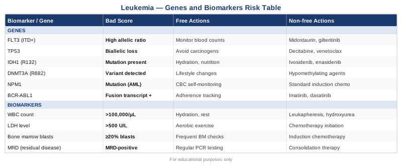
· UpdatedLeukemia Genes and Biomarkers - 8 Genes And 7 Biomarkers To Track
Living with a leukemia diagnosis — whether your own or a loved one's — puts you in a position most people are completely unprepared for: suddenly, you are expected to make sense of a dense stream of lab results, genetic reports, and clinical recommendations. The emotional weight is enormous, but what often makes it worse is the feeling that you are not quite sure which numbers actually matter and why. That gap is real, and it deserves a direct answer.
Leukemia is not a single disease. Acute myeloid leukemia (AML), acute lymphoblastic leukemia (ALL), chronic myeloid leukemia (CML), and chronic lymphocytic leukemia (CLL) each have distinct genetic drivers, different disease trajectories, and separate prognostic markers. Advice that applies well to CML may be largely irrelevant to CLL, and the biomarkers that guide AML management look very different from those used to monitor CLL. Generic reassurances about "staying positive" skip over the part where knowing your FLT3 status or your MRD result could change your treatment path entirely.
This article takes a different approach. It focuses on two specific levels of understanding: the biomarkers — measurable values in blood, bone marrow, and specialized tests — that give the clearest real-time signal about disease activity and treatment response; and the genetic markers that increasingly determine how a leukemia behaves, how aggressively it should be treated, and which targeted therapies have a chance of working. Both levels are actionable in the sense that they inform decisions, open conversations with your care team, and in some cases guide lifestyle and supportive interventions with genuine evidence behind them.
The goal is not false hope, and this article does not promise cures. What it does offer is a more precise map: seven biomarkers to track with clear guidance on measurement, interpretation, and what to do if results move in the wrong direction, plus eight genetic markers that are reshaping how oncologists understand and treat leukemia. Better information, read carefully and discussed with qualified specialists, leads to better decisions. That is where genuine hope lives.
7 Biomarkers That Define Your Leukemia Landscape
Biomarkers in leukemia management serve three distinct roles: they help confirm diagnosis, they track treatment response, and they identify complications before they become crises. The seven below are not the only relevant markers, but they represent the most consistently informative ones across the major leukemia subtypes. Each has a measurement method, a cost range, and a concrete plan when results are off-target — both with and without supplements or specialized interventions.
1. Complete Blood Count with Differential
Why it matters: The complete blood count with differential is the most fundamental test in leukemia monitoring. It measures red blood cells, white blood cells, and platelets, while the differential component specifies which types of white cells are present — including abnormal blasts that signal disease. In active AML or ALL, a high white blood cell count with elevated blast percentage is a direct indication of disease burden. During and after treatment, the recovery of absolute neutrophil count (ANC) above 1,000/µL and platelets above 100,000/µL defines complete hematologic remission and clearance from aplasia.
How to measure: Standard venous blood draw at any clinical or hospital laboratory. Typically ordered every 1–2 weeks during induction chemotherapy, then monthly in stable remission, or as directed. The NCI's leukemia overview notes that serial CBCs remain the backbone of clinical follow-up regardless of leukemia subtype. Cost: approximately $25–$80 with insurance, $100–$200 without.
If the score is bad — the plan without supplements: When neutrophil counts are dangerously low (ANC below 500/µL), the most important free actions are behavioral and hygienic: rigorous handwashing, N95 masks in crowded settings, avoidance of raw or undercooked food, and immediate reporting of any fever above 38°C (100.4°F). For patients with CLL or CML in chronic phase whose WBC is elevated but blasts are absent, moderate aerobic walking (20–30 minutes daily at comfortable pace) supports immune tone without stressing recovering marrow. Adequate sleep — 7–9 hours — is not trivial here; disrupted sleep measurably impairs immune surveillance.
If the score is bad — the plan with supplements or equipment: Granulocyte colony-stimulating factor (G-CSF, e.g., filgrastim) is a prescription medication used in clinical practice to accelerate neutrophil recovery after chemotherapy — discuss timing precisely with your oncologist, as it is not appropriate in all protocols. For anemia not related to iron deficiency, erythropoiesis-stimulating agents may be considered in CLL or MDS-related contexts. Protein intake of 1.2–1.5g/kg/day supports marrow recovery; whey or pea protein supplementation is a reasonable adjunct when dietary intake is limited by nausea or appetite loss. Iron supplementation should only follow confirmed iron deficiency (check ferritin first — iron given unnecessarily to a patient with elevated ferritin from inflammation can cause harm).
2. Lactate Dehydrogenase (LDH)
Why it matters: LDH is an intracellular enzyme released into the bloodstream when cells are rapidly destroyed or proliferating. In leukemia, an elevated LDH reflects high tumor burden and accelerated cellular turnover. It is a reliable prognostic signal in aggressive leukemias and lymphomas — multiple large cohort studies have confirmed that high baseline LDH correlates with shorter remission duration and lower overall survival in AML and aggressive ALL. It is also the first laboratory sign of tumor lysis syndrome (TLS), the potentially fatal metabolic emergency that can follow effective treatment when large numbers of malignant cells die simultaneously, releasing potassium, phosphate, and uric acid into the bloodstream.
How to measure: Often included in a comprehensive metabolic panel (CMP) or ordered standalone. Cost: $20–$60. Reference range: approximately 140–280 U/L, though laboratory-specific ranges vary. Trends matter more than single values — a rising LDH during treatment warrants immediate clinical attention.
If the score is bad — the plan without supplements: The most critical free intervention is aggressive hydration: 2–3 liters of fluid per day (unless cardiac or renal function restricts this) reduces the risk of uric acid crystallization and acute kidney injury during periods of high cell turnover. Avoid NSAIDs during high-LDH periods, as they reduce renal perfusion and can accelerate nephrotoxicity. Immediately report dark urine, reduced urination, muscle cramps, or unusual fatigue — these are early signals of TLS or kidney stress.
If the score is bad — the plan with supplements or equipment: Allopurinol (a xanthine oxidase inhibitor) is standard medical prophylaxis for TLS in at-risk patients — it is prescription-only and must be started before chemotherapy, not during crisis. Rasburicase is used in high-risk TLS when uric acid is already dangerously elevated. From a dietary angle, reducing purine-rich foods (organ meats, sardines, anchovies, shellfish) and fructose-containing beverages during treatment cycles is a low-risk, modest adjunct. Sodium bicarbonate IV may be used to alkalinize urine in hospital-managed TLS — this is not a home intervention.
3. Beta-2 Microglobulin (B2M)
Why it matters: Beta-2 microglobulin is a small protein found on the surface of most nucleated cells, including lymphocytes. When lymphocytes are proliferating rapidly or being destroyed, B2M is shed into the circulation. Elevated serum B2M is one of the strongest independent prognostic markers in CLL, outperforming traditional clinical staging in several analyses. High B2M at diagnosis predicts shorter time to first treatment and lower overall survival. A 2019 study in Blood demonstrated that B2M combined with TP53 mutational status and IGHV mutation status provided superior risk stratification compared to Rai or Binet staging alone in newly diagnosed CLL patients.
How to measure: Simple venous blood draw; available through LabCorp, Quest Diagnostics, and most hospital labs as a standalone test. Cost: $40–$120. B2M can also be measured in 24-hour urine when renal assessment is needed. Normal serum range: under 2.5 mg/L in most labs.
If the score is bad — the plan without supplements: Elevated and rising B2M is a signal to review with your hematologist whether the watch-and-wait strategy remains appropriate — it may indicate disease progression toward treatment threshold. Aerobic exercise (targeting 150 minutes per week at moderate intensity) has been shown in multiple cancer cohorts to reduce systemic inflammation markers, which may slow the inflammatory milieu driving lymphocyte turnover. This is supportive biology, not a cure, but the signal is consistent enough to take seriously.
If the score is bad — the plan with supplements or equipment: No supplement directly targets B2M as a mechanism. However, interventions that dampen chronic inflammation — vitamin D optimization (target serum level 50–70 ng/mL), omega-3 fatty acids at 2–4g EPA+DHA daily, and removal of excess refined carbohydrates that drive insulin/mTOR-mediated inflammation — are biologically rational adjuncts, even if direct B2M-lowering RCTs in CLL are not yet available. Discuss these with your oncology team in the context of your specific treatment plan.
4. Minimal Residual Disease (MRD)
Why it matters: MRD testing is the most sensitive biomarker currently in clinical use for leukemia. It detects whether any cancer cells remain after treatment — even when a patient is in apparent remission by all conventional criteria. MRD negativity (no detectable leukemia cells at sensitivities of one in 10,000 to one in one million normal cells) is now considered the strongest single predictor of long-term remission-free survival in ALL and AML. In CLL, achieving undetectable MRD after treatment correlates with significantly longer progression-free survival, and this metric is now shaping treatment duration decisions in trials with venetoclax-based regimens. MRD is not just a number — it is increasingly a clinical decision point.
How to measure: Two main technologies are used: multiparameter flow cytometry (MFC, which identifies leukemic cells by surface protein patterns) and molecular testing by PCR or next-generation sequencing (which detects leukemia-specific gene fusions or mutations, including BCR-ABL1 transcripts in CML). Bone marrow is the standard sample for most leukemia types, though peripheral blood is used for CML BCR-ABL1 monitoring and increasingly for CLL MRD. Measured at defined treatment milestones. Cost: $300–$1,200+ depending on method and lab; typically covered when ordered as part of active leukemia management.
If the score is bad (MRD positive when target is negative): This is a clinical signal requiring immediate discussion with your oncologist about next steps — which may include treatment intensification, a change of regimen, or evaluation for stem cell transplant or CAR-T cell therapy. The NCI overview of targeted therapies describes how MRD-directed adjustments are increasingly integrated into modern protocols.
If the score is bad — supportive actions: No lifestyle intervention has been shown in human trials to directly clear MRD. However, supporting the immune system's own surveillance capacity is legitimate biology: adequate sleep (7–9 hours consistently) supports T-cell function and NK cell activity, both of which contribute to leukemia cell clearance. Protein adequacy (1.2–1.5g/kg/day) supports immune cell production. Psychological stress reduction is not trivial here — chronic psychological stress elevates cortisol, which has documented immunosuppressive effects on lymphocyte function. Mindfulness-based stress reduction (discussed in the complementary section) has RCT evidence in cancer patients.
5. Ferritin
Why it matters: Ferritin plays two clinically distinct roles in leukemia. At the low end, iron deficiency is common in patients with impaired red blood cell production (as in AML), and depleted ferritin directly explains much of the fatigue, cognitive fog, and exercise intolerance that patients attribute entirely to cancer or chemotherapy. At the high end, hyperferritinemia (ferritin above 1,000–2,500 ng/mL) is an inflammatory marker associated with macrophage activation syndrome and cytokine release states seen in some acute leukemia presentations — and after stem cell transplant. Elevated ferritin in the context of transfusion overload (common in patients requiring multiple red cell transfusions) also signals iron loading that can damage the liver, heart, and endocrine organs over time.
How to measure: Standard blood draw, often included in an iron panel alongside serum iron and TIBC. Cost: $20–$60. Reference ranges: approximately 12–300 ng/mL for men, 12–150 ng/mL for women. In leukemia management, both ends of the range are actively monitored.
If the score is bad (low ferritin — below 12 ng/mL) — without supplements: Prioritize dietary iron from heme sources (red meat, lamb, dark poultry) and non-heme sources (legumes, spinach, tofu), always paired with vitamin C to maximize absorption. Avoid tea and calcium-rich foods within 1–2 hours of iron-containing meals, as they competitively inhibit absorption.
If the score is bad (low ferritin) — with supplements or medical intervention: Oral ferrous sulfate (325mg, providing 65mg elemental iron) taken every other day has been shown in randomized trials to produce equivalent or better absorption than daily dosing with fewer GI side effects — a finding highlighted by Thomas Dayspring in discussions of practical iron repletion. IV iron infusion (ferric carboxymaltose or iron sucrose) is indicated when oral supplementation is insufficient or when GI side effects limit adherence, particularly before chemotherapy cycles. Always recheck ferritin and CBC at 6–8 weeks.
If the score is bad (high ferritin/inflammatory hyperferritinemia) — medical management: Iron chelation with deferasirox (oral, once daily) or deferoxamine (subcutaneous infusion) is used in transfusion-dependent patients with iron overload. Discuss initiation threshold (often ferritin consistently above 1,000 ng/mL with multiple transfusions) with your hematologist. Anti-inflammatory dietary patterns (Mediterranean diet, omega-3 supplementation) are reasonable adjuncts to reduce inflammatory contribution to ferritin elevation.
6. C-Reactive Protein and Interleukin-6
Why it matters: Chronic systemic inflammation is no longer viewed as merely a consequence of cancer — it is increasingly understood as a driver. IL-6, in particular, activates the JAK-STAT3 signaling pathway that promotes survival and proliferation in multiple leukemia subtypes. Elevated high-sensitivity CRP (hsCRP) and IL-6 correlate with disease activity in CLL and have been linked to treatment intolerance in AML. Tracking these markers also serves a critical practical function: during chemotherapy, a rising CRP may reflect a bacterial infection that needs urgent treatment, while a stable CRP in a febrile patient might suggest drug fever rather than sepsis — a clinically critical distinction.
How to measure: hsCRP by standard venous blood draw: $15–$50, widely available. IL-6 is less routinely ordered but available at major reference labs and is increasingly used in transplant and cytokine release syndrome monitoring. Cost for IL-6: $80–$200. Normal hsCRP: below 1 mg/L (low cardiovascular risk range); values above 3 mg/L indicate significant systemic inflammation.
If the score is bad — the plan without supplements: The highest-yield free interventions for systemic inflammation reduction are: elimination of smoking (which raises CRP independently of any cancer process), consistent sleep schedule (sleep disruption acutely and dose-dependently raises IL-6), and moderate aerobic exercise 3–5 times per week. A dietary shift away from ultra-processed foods, refined carbohydrates, and industrial seed oils reduces dietary inflammatory load. These changes do not replace oncologic treatment, but they reduce the inflammatory burden your immune system is operating under.
If the score is bad — the plan with supplements: Omega-3 fatty acids at 2–4g combined EPA+DHA daily have the strongest evidence base among supplements for lowering CRP in inflammatory conditions (multiple meta-analyses confirm this). Curcumin (1,000–2,000mg/day standardized extract, with piperine for bioavailability) has shown anti-inflammatory effects in human trials and pre-clinical anti-leukemic properties at high concentrations; cycle 8 weeks on, 2 weeks off, and discuss with your oncology team before initiating during active chemotherapy, as interactions with drug metabolism exist. Vitamin D normalization (targeting 50–70 ng/mL) is consistently associated with reduced inflammatory marker levels in deficient populations.
7. Bone Marrow Blast Percentage
Why it matters: The percentage of blast cells (immature, abnormal cells) in the bone marrow is the single most direct measure of leukemic disease burden and treatment response. Greater than 20% blasts by WHO criteria defines AML, distinguishing it from myelodysplastic syndrome. After induction chemotherapy, achieving less than 5% blasts — the threshold for morphological complete remission — is the primary treatment goal in AML. Even within remission, residual blast levels below 5% predict very different outcomes depending on whether MRD is negative or positive at the molecular level. In MDS-related leukemia, blast trajectories over time are more informative than any single measurement.
How to measure: Bone marrow aspiration and trephine biopsy, performed by a hematologist under local anesthesia at the posterior iliac crest. Typically done at diagnosis, post-induction, at relapse, and before transplant. More uncomfortable than blood draws, but essential information. Cost: $2,000–$5,000+ for the procedure; typically covered when medically indicated in active leukemia management.
If the score is bad — clinical priority: High or rising blast percentage represents an emergency in AML and a treatment-threshold event in MDS/AML. The primary action is oncology management — salvage chemotherapy, transplant evaluation, or clinical trial enrollment. From a supportive standpoint, maintaining adequate nutritional status through treatment preserves eligibility for aggressive therapies. Patients with severe protein-calorie malnutrition are at higher risk of transplant-related mortality. A registered dietitian with oncology experience is a valuable adjunct to the care team for patients with impaired intake.
If the score is bad — supportive plan with supplementation: High-calorie oral nutritional supplements (such as Ensure or similar products, or protein-enriched smoothies) support caloric and protein goals when appetite is suppressed. Glutamine supplementation (10–30g/day) has some evidence supporting mucositis reduction during high-dose chemotherapy, which in turn supports caloric intake by reducing mouth pain and swallowing difficulty. Evidence quality is moderate; always check with your oncology pharmacist for compatibility with your specific regimen.
The seven biomarkers above create a practical monitoring framework. Each reflects a different dimension of leukemia biology, and together they give a more complete picture than any single value could provide. The next layer of understanding goes deeper — to the genetic level — where the mutations present at diagnosis increasingly determine not just prognosis, but which treatments are most likely to work.
8 Genetic Markers That Shape Leukemia Biology
Genetic testing in leukemia has transformed from a research curiosity into a clinical necessity. The mutations found in leukemic cells at diagnosis determine risk stratification, guide treatment selection, and — in an expanding number of cases — are directly targeted by approved drugs. Understanding your mutation profile, or a loved one's, is no longer optional information. These eight genetic markers are among the most clinically consequential.
BCR-ABL1 — The Philadelphia Chromosome
What it is: The BCR-ABL1 fusion gene results from a translocation between chromosomes 9 and 22, creating the Philadelphia chromosome. It encodes a constitutively active tyrosine kinase that drives uncontrolled cell proliferation. BCR-ABL1 is present in virtually all CML patients and in approximately 25–30% of adult ALL cases, where its presence historically predicted a poor outcome.
Why it matters now: BCR-ABL1 is the defining example of targeted therapy success in oncology. Imatinib (Gleevec), approved in 2001, was the first targeted kinase inhibitor to demonstrate survival benefit in cancer — transforming CML from a disease with median survival of 3–5 years into one where patients can now achieve treatment-free remission and essentially normal life expectancy with continued therapy. Second- and third-generation TKIs (dasatinib, nilotinib, ponatinib, asciminib) have been developed for imatinib-resistant disease.
If the gene is detected — plan without supplements: Strict adherence to TKI therapy is the most important free action — missed doses are the primary cause of treatment failure and resistance development. Regular BCR-ABL1 PCR monitoring (every 3 months in CML on treatment) should not be skipped. Exercise (particularly aerobic activity) during TKI therapy is safe, reduces fatigue, and supports cardiovascular health — important because some TKIs carry cardiovascular risks with long-term use.
If the gene is detected — plan with supplementation or equipment: Grapefruit and Seville orange should be avoided with some TKIs (particularly nilotinib) due to CYP3A4 inhibition that can raise drug levels. Vitamin D deficiency is common in CML patients and associated with inferior outcomes in some analyses — repletion to 50–70 ng/mL serum level is a low-risk adjunct. Omega-3 supplementation may mitigate some of the cardiovascular effects associated with second-generation TKIs, though this has not been studied in a dedicated RCT.
FLT3 — The Prognostic and Therapeutic Target in AML
What it is: Mutations in the FLT3 gene occur in approximately 25–30% of AML cases and come in two forms: internal tandem duplications (FLT3-ITD), which carry a significantly worse prognosis, and tyrosine kinase domain (TKD) mutations, with more variable implications. FLT3-ITD drives constitutive kinase signaling and promotes blast proliferation.
Why it matters: FLT3-ITD positivity was historically associated with high relapse rates even after achieving remission. Multiple approved FLT3 inhibitors now exist: midostaurin (in combination with standard chemotherapy for newly diagnosed FLT3-mutated AML) and gilteritinib (for relapsed/refractory FLT3-mutated AML). Allelic ratio — the proportion of FLT3-ITD relative to wild-type FLT3 — provides additional prognostic granularity.
If the gene is mutant — plan without supplements: Enroll at a center with clinical trial access if possible — FLT3 is one of the most active areas of AML drug development. Maintain a healthy weight during and after treatment; adipose tissue expresses FLT3 ligand and may sustain residual leukemic niches, though this is based on pre-clinical data.
If the gene is mutant — plan with supplementation: No supplement directly inhibits FLT3. Anti-inflammatory diet patterns (Mediterranean diet) and omega-3 supplementation reduce the inflammatory background that FLT3 signaling intersects with. Vitamin D receptor signaling has shown pre-clinical inhibitory effects on FLT3-expressing leukemic cells, providing a biologically plausible rationale for vitamin D optimization during remission maintenance — though clinical trials are needed to confirm this.
NPM1 — A Favorable Marker with Surveillance Implications
What it is: NPM1 mutations (most commonly a 4-base insertion in exon 12) occur in approximately 30% of AML cases and are typically associated with favorable prognosis when FLT3-ITD is absent. NPM1-mutated AML without concurrent FLT3-ITD high allelic ratio is classified as a favorable-risk subgroup by ELN 2022 guidelines — one of the few AML subtypes where intensive chemotherapy alone, without transplant, may achieve durable remission.
Why MRD monitoring is critical: NPM1 mutations create a patient-specific molecular marker ideal for MRD monitoring by PCR. Rising NPM1 transcript levels in blood or marrow during remission reliably predict relapse weeks to months before clinical signs appear — a window where intervention may still be effective.
If the gene is mutant — plan: NPM1 mutation is often genuinely good news in AML without concurrent high-ratio FLT3-ITD. The priority is rigorous MRD monitoring every 3 months during remission. Lifestyle priorities are identical to those in the biomarker section: adequate sleep, consistent nutrition, and systemic inflammation reduction. No approved supplements modify NPM1 function.
TP53 — The Most Feared Mutation in Leukemia
What it is: TP53 encodes the p53 protein, the cell's master guardian against malignant transformation. TP53 mutations or deletions (particularly del(17p) in CLL) are associated with resistance to most conventional chemotherapy, rapid disease progression, and significantly reduced overall survival across nearly all leukemia subtypes. In AML, TP53-mutated disease has one of the lowest complete remission rates with standard induction chemotherapy.
Recent progress: Venetoclax (a BCL-2 inhibitor) and azacitidine combinations have shown activity in TP53-mutated AML when standard induction is not feasible. Eprenetapopt (APR-246), a small molecule that restores p53 function, has shown early promise in clinical trials specifically in TP53-mutated MDS and AML. Clinical trial enrollment is arguably the most important action for TP53-mutated patients.
If the gene is mutant — plan without supplements: Minimize any additional genotoxic stress: avoid tobacco entirely (compounds UV-induced DNA damage), limit unnecessary radiation exposure (unnecessary CT scans etc.), and discuss with your oncologist whether any medications you take have known p53 pathway interactions. Seek a second opinion at a comprehensive cancer center with TP53-specific trial experience.
If the gene is mutant — plan with supplementation: No supplement restores TP53 function. However, maintaining immune competence through adequate nutrition, vitamin D, and omega-3 status remains relevant, as immune surveillance is even more important when intrinsic tumor suppression is impaired. Avoid prolonged or high-dose zinc supplementation without monitoring — zinc affects p53 binding to DNA and, in excess, has been shown to have pro-oxidant effects in some cancer models.
IDH1 and IDH2 — Actionable Targets for Metabolic Reprogramming
What they are: Isocitrate dehydrogenase 1 and 2 (IDH1 and IDH2) are metabolic enzymes that, when mutated, produce an oncometabolite called 2-hydroxyglutarate (2-HG). 2-HG competitively inhibits α-KG-dependent enzymes involved in DNA demethylation and histone modification, locking cells in an undifferentiated state — a key step in leukemogenesis. IDH1 mutations occur in approximately 6–10% of AML, IDH2 in 8–15%.
Why they matter — targeted therapy: Ivosidenib (IDH1 inhibitor) and enasidenib (IDH2 inhibitor) are approved for relapsed/refractory IDH-mutated AML and, in combination with azacitidine, for newly diagnosed IDH-mutated AML in patients ineligible for intensive chemotherapy. These drugs work by forcing leukemic cells to differentiate rather than killing them directly — a mechanism called differentiation syndrome (an early side effect requiring awareness).
If the gene is mutant — plan without supplements: In addition to standard treatment, metabolic health interventions are specifically relevant here because IDH mutations alter cellular metabolism. A low-glycemic diet that reduces substrate availability for the mutant enzyme's overactive pathway is biologically rational. Intermittent caloric restriction has shown pre-clinical effects on 2-HG production in IDH-mutant cells.
If the gene is mutant — plan with supplementation: Alpha-ketoglutarate (α-KG) supplementation has been explored in pre-clinical IDH-mutant cancer models as a way to competitively overcome 2-HG-mediated enzyme inhibition. Human data in leukemia are not yet available. Magnesium and B-vitamin adequacy (particularly B12 and folate) support methylation pathways that are disrupted by 2-HG. These are supportive measures, not treatments.
DNMT3A — An Epigenetic Driver With Deep Roots
What it is: DNMT3A encodes a DNA methyltransferase responsible for de novo DNA methylation — a fundamental epigenetic control mechanism. DNMT3A mutations occur in approximately 18–22% of AML cases and are one of the earliest genetic events in clonal hematopoiesis of indeterminate potential (CHIP), often preceding leukemia by years or decades. DNMT3A-mutated AML is classified as intermediate risk and is associated with chemotherapy sensitivity, though co-occurring mutations (particularly FLT3-ITD or TP53) worsen this.
Plan without supplements: Lifestyle factors that reduce CHIP progression are relevant: smoking cessation (smoking has been directly linked to accelerated CHIP expansion in large population studies), alcohol moderation (ethanol disrupts DNA methylation patterns), and cardiovascular health optimization (CHIP is also a cardiovascular risk amplifier). Annual CBC monitoring in patients known to have CHIP with DNMT3A mutations can detect early signs of progression.
Plan with supplementation: B vitamins (particularly folate, B6, and B12) are essential cofactors for DNA methylation cycles. Deficiency in any of these, particularly in older adults, can worsen epigenetic drift. Supplementing these to ensure adequacy — not excess — is a reasonable adjunct. SAM-e (S-adenosylmethionine), the universal methyl donor, has been studied for methylation support but should be used cautiously in active cancer settings given its role in cellular growth.
CEBPA — A Biallelic Mutation That Changes the Story
What it is: CEBPA encodes a transcription factor critical for granulocyte differentiation. Single CEBPA mutations confer intermediate risk in AML. However, biallelic (double) CEBPA mutations — affecting both alleles — define one of the most favorable-risk AML subgroups with high complete remission rates and potentially curable outcomes with intensive chemotherapy alone without transplant. This distinction — single versus double mutation — is clinically critical and must be confirmed by molecular testing, not just detection of any CEBPA alteration.
Plan: If biallelic CEBPA-mutated AML is confirmed with no co-occurring adverse mutations, the evidence supports intensive chemotherapy without automatic referral for transplant in first remission. Discuss this explicitly with your oncology team, as the data on transplant-free management in this subgroup is now robust. MRD monitoring by flow cytometry during remission is recommended. No specific supplemental strategies modify CEBPA function.
RUNX1 — Implications for Both AML and Germline Risk
What it is: RUNX1 (also called AML1) encodes a transcription factor essential for normal hematopoiesis. Somatic RUNX1 mutations in AML are classified as adverse risk by ELN 2022 guidelines and are associated with resistance to standard induction chemotherapy and higher relapse rates. Additionally, germline RUNX1 mutations cause RUNX1 familial platelet disorder with predisposition to AML — a rare but increasingly recognized inherited cancer predisposition syndrome.
Plan without supplements: RUNX1-mutated AML warrants upfront consideration of allogeneic stem cell transplant in first remission given adverse-risk classification. Family members of patients with confirmed germline RUNX1 mutations should undergo genetic counseling and surveillance. Physically maintaining conditioning status throughout treatment improves transplant eligibility.
Plan with supplementation: Physical conditioning protocols (structured exercise rehabilitation) during and after induction chemotherapy have shown in several clinical trials to preserve transplant eligibility and reduce post-transplant complications. This is distinct from casual exercise — structured programs at transplant centers, guided by physiotherapists, provide the most evidence-based benefit.
What "The Cancer Code" Reveals About Leukemia Biology
Dr. Jason Fung's 2020 book The Cancer Code: A Revolutionary New Understanding of a Medical Mystery represents one of the most coherent attempts to reconcile the competing theories of cancer causation — genetic, metabolic, and evolutionary — into a unified framework. While the book does not focus on leukemia specifically, its core arguments about the cancer microenvironment, insulin/IGF-1 signaling, and epigenetic programming apply with striking relevance to leukemia biology. Here are the ten most impactful ideas from the book for anyone tracking a leukemia diagnosis.
1. Cancer Is Not Just a Genetic Disease — It Is Also a Cellular Program
Fung argues that cancer does not arise purely from random genetic mutations but represents the activation of an ancient, conserved survival program — a reversal to a more primitive cellular state. This reframes leukemia: the mutations identified (FLT3, NPM1, IDH1/2) are not causes so much as triggers that unlock a growth program already encoded in the genome. This matters because it suggests that metabolic interventions may modify the environment in which that program runs, even if they cannot reverse the mutation itself.
2. Insulin and IGF-1 Are the Two Most Important Cancer Growth Signals in Modern Diets
Fung presents substantial human evidence that chronically elevated insulin and IGF-1 — driven by high-carbohydrate diets, sedentary behavior, and excess body fat — function as powerful cancer growth promoters. Leukemic blasts express insulin receptors and IGF-1 receptors and respond to these signals by activating mTOR and PI3K pathways. Reducing dietary glycemic load and managing body composition are not luxury interventions — they target the signaling environment feeding leukemic cell growth.
3. Intermittent Fasting and Time-Restricted Eating Reduce Key Cancer Promoters
Fung reviews human evidence on fasting showing consistent reductions in insulin, IGF-1, and mTOR activity during caloric restriction and intermittent fasting protocols. While no randomized trial in leukemia patients specifically has tested fasting as an adjunct to chemotherapy, mechanistic and epidemiological data are compelling. Importantly, fasting during chemotherapy (12–24 hours around treatment) has been studied in other cancers and may protect normal cells while sensitizing cancer cells — discuss this with your oncologist before attempting.
4. Obesity Is an Independent Risk Factor for Most Leukemia Subtypes
The book reviews epidemiological data showing obesity increases risk of AML, CLL, and ALL. The mechanism involves chronic adipokine signaling (particularly leptin elevation and adiponectin reduction), inflammatory cytokine production from visceral fat, and hyperinsulinemia. For patients already diagnosed, excess body fat continues to fuel the inflammatory and signaling environment discussed above — weight management is not cosmetic; it is metabolic oncology.
5. The Tumor Microenvironment Is as Important as the Tumor Cell
Fung devotes substantial attention to the idea that cancer cells do not exist in isolation — they actively reshape surrounding tissue to support their growth. In leukemia, the bone marrow niche is the primary microenvironment, and it is increasingly understood to provide survival signals to leukemic blasts through direct cell contact and paracrine cytokine signaling. Interventions that disrupt niche-leukemia cell interaction are an active area of drug development. From a patient perspective, this reinforces the importance of the systemic interventions above.
6. Epigenetics Is the Bridge Between Lifestyle and Cancer Gene Expression
One of Fung's most useful contributions is explaining how epigenetic modifications — DNA methylation, histone acetylation — translate lifestyle factors into gene expression changes that either suppress or activate cancer-related pathways. This is directly relevant to DNMT3A and IDH-mutated leukemias, where epigenetic dysregulation is the central mechanism. Lifestyle factors shown to favorably modify the epigenome include: consistent aerobic exercise, adequate sleep, stress reduction, and dietary methyl donors (folate, B12, betaine).
7. Dietary Fructose Specifically Fuels mTOR-Driven Cancer Growth
Fung presents a specific case against fructose (from added sugars, high-fructose corn syrup) as a uniquely problematic cancer fuel — not because it raises insulin as dramatically as glucose, but because it directly activates lipogenesis and mTOR via independent pathways. Eliminating sugar-sweetened beverages and processed foods containing added fructose is one of the clearest, most specific dietary recommendations that emerges from the metabolic cancer literature.
8. Stress Hormones Directly Impair the Immune Response to Cancer
The cortisol and catecholamine response to chronic psychological stress suppresses NK cell and T-cell activity — the immune cells most responsible for clearing residual cancer cells after treatment. Fung reviews this evidence and connects it to the clinical observation that patients who experience high psychosocial distress have measurably worse cancer outcomes across tumor types. This is not a "think positive" argument — it is mechanistic immunology with clear implications for stress management practices.
9. Metformin Has Genuine Cancer-Modifying Properties Independent of Diabetes
Fung reviews the growing epidemiological and mechanistic evidence that metformin — a widely prescribed diabetes drug — reduces cancer incidence and may improve outcomes in several cancer types, including some leukemia contexts. Metformin activates AMPK and inhibits mTOR, directly countering the growth signals discussed throughout the book. While not yet standard of care in leukemia, several clinical trials are active. This is a meaningful discussion to have with your oncologist if you have diabetes or prediabetes.
10. The Two-Hit Model of Cancer Requires Both Initiating Mutations and Permissive Environments
Fung returns throughout the book to the argument that a mutation alone is not sufficient to produce cancer — it requires a permissive cellular environment. This is supported by the observation that many people carry somatic CHIP mutations (including DNMT3A and TET2) without ever developing leukemia. The permissive environment — driven by inflammation, metabolic dysfunction, immune suppression — is modifiable. This is the book's most important practical message: you cannot change the mutations that initiated the disease, but you can work on the environment in which residual cells are trying to survive.
Complementary Approaches Supported by Clinical Evidence
The following four modalities have meaningful human clinical evidence in cancer patients broadly, or in leukemia specifically. None replaces conventional oncologic treatment. Each is included because it addresses a distinct aspect of the leukemia patient experience with evidence above anecdote.
Mindfulness-Based Stress Reduction (MBSR)
MBSR is an 8-week structured program developed by Jon Kabat-Zinn that combines body scan meditation, seated mindfulness practice, mindful movement, and didactic content on stress and perception. In cancer patients, it directly addresses the chronic psychological stress burden that, as discussed above, has measurable immunosuppressive effects — including on NK cell and T-cell populations critical for residual leukemia cell clearance.
A 2014 randomized controlled trial published in Psychooncology involving 271 cancer patients (mixed tumor types including hematologic malignancies) found that MBSR produced significant reductions in depression, anxiety, fatigue, and global distress compared to wait-list controls, with moderate-to-large effect sizes. In hematologic malignancy patients specifically, a 2018 RCT at the University of Calgary demonstrated that MBSR reduced cortisol levels and improved immune parameters compared to active control conditions, though leukemia-specific enrollment was limited.
For leukemia patients: MBSR programs are available in hospital-affiliated integrative oncology departments, online (MBSR Online from the UMass Center for Mindfulness, the program's origin), and through apps (Insight Timer, Calm). The 8-week structured program is more effective than casual meditation. Start during a period of stable disease or early remission rather than at crisis — the skills are most usable when built in advance of high-stress periods like transplant preparation.
Qigong
Qigong is a traditional Chinese movement and breathing practice that combines slow physical movement, coordinated breath, and meditative intention. In the cancer context, it is studied primarily for its effects on fatigue, quality of life, and immune parameters. The physiological mechanisms proposed include: parasympathetic nervous system activation, reduced cortisol and pro-inflammatory cytokines, and improved lymphatic circulation.
A 2019 systematic review and meta-analysis in Cancer Medicine (analyzing 22 RCTs with 1,751 cancer patients) found that qigong practice significantly reduced cancer-related fatigue, improved quality of life, and reduced anxiety and depression compared to usual care or waitlist controls. One RCT specifically in leukemia and lymphoma patients (conducted in China, 2016) demonstrated improved NK cell activity and reduced IL-6 and TNF-alpha levels after 12 weeks of qigong practice compared to non-practicing controls.
For leukemia patients: Qigong is exceptionally accessible for patients with low physical tolerance — sessions of 15–30 minutes can be adapted to seated practice during periods of fatigue or neutropenia. The Spring Forest Qigong or Lee Holden programs are widely available as DVDs and streaming content. Twice-daily 15-minute sessions appear to produce the most consistent evidence-based benefits in oncology populations. Avoid group classes during periods of immunosuppression; home practice is equivalent in most trial comparisons.
Music Therapy
Music therapy, as delivered by a credentialed music therapist, involves active or receptive music engagement (listening, singing, instrument use, composition) tailored to the clinical context. In oncology settings — particularly inpatient chemotherapy units — it is used to reduce procedural anxiety, chemotherapy-related nausea and distress, and pain during bone marrow biopsies and other invasive procedures.
A Cochrane systematic review on music therapy for cancer patients (Bradt et al., updated 2021, including 52 trials) found moderate-quality evidence for significant reductions in anxiety, pain, fatigue, and improvements in quality of life compared to usual care. Several of the included trials specifically involved hematologic malignancy patients undergoing stem cell transplant preparation — the most psychologically demanding phase of leukemia treatment — and found reductions in procedural distress and anticipatory nausea.
For leukemia patients: Ask your treatment center whether a credentialed music therapist is available — many comprehensive cancer centers and transplant units now include music therapy as part of integrative oncology services, often at no additional charge. During outpatient chemotherapy or when home-based practice is more feasible, personalized playlists in the 60–80 bpm range have shown measurable anxiolytic effects via audio entrainment in controlled studies. This is one of the lowest-risk, highest-accessibility interventions available.
Microbiome-Directed Therapies
The gut microbiome is no longer viewed as incidental to leukemia — it is an active participant in immune function, treatment response, and toxicity. In allogeneic stem cell transplant recipients (a major therapeutic pathway in AML and ALL), microbiome diversity at the time of transplant is now established as an independent predictor of overall survival and graft-versus-host disease (GVHD) risk. A landmark study from Memorial Sloan Kettering Cancer Center (Peled et al., New England Journal of Medicine, 2020) demonstrated that lower gut microbiome diversity at engraftment predicted significantly higher transplant-related mortality, independent of other clinical variables.
Specific microbiome-directed interventions with clinical evidence include: fecal microbiota transplant (FMT) for antibiotic-resistant GVHD management (approved in some contexts), prophylactic use of butyrate-producing probiotics during antibiotic exposure, and dietary fiber interventions that support Bifidobacterium and Lactobacillus populations. The NEJM study referenced above found that patients with higher intestinal abundance of the butyrate-producing genus Blautia had lower GVHD mortality.
For leukemia patients: If stem cell transplant is planned, microbiome optimization before transplant is an evidence-adjacent strategy worth discussing with your team. A high-fiber, plant-rich diet in the months before transplant supports microbiome diversity, though antibiotic prophylaxis protocols will inevitably reduce diversity during the transplant period itself. Probiotic use during active chemotherapy requires oncology team sign-off — in neutropenic patients, live bacterial preparations carry theoretical infection risk. Post-treatment microbiome rehabilitation with fiber-rich food and, when appropriate, well-characterized probiotic formulations (Lactobacillus rhamnosus GG has the broadest safety data) is a reasonable recovery strategy.
Conclusion
Navigating leukemia with better information is not the same as navigating it alone — it means arriving at each clinical conversation with more precise questions, a clearer sense of what your results mean, and a map of the modifiable factors within your control.
The seven biomarkers outlined here — CBC with differential, LDH, beta-2 microglobulin, MRD, ferritin, CRP/IL-6, and bone marrow blast percentage — give you a monitoring framework that spans diagnosis, treatment response, and remission surveillance. The eight genetic markers — BCR-ABL1, FLT3, NPM1, TP53, IDH1/IDH2, DNMT3A, CEBPA, and RUNX1 — define the biological terrain of your specific disease and increasingly point toward therapies designed for exactly that terrain.
The next smart step is to review your most recent laboratory and genetic reports with your oncology team, identify which of these markers have been tested and which have not, and ask directly how your results inform your current risk classification and treatment plan. If you have not yet had comprehensive molecular profiling (including an NGS panel), that conversation is worth initiating. Bring this information as a starting point, not a conclusion — and work alongside qualified specialists who know your full clinical picture.
Cancer & Oncology Mental Health Endocrine & Metabolic
Autoimmune: Inflammatory Conditions
Cancer & Oncology: Blood Cancer