This article contains AI generated content.
· UpdatedLeukocytoclastic Vasculitis Genes Biomarkers – 6 Genes And 7 Biomarkers To Track
Introduction
Leukocytoclastic vasculitis — the name alone tells you something important. Leukocytoclasis refers to the destruction of white blood cells within vessel walls, leaving behind nuclear debris visible under a microscope. It is a precise histological description of a process that can arise from dozens of different triggers: a drug reaction, an unresolved viral infection, an autoimmune disease quietly progressing beneath the surface, or — in roughly half of cases — no identifiable cause at all. Two people can receive the same biopsy report and be in entirely different situations.
That gap between what shows up on a skin biopsy and what is actually driving the disease is exactly where most standard management falls short. The typical approach — remove the trigger if you can find it, prescribe corticosteroids, and watch — leaves many patients in a frustrating cycle of flares with no clear understanding of what makes them personally vulnerable. It is a strategy built for the average case, not for the individual one.
What has emerged from research over the past two decades is a more granular picture. Certain genetic variants shape how vigorously the immune system responds to immune complexes and how efficiently those complexes get cleared. Specific biomarkers, tracked serially rather than as one-off snapshots, reveal patterns that a single clinic visit can never expose. Neither layer gives a complete picture alone, but together they start to explain why LCV behaves so differently from person to person — and where the highest-leverage interventions sit.
This article walks through both layers. The core focus is on the seven biomarkers most worth tracking: the ones that separate cutaneous-only disease from systemic involvement, catch the frequently missed secondary causes, and reflect whether the underlying process is resolving or escalating. A second section covers the six genetic variants with the clearest relevance to LCV susceptibility and severity, with practical plans for each. Additional sections draw on Andrew Huberman's evidence-based immune regulation research, the Autoimmune Protocol from Sarah Ballantyne, and three other complementary approaches backed by human clinical data. No miracle claims, no cure promises — just a more precise map that leads to better decisions.
Summary
This article covers 7 key biomarkers and 6 genetic variants directly relevant to leukocytoclastic vasculitis. In the biomarker section, you will discover why complement levels (C3 and C4) are the single most informative early test, how to interpret ANCA results without jumping to the wrong conclusions, why cryoglobulins are missed far more often than they should be, and what elevated IgA means for kidney risk and prognosis. Each biomarker comes with a clear protocol covering what to do when results are abnormal — with and without supplements, with specific frequencies, cycling guidance, and side effect notes. The genetics section examines the HLA variants that raise baseline immune reactivity, the TNF-alpha and IL-6 polymorphisms that determine how aggressively your body amplifies an inflammatory trigger, and the FcγRIIIa variant that governs how efficiently immune complexes get cleared before they deposit in vessel walls. Following those two sections, you will find a condensed synthesis of Andrew Huberman's immune regulation research applied to LCV, a practical summary of the Autoimmune Protocol, and three additional evidence-backed complementary approaches. If you have been looking for a more precise framework than "avoid triggers and wait," this is it.
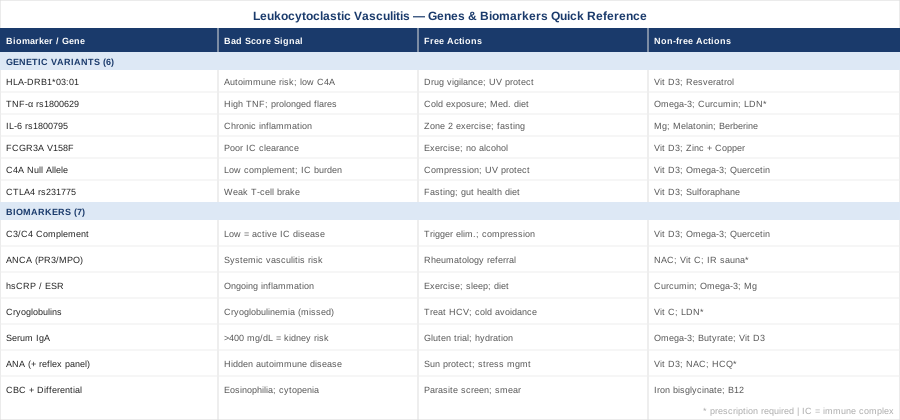
7 Biomarkers to Track in Leukocytoclastic Vasculitis
Biomarkers in LCV serve three distinct purposes: identifying the underlying cause, assessing whether disease is purely cutaneous or systemically involved, and tracking whether the inflammatory process is resolving or progressing. The seven markers below address all three. Not every patient needs all seven at every visit, but understanding what each one reveals — and what it misses — gives you and your physician a framework for making smarter decisions over time.
1. Complement C3 and C4 — The Most Important First Test
Why it matters
Leukocytoclastic vasculitis is fundamentally an immune complex-mediated disease. Circulating immune complexes deposit in small vessel walls, activate the complement cascade, and trigger the neutrophil recruitment responsible for the characteristic tissue destruction. Complement proteins C3 and C4 are consumed in this process — which means low levels provide direct evidence that complement-activating immune complexes are actively at work. When both C3 and C4 are depressed simultaneously, this strongly suggests systemic immune complex disease. When only C4 is low, it points more specifically toward classical pathway activation, as seen in lupus, hepatitis C-associated cryoglobulinemia, and hereditary C4 deficiency.
Complement also functions as a risk stratifier. Research in cutaneous vasculitis cohorts has consistently found that hypocomplementemia at presentation associates with significantly higher rates of systemic involvement — renal and joint manifestations in particular — and with more frequent relapse. A patient with normal complement and isolated skin findings has a very different prognosis from one with depressed C3 and C4, even if their purpura looks identical.
How to measure it
Standard serum C3 and C4 are ordered as part of a complement panel. CH50 (total hemolytic complement) can be added to screen for complete deficiency of individual complement components. Cost: approximately $30–80 for C3 and C4 together at most commercial labs; $50–120 for CH50. Timing matters: complement levels fluctuate with disease activity. Serial measurements every 4–8 weeks during active disease, and every 3–6 months during remission, give a far more useful picture than a one-time snapshot at diagnosis.
If the score is bad — the plan without supplements
Low complement in LCV usually reflects active immune complex consumption, not a primary complement deficiency. The most effective non-supplemental interventions start with source reduction:
- Systematic trigger elimination: The most common reversible causes are drugs (NSAIDs, sulfonamides, diuretics, penicillin-class antibiotics, TNF inhibitors), acute infections (streptococcus, hepatitis B and C, HIV), and secondary autoimmune diseases (lupus, rheumatoid arthritis, Sjögren's). A thorough drug review and targeted infection panel should precede any other intervention. - Anti-inflammatory diet: A whole-food diet low in refined carbohydrates and seed oils reduces dietary antigen load and decreases systemic immune complex production. The effect on complement levels is indirect but meaningful over 8–12 weeks. - Compression stockings (20–30 mmHg): Dependent pooling worsens pericapillary immune complex deposition in the legs. Compression is one of the few purely mechanical interventions with clinical evidence in cutaneous LCV and costs nothing beyond the stockings.
If the score is bad — the plan with supplements or equipment
- Vitamin D3 (2000–5000 IU/day, ongoing): Vitamin D directly upregulates complement regulatory proteins including Factor H, reducing uncontrolled complement activation. Target 25-OH-D: 40–70 ng/mL. Monitor calcium annually at higher doses. - Omega-3 fatty acids (EPA+DHA combined, 2–4 g/day with meals, ongoing): Reduce immune complex-driven inflammation through resolvin and protectin pathways. Side effects: mild GI discomfort at higher doses; blood-thinning relevant if on anticoagulants. - Quercetin (500–1000 mg/day with food, cycle 8 weeks on / 2 weeks off): Inhibits complement-mediated leukotriene release and reduces neutrophil activation downstream of immune complex deposition. Generally well-tolerated; rare headache.
2. ANCA — Ruling In and Ruling Out Systemic Vasculitis
Why it matters
Anti-neutrophil cytoplasmic antibodies (ANCA) represent the single most important test for distinguishing idiopathic cutaneous LCV from ANCA-associated systemic vasculitis (AAV) — a group that includes granulomatosis with polyangiitis (GPA), microscopic polyangiitis (MPA), and eosinophilic granulomatosis with polyangiitis (EGPA). All of these can present initially with cutaneous purpura that is histologically indistinguishable from simple cutaneous LCV.
Positive c-ANCA (directed against PR3) is strongly associated with GPA. Positive p-ANCA (directed against MPO) is more associated with MPA and drug-induced vasculitis. The distinction carries enormous clinical weight: AAV requires immunosuppression with cyclophosphamide or rituximab, not just corticosteroids. A missed ANCA in a patient who appears to have simple cutaneous LCV can delay organ-saving treatment. ANCA testing is warranted in all LCV patients with any systemic feature — fever, arthralgia, hematuria, pulmonary symptoms — and in those with no identified trigger after initial workup.
How to measure it
ANCA is measured by indirect immunofluorescence (IIF) followed by confirmatory ELISA for anti-PR3 and anti-MPO. The ELISA is more specific than IIF alone and should be performed whenever IIF is positive. Cost: approximately $100–250 for the combined panel at most immunology labs. Serial testing in known ANCA-positive patients every 3–6 months during remission can provide early warning of relapse before symptoms recur.
If the score is bad — the plan without supplements
A positive ANCA result changes the management framework entirely:
- Immediate referral to a rheumatologist with vasculitis experience — this is not optional. - Urine microscopy and creatinine measurement: even microscopic hematuria or proteinuria changes urgency and may indicate active glomerulonephritis. - Chest imaging (HRCT if indicated) to screen for pulmonary involvement. - If drug-induced ANCA is suspected (hydralazine, propylthiouracil, minocycline, levamisole-adulterated cocaine): discontinue the drug. ANCA levels typically fall and vasculitis resolves over weeks to months following drug withdrawal.
If the score is bad — the plan with supplements or equipment
In AAV, supplements are adjunctive to disease-modifying therapy, not alternatives. During remission maintenance, after rheumatology consultation:
- N-acetylcysteine (NAC, 600 mg twice daily, ongoing): Reduces oxidative burst in neutrophils, directly relevant to ANCA-stimulated MPO release. Mostly mechanistic evidence; clinical trials in vasculitis are limited. Well-tolerated; occasional GI discomfort. - Vitamin C (500–1000 mg/day, ongoing): Supports endothelial repair and reduces leukocyte-endothelium adhesion. GI upset at higher doses; otherwise well-tolerated. - Infrared sauna (15–20 minutes at 65–75°C, 3–4x/week, during confirmed remission only): Emerging evidence suggests regular mild heat exposure modulates regulatory T cell activity and reduces inflammatory cytokines. Stay well-hydrated; do not use during active flares or systemic symptoms.
3. High-Sensitivity CRP and ESR — Tracking the Trajectory, Not the Snapshot
Why it matters
CRP and ESR are non-specific markers of systemic inflammation, but their value in LCV lies in serial tracking rather than single values. An ESR of 45 mm/hr at diagnosis that normalizes within four weeks after drug withdrawal tells a completely different story than an ESR climbing from 45 to 80 mm/hr despite treatment removal.
High-sensitivity CRP (hsCRP) is preferable to standard CRP because of its sensitivity at lower levels of chronic inflammation — exactly the range where the boundary between active and quiescent disease lives. Peter Attia consistently includes hsCRP in his core inflammatory surveillance panel, emphasizing that values below 0.5 mg/L represent genuinely low systemic inflammatory burden, while values above 3 mg/L indicate ongoing inflammatory activity worth investigating regardless of symptoms. In LCV specifically, persistently elevated hsCRP despite apparent skin healing may signal ongoing subclinical systemic vasculitis or an unresolved secondary cause that the skin has stopped advertising.
How to measure it
hsCRP: $20–50 at most commercial labs; included in most comprehensive metabolic panels. ESR: similarly inexpensive at $15–40. Both should be measured at baseline, then every 4–6 weeks during active disease, and every 3–6 months during monitoring phases.
If the score is bad — the plan without supplements
- Elimination of high-glycemic foods and industrial seed oils — both drive prostaglandin E2 and leukotriene production independently of immune complex activity - Identification and resolution of any infectious or drug trigger - Regular moderate aerobic exercise: consistent evidence across inflammatory conditions shows 30 minutes of moderate-intensity exercise five times weekly reduces CRP by 20–30% over 12 weeks - Sleep duration and quality: 7–9 hours of sleep reduces IL-6 and CRP independently of all other interventions; this is not soft advice — the effect size rivals many pharmacological anti-inflammatory agents at low doses
If the score is bad — the plan with supplements or equipment
- Curcumin with piperine (500–1000 mg/day, cycle 8–12 weeks on / 2 weeks off): Multiple RCTs in inflammatory conditions demonstrate reductions in CRP and ESR. Use a phospholipid or piperine-enhanced formulation for absorption. Avoid high doses in gallbladder disease; occasional GI discomfort. - Omega-3 EPA+DHA (2–4 g/day with meals, ongoing): hsCRP reductions detectable at 6–8 weeks. Well-established mechanism and consistent effect across trials. - Magnesium glycinate (300–400 mg/day at bedtime, ongoing): Magnesium deficiency independently associates with elevated CRP; repletion reduces inflammatory markers. Start low and titrate; loose stools at higher doses.
4. Cryoglobulins — The Most Frequently Missed Diagnosis
Why it matters
Cryoglobulinemia is one of the most underdiagnosed causes of LCV — particularly in patients with no obvious drug trigger or identifiable autoimmune disease. Cryoglobulins are immunoglobulins that precipitate below body temperature and redissolve upon warming. This property is also what makes the test technically demanding: blood must be collected in a pre-warmed tube, kept at 37°C until serum separation, and processed within 30 minutes at that temperature before refrigeration. Many labs, including experienced ones, handle this incorrectly, producing false negatives that send patients down years of unexplained recurrences.
Type II and Type III mixed cryoglobulinemia are the types most strongly associated with LCV. Hepatitis C infection is the cause in up to 90% of mixed cryoglobulinemia cases — and because hepatitis C can be clinically silent for years or decades, prior negative tests from years ago do not rule it out. Other associations include hepatitis B, HIV, lymphoma, and autoimmune conditions including Sjögren's syndrome and lupus.
How to measure it
The cryoglobulin test requires a warm-collection protocol that you should confirm with your lab before the blood draw. Cost: $80–150; specialized collection required. In patients with unexplained or recurrent LCV, simultaneous testing for hepatitis C antibody and RNA (not just antibody alone) is essential and inexpensive.
If the score is bad — the plan without supplements
- Screen for and treat hepatitis C: Current direct-acting antiviral (DAA) therapy achieves cure rates above 95%. Cryoglobulinemic vasculitis resolves in the majority of treated patients within months of viral eradication. This is curative, not suppressive. - Strict cold avoidance: Cold temperatures precipitate cryoglobulins in vivo, triggering complement activation and new purpuric crops. Warm clothing, avoiding cold environments, insulated gloves in winter, and avoiding cold beverages are directly relevant interventions for this subtype. - Plasmapheresis: In severe acute cryoglobulinemic vasculitis, plasmapheresis rapidly removes circulating cryoglobulins. This is a hospital-based intervention but important to know about and request if flares are severe.
If the score is bad — the plan with supplements or equipment
- Vitamin C (1000 mg/day, ongoing): Antioxidant support for endothelial damage caused by cryoprecipitate deposition and complement activation. Well-tolerated. - Low-dose naltrexone (LDN, 1.5–4.5 mg at bedtime, requires prescription): Emerging data from autoimmune case series demonstrates immune modulation through transient opioid receptor blockade, upregulating endogenous endorphins and modulating macrophage inflammatory activity. Titrate: start at 1.5 mg, increase by 1.5 mg every 2 weeks to target dose. Side effects: vivid dreams for the first 2–3 weeks, which typically resolve.
5. Immunoglobulin A — When the Kidneys Are at Risk
Why it matters
Elevated serum IgA — particularly polymeric IgA1 — is the defining feature of IgA vasculitis (formerly Henoch-Schönlein purpura), the most common systemic vasculitis in children but also occurring in adults, where it tends to run a more severe course. Adults with IgA vasculitis have higher rates of renal involvement (IgA nephropathy), arthritis, and abdominal pain than children, and renal disease can develop silently even while the skin findings are resolving.
The prognostic significance of elevated IgA at presentation is well established in registry data: higher IgA levels correlate with greater renal involvement and slower remission. Tracking IgA levels over time, combined with urinalysis and urine protein-to-creatinine ratio at every visit, provides early warning of nephritis developing beneath the surface before it becomes symptomatic.
How to measure it
Serum IgA: $30–60; included in most immunoglobulin panels. Normal adult range: 70–400 mg/dL. Values above 400 mg/dL in the context of LCV warrant a kidney-focused workup. Pair with urine dipstick and microscopy at every follow-up visit.
If the score is bad — the plan without supplements
- Gluten elimination trial (minimum 3 months): IgA antibody production is driven partly by gut mucosal immune activation. Dietary gluten in genetically susceptible individuals stimulates mucosal IgA responses and has been associated with IgA nephropathy flares. A systematic gluten elimination trial with before/after IgA measurement provides objective data on relevance in your case. - Prompt treatment of upper respiratory infections: Mucosal IgA responses are strongly amplified by respiratory infections, which frequently trigger IgA vasculitis flares. Early antibiotic treatment when indicated and appropriate vaccination reduce the frequency and magnitude of IgA production spikes. - Hydration and blood pressure control: Both directly protect kidney function in IgA nephropathy and are the cornerstone of non-pharmacological renal protection.
If the score is bad — the plan with supplements or equipment
- Omega-3 EPA+DHA (4 g/day with meals, ongoing): Specifically supported for IgA nephropathy by the OMEGA trial and subsequent meta-analyses showing reduced proteinuria and slowed GFR decline. Most relevant when renal involvement is present. - Sodium butyrate (600–1200 mg/day, cycle 8–12 weeks, reassess): Short-chain fatty acids from butyrate-producing gut bacteria suppress mucosal IgA overproduction by inducing regulatory T cells in the gut-associated lymphoid tissue. Initial GI adjustment in the first 1–2 weeks is common. - Vitamin D3 (3000–5000 IU/day, ongoing): Downregulates IgA class-switching by reducing BAFF and APRIL — key cytokines driving IgA overproduction. Target 25-OH-D: 50–70 ng/mL.
6. Antinuclear Antibodies — Screening for Hidden Autoimmune Disease
Why it matters
Approximately 20–30% of LCV cases are associated with systemic autoimmune diseases, most commonly systemic lupus erythematosus, rheumatoid arthritis, and Sjögren's syndrome. An ANA screen with reflex testing (anti-dsDNA, anti-Sm, anti-Ro/La, anti-Scl70, anti-Jo-1) provides a structured roadmap for this evaluation. A positive ANA in the context of LCV is not diagnostic of SLE, but it is a strong signal to look further. Anti-dsDNA antibodies are the most specific for SLE and correlate with disease activity, particularly renal nephritis; their levels often rise weeks before clinical flares. Tracking ANA titers over time in known-positive patients gives advance warning before symptoms recur.
How to measure it
ANA by immunofluorescence: $40–90. Positive result at or above 1:80 triggers reflex antibody testing at most labs. Full panel including all subtype antibodies: $150–300. Recommended at baseline for all LCV patients without an identified trigger.
If the score is bad — the plan without supplements
- Rheumatology referral if ANA is at or above 1:160 with relevant specific antibodies - Strict sun protection: UV radiation is a documented trigger for ANA-positive LCV and SLE flares. SPF 50+ applied daily, broad-brimmed hat and long sleeves outdoors — this is not cosmetic advice in this context. - Stress management: Cortisol dysregulation amplifies the Th2/Th17 immune skewing that drives autoantibody production. Evidence-based techniques (breathing, MBSR, regular sleep) have measurable effects on ANA titers in low-grade autoimmunity.
If the score is bad — the plan with supplements or equipment
- Vitamin D3 (3000–5000 IU/day, ongoing): Several RCTs in SLE-associated conditions demonstrate direct reductions in anti-dsDNA antibody levels with vitamin D repletion. Monitor calcium annually. - NAC (1200–2400 mg/day in divided doses, cycle 3 months, reassess): In a small RCT of SLE patients, high-dose NAC reduced mTOR activity in T cells and improved disease activity scores. GI side effects possible at higher doses; start lower and titrate. - Hydroxychloroquine: This prescription medication deserves mention specifically here. In ANA-positive LCV with any suggestion of evolving lupus, hydroxychloroquine reduces flare frequency, lowers anti-dsDNA titers, and has a favorable long-term safety profile. The decision requires a rheumatologist, but the conversation is worth initiating.
7. CBC with Differential — What the Blood Count Reveals Beyond Anemia
Why it matters
The complete blood count with differential is inexpensive and consistently underused in LCV workups beyond checking for obvious infection or anemia. Two specific findings deserve focused attention: eosinophilia and thrombocytopenia.
Eosinophilia (absolute eosinophil count above 500/μL) in the context of LCV points strongly toward EGPA (formerly Churg-Strauss syndrome), drug hypersensitivity, or parasitic infection. EGPA is an ANCA-associated vasculitis with cardiac and neurological involvement — recognizing it changes management from supportive to aggressive immunosuppression. Thrombocytopenia in LCV may signal antiphospholipid syndrome, thrombotic thrombocytopenic purpura, or hepatitis C-related consumption. Leukopenia with lymphopenia is a classic finding in SLE. None of these will be caught without the differential.
How to measure it
CBC with 5-part differential: $15–50 at most labs. A standard CBC without differential misses eosinophilia. Serial measurement every 4–8 weeks during active disease is appropriate.
If the score is bad — the plan without supplements
- Eosinophilia: Screen for parasites (stool ova and parasites, serology for Toxocara and Strongyloides), review all medications, and evaluate for EGPA. Chest X-ray and spirometry are reasonable first steps if pulmonary involvement is a concern. - Thrombocytopenia: Confirm with peripheral smear. Urgent testing for hepatitis C RNA, antiphospholipid antibodies, and if platelet count is below 50,000, expedited hematology consultation. - Iron deficiency anemia: Common in chronic inflammation; confirm with iron studies, ferritin, and transferrin saturation before treating — anemia of chronic inflammation and iron deficiency have different management.
If the score is bad — the plan with supplements or equipment
- Iron bisglycinate (25–50 mg elemental iron, every other day with vitamin C, until repletion confirmed): If iron deficiency is confirmed by labs. Every-other-day dosing improves absorption versus daily by reducing hepcidin upregulation (Moretti et al., American Journal of Clinical Nutrition, 2015). Avoid in hemochromatosis; GI side effects if taken on an empty stomach. - Methylcobalamin (1000 mcg/day sublingual, ongoing): If B12 deficiency is confirmed. Sublingual bypasses absorption variability from intrinsic factor. No meaningful upper limit at this dose.
Genetics and Epigenetics: 6 Variants That Shape LCV Risk and Severity
While biomarkers reflect what is happening in your body right now, genetic variants define the terrain on which it happens. They explain, in part, why some people develop severe, recurrent LCV from a medication that thousands of others tolerate without any reaction. Understanding your genetic profile does not change the diagnosis, but it can inform the intensity of monitoring, the priority of specific preventive strategies, and the biological rationale behind lifestyle interventions that might otherwise seem arbitrary. Most of the following variants can be identified through direct-to-consumer raw genetic data (23andMe or AncestryDNA analyzed through tools like SelfDecode or Genetic Genie) or through clinical immunogenetic panels.
HLA-DRB1*03:01 — The Autoimmune Risk Allele
HLA-DRB1*03:01 (part of the 8.1 ancestral haplotype in European populations, often co-inherited with HLA-B*08) is the most consistently replicated genetic risk factor across multiple autoimmune diseases — including SLE, Sjögren's syndrome, and drug-induced autoimmune reactions — all of which can manifest as LCV. The mechanism operates through antigen presentation: this HLA variant presents certain antigens, including drug-hapten complexes and modified self-antigens, in configurations that promote aberrant T helper cell activation, lowering the threshold for autoimmune responses to triggering events.
Ali Torkamani's polygenic risk work and Gary Brecka's discussions of HLA-driven autoimmune susceptibility both point to the same practical conclusion for carriers: trigger vigilance matters more for this genotype than for average-risk individuals, because the threshold between "tolerated exposure" and "active vasculitis" is meaningfully lower.
If the allele is present — plan without supplements: Heightened drug vigilance (particularly sulfonamides, NSAIDs, diuretics, and TNF inhibitors); aggressive infection treatment; strict UV protection (UV-induced apoptosis generates neo-antigens more likely to trigger autoimmune responses in HLA-DR3-positive individuals); vitamin D testing twice yearly given this haplotype's association with low C4A complement.
If the allele is present — plan with supplements: Vitamin D3 (3000–5000 IU/day, ongoing, target 50–70 ng/mL) directly suppresses HLA class II-driven T helper cell activation; resveratrol (500 mg/day with a fat-containing meal, cycle 3 months on / 1 month off) activates SIRT1-mediated NF-κB suppression. Well-tolerated; mild blood-thinning at higher doses of resveratrol.
TNF-Alpha (-308 G>A, rs1800629) — The Inflammatory Amplifier
The TNF-alpha -308 A allele (rs1800629) increases transcriptional activity at the TNF promoter. Carriers produce more TNF-alpha for any given inflammatory stimulus. In LCV, TNF-alpha amplifies neutrophil activation, increases endothelial permeability, and sustains the inflammatory response long after the triggering event has been removed. The A allele is present in approximately 15–20% of people of European ancestry and has been associated with increased severity and duration of autoimmune-related vasculitis in multiple cohorts.
Gary Brecka has specifically discussed TNF polymorphisms in the context of inflammatory load and methylation — the key insight being that this variant does not cause disease on its own, but it dramatically amplifies any existing trigger. This means trigger elimination matters even more for A-allele carriers, and the anti-inflammatory lifestyle strategies below have a higher return on investment for this genotype.
If the allele is present — plan without supplements: Zero tolerance for known LCV triggers (a trigger that causes a mild reaction in a low-risk individual causes a severe, prolonged one in A-allele carriers); Mediterranean or Wahls-type diet specifically reduces TNF-alpha production — measurable reductions in 8–12 weeks; brief cold water exposure (2–3 minutes at or below 15°C, 3–4x/week) activates norepinephrine, which suppresses TNF-alpha gene expression for hours, as demonstrated in Søberg et al. (2021, Cell Metabolism).
If the allele is present — plan with supplements: Omega-3 EPA+DHA (3–4 g/day, ongoing) directly inhibits TNF-alpha synthesis through competition with arachidonic acid pathways; curcumin with piperine (1000 mg/day, cycle 8 weeks on / 2 weeks off) inhibits NF-κB upstream of TNF-alpha transcription — particularly relevant here because the -308 variant increases TNF at the transcriptional level; low-dose naltrexone (1.5–4.5 mg/night, prescription required) has demonstrated TNF-alpha suppression in multiple autoimmune case series.
IL-6 (-174 G>C, rs1800795) — The Chronicity Driver
IL-6 is the cytokine most responsible for converting acute inflammation into chronic, systemic inflammation. The -174 G allele produces higher basal IL-6 levels by increasing promoter activity. In LCV, elevated IL-6 prolongs the inflammatory response, sustains neutrophil recruitment, and drives acute phase protein production — CRP, fibrinogen, serum amyloid A — which is why G-allele carriers tend to show elevated CRP and ESR for longer after a triggering event than expected. Ali Torkamani's work on polygenic inflammatory scores consistently identifies IL-6 pathway genes as the highest-leverage targets for lifestyle modification, meaning this variant responds well to environmental interventions.
If the allele is present — plan without supplements: Sleep quality is the most impactful single intervention — IL-6 production spikes with sleep deprivation, and this genotype amplifies the consequence; Zone 2 aerobic exercise (30–45 minutes, 5x/week at conversational pace) specifically reduces chronic pro-inflammatory IL-6 while avoiding the transient acute IL-6 spike from high-intensity training; time-restricted eating (14–16 hour fasting window) targets visceral adipose tissue, which is a major IL-6 source, and activates AMPK-mediated NF-κB suppression.
If the allele is present — plan with supplements: Magnesium glycinate (400 mg/day at bedtime, ongoing) — low magnesium independently elevates IL-6; repletion reduces it; melatonin at physiological doses (0.5–1 mg, 30 minutes before sleep, ongoing) suppresses IL-6 production via NF-κB inhibition — avoid the 5–10 mg pharmacological doses that may suppress endogenous production over time; berberine (500 mg twice daily with meals, cycle 8 weeks on / 4 weeks off) activates AMPK, directly suppressing NF-κB and IL-6 transcription — note potential interaction with metformin.
FCGR3A (V158F, rs396991) — Impaired Immune Complex Clearance
FcγRIIIa, encoded by FCGR3A, is an Fc receptor on NK cells, macrophages, and neutrophils that binds IgG-containing immune complexes for clearance and destruction. The F158 variant (phenylalanine at position 158) has lower binding affinity for IgG1 and IgG3 than the V158 variant — meaning F158 homozygotes are less efficient at clearing immune complexes from the circulation. In a disease fundamentally driven by immune complex deposition in vessel walls, this is directly consequential: FF genotype individuals accumulate immune complexes more readily, and those complexes persist longer in vessel walls before being cleared, sustaining inflammation even after the triggering antigen is no longer present.
If the F158 variant is present — plan without supplements: Aggressive trigger elimination (since clearance is impaired, reducing immune complex production is the most direct compensation); regular moderate exercise (which increases FcγRIII expression on circulating NK cells); strict alcohol avoidance during active disease (alcohol reduces macrophage phagocytic efficiency, compounding the clearance impairment this variant creates).
If the F158 variant is present — plan with supplements: Vitamin D3 (4000–5000 IU/day, ongoing) upregulates Fc receptor expression on monocytes and macrophages, partially compensating for reduced FcγRIII binding affinity; zinc (15–30 mg/day with food, ongoing) is required for phagocyte function — deficiency impairs macrophage Fc receptor activity. Important: take zinc away from copper, and supplement copper (1–2 mg/day) if zinc is taken long-term to maintain the appropriate ratio.
C4A Null Allele — Structural Complement Deficiency
Complement C4 is encoded by two structurally similar genes, C4A and C4B, that vary widely in copy number across individuals. C4A null alleles — where one or both copies of the C4A gene are deleted or non-functional — reduce C4A protein levels and impair the opsonization of immune complexes. This is why C4A null alleles are strongly associated with SLE (which is fundamentally an immune complex disease) and with secondary LCV in SLE patients. C4A null alleles occur in approximately 10–15% of Europeans and are a component of the same 8.1 ancestral haplotype that includes HLA-DR3, which explains why low serum C4 and LCV co-occur so frequently in this population group.
If the null allele is present — plan without supplements: Aggressive immune complex source reduction remains the primary strategy, since C4A itself cannot be supplemented — remove triggers, treat infections early, use compression, and monitor C4 serially (baseline levels will be structurally lower, so any additional drop is a meaningful signal of active disease); strict UV protection; track complement levels at increased frequency (every 6–8 weeks during monitoring phases).
If the null allele is present — plan with supplements: The approach parallels the complement biomarker strategy above: Vitamin D3, omega-3, and quercetin to reduce complement activation burden rather than attempt to increase production. The logic is different — here the goal is reducing the rate at which a structurally limited complement system gets consumed.
CTLA4 (+49 A>G, rs231775) — A Weaker Brake on T Cell Activation
CTLA4 encodes the co-inhibitory receptor that turns off T cell activation after an immune response, functioning as a natural brake on the immune system. The +49 A allele produces a CTLA4 protein with reduced cell surface expression — meaning that brake engages less effectively. Carriers show a persistently lower threshold for T cell activation in response to antigenic stimulation, and are more likely to develop autoimmune conditions that depend on sustained T cell help, including SLE, type 1 diabetes, Graves' disease, and autoimmune-associated LCV. The A allele frequency is approximately 45–50% in Europeans — it is common enough that its contribution to LCV susceptibility may be significantly underappreciated.
If the A allele is present — plan without supplements: Reduce systemic antigen load — this is the most direct way to compensate for an inefficient T cell brake. Dietary interventions (gluten and casein elimination trials, addressing intestinal permeability) reduce the pool of non-self antigens reaching systemic immune compartments; intermittent fasting (16:8 or occasional 24-hour fasts) induces autophagy that degrades the misfolded proteins driving autoreactive T cell populations.
If the A allele is present — plan with supplements: Vitamin D3 (3000–5000 IU/day, ongoing) directly upregulates CTLA4 expression on regulatory T cells, partially compensating for the reduced expression from this variant; sulforaphane from broccoli sprout extract (30–60 mg/day, cycle 8 weeks on / 2 weeks off) activates Nrf2, which induces regulatory T cell function and enhances CTLA4-mediated suppression. Possible thyroid interaction at very high doses; stay within recommended range.
What Andrew Huberman's Research on Immune Regulation Reveals About LCV
Andrew Huberman, a neuroscientist at Stanford University, has synthesized a substantial body of peer-reviewed research on immune regulation, inflammation, and the nervous system's bidirectional control over immune function across multiple podcast episodes. While none of his material addresses leukocytoclastic vasculitis directly, the cellular and molecular mechanisms he covers map precisely onto the pathophysiology of immune complex-mediated small vessel inflammation. The following ten insights represent the most relevant practical applications of his work for LCV patients.
1. Sickness Behavior Is a Protective Signal, Not Weakness
Huberman explains that fatigue, social withdrawal, and appetite suppression during active inflammation are not incidental — they are orchestrated by the immune system via IL-1β, IL-6, and TNF-alpha acting on the vagal afferents and hypothalamus. These symptoms signal that metabolic resources are being redirected toward immune activity. Fighting through active vasculitis flares with stimulants or forced activity competes with the immune system for those resources. Rest during active disease is mechanistically justified and not optional.
2. Slow-Wave Sleep Is When Regulatory T Cells Are Deployed
Growth hormone released during slow-wave sleep activates regulatory T cells (Tregs) that suppress autoreactive immune activity. For LCV with an autoimmune component, achieving 7–9 hours of sleep — specifically sleep that reaches adequate slow-wave stages — is the most evidence-supported Treg-enhancing intervention available without a prescription. Huberman recommends a consistent wake time, a cool and fully dark sleeping environment, and no bright light exposure after 10 pm as the foundation.
3. Morning Sunlight Calibrates the Inflammatory Set Point
Two to ten minutes of outdoor light exposure within the first hour of waking synchronizes circadian cortisol timing. Properly timed cortisol is acutely anti-inflammatory. Dysregulated cortisol — blunted morning peak, elevated nocturnal levels from stress or artificial light exposure — drives chronic low-grade inflammation. In individuals with complement or Fc receptor genetic vulnerabilities, this basal inflammatory elevation sustains the immune complex deposition cycle. The intervention here is not about UV dose; it is about timing.
4. Brief Cold Exposure Suppresses TNF-Alpha at the Gene Level
Huberman references the Søberg et al. (2021, Cell Metabolism) study demonstrating that deliberate cold exposure increases norepinephrine by 200–300% and reduces TNF-alpha and IL-6 gene expression for hours afterward. For LCV patients with TNF-alpha risk variants or persistently elevated inflammatory markers, this is an accessible, free daily intervention with measurable anti-inflammatory effects. Protocol: 2–3 minutes of cold water immersion or cold shower at or below 15°C, 3–4 times per week. Use during remission only — not during active skin lesions or open purpuric lesions.
5. Nasal Breathing Delivers Nitric Oxide to Small Vessels
The nasal sinuses produce nitric oxide (NO), which is inhaled into the lungs and enters systemic circulation. NO has direct vasodilatory and anti-inflammatory effects on small vessels — precisely the vessels affected in LCV. Mouth breathing bypasses this NO delivery system entirely. Nasal breathing during sleep (mouth tape if required), during exercise, and throughout daily activity is a structurally sound vasculoprotective habit for any LCV patient.
6. The Gut-Immune Axis Is Mechanistically Central, Not Metaphorical
Roughly 70% of immune cells reside in gut-associated lymphoid tissue. Huberman has discussed research showing that gut dysbiosis directly raises serum IgA, reduces secretory IgA, and impairs Treg education in the intestinal lamina propria. For LCV driven by IgA vasculitis or any autoimmune mechanism, gut microbiome composition is not adjacent to the treatment question — it is part of it. The simplest metric is dietary diversity: 30 or more distinct plant foods per week is associated with measurably higher microbiome diversity and lower systemic inflammatory markers.
7. Zone 2 Cardio Is the Anti-Inflammatory Exercise Dose
High-intensity exercise transiently elevates CRP and acute-phase reactants. Moderate aerobic exercise at Zone 2 intensity (conversational pace, approximately 60–70% of maximum heart rate, 30–45 minutes) specifically reduces IL-6, TNF-alpha, and CRP over 8–12 weeks of consistent training without triggering the pro-inflammatory cascade that HIIT produces. Huberman emphasizes 150–200 minutes of Zone 2 per week as the evidence-based anti-inflammatory exercise dose — and this distinction between intensity levels is particularly relevant during LCV management.
8. Psychological Stress Opens Intestinal Tight Junctions Within Hours
Stress-activated corticotropin-releasing hormone directly disrupts intestinal tight junctions by activating mast cells in the gut wall, increasing mucosal permeability within hours of a stress exposure. This floods systemic circulation with bacterial lipopolysaccharide and dietary antigens that stimulate immune complex formation. The mechanism connecting chronic stress to LCV flares is specific and molecular, not vaguely psychosomatic. Huberman's evidence-based stress reduction protocols — cyclic physiological sighing (two quick nasal inhales followed by a long mouth exhale, practiced for 5 minutes) and non-sleep deep rest (NSDR/Yoga Nidra) — directly reduce this permeability-increasing pathway.
9. Time-Restricted Eating Suppresses NF-κB Without Caloric Restriction
A 14–16 hour fasting window activates SIRT1 and AMPK, which suppress NF-κB — the master transcription factor driving TNF-alpha, IL-6, and the entire complement-activating inflammatory cascade relevant to LCV. Huberman's practical recommendation: finish eating 3 hours before sleep, start eating 2–3 hours after waking. This creates the fasting window without requiring caloric restriction and produces measurable anti-inflammatory effects within 4–6 weeks of consistent practice.
10. Red and Near-Infrared Light Reduces Endothelial Inflammation
Research Huberman has covered shows that 630–670 nm red light and 810–850 nm near-infrared light delivered to the skin activate cytochrome c oxidase in mitochondria of endothelial cells, reducing local inflammatory cytokine production and supporting mitochondrial repair. For LCV, where small vessel endothelial inflammation is the pathological core, this represents a mechanistically coherent adjunct. Evidence in LCV specifically is preliminary; evidence in inflammatory skin conditions and vascular function more broadly is stronger. Protocol: a clinical-grade panel device, 10–15 minutes daily at 10–15 cm distance. Avoid over active, open purpuric lesions.
Complementary Approaches Backed by Human Evidence
The following four modalities have meaningful human clinical evidence relevant to LCV's underlying mechanisms: immune dysregulation, gut permeability, systemic inflammation, and stress-driven flares. They are adjuncts to medical management, not replacements, and are selected specifically for the pathophysiology of this condition rather than for general wellness appeal.
The Autoimmune Protocol — Sarah Ballantyne
The Autoimmune Protocol (AIP), developed by Dr. Sarah Ballantyne, a research scientist with a doctorate in medical biophysics, is a dietary and lifestyle framework specifically designed to modulate gut permeability and the gut-immune axis in autoimmune conditions. The elimination phase removes foods with the highest potential to drive immune reactivity — grains, legumes, nightshades, eggs, dairy, seeds and nuts, and seed oils — while prioritizing nutrient density in the micronutrients most commonly depleted in autoimmune disease: zinc, vitamin A, vitamin D, magnesium, and long-chain omega-3 fats. Given that LCV frequently has autoimmune drivers — SLE, IgA vasculitis, drug-induced autoimmunity — the AIP's mechanistic target (reducing mucosal immune activation that generates the immune complexes depositing in vessel walls) is directly relevant.
A pilot randomized controlled trial (Konijeti et al., Inflammatory Bowel Diseases, 2017) demonstrated that the AIP elimination phase produced significant reductions in CRP and disease activity scores in inflammatory bowel disease over 6 weeks. The evidence base is stronger in IBD than in vasculitis directly, but the shared gut-immune mechanism — dysbiosis, mucosal immune activation, IgA overproduction, impaired Treg education — makes the extrapolation scientifically reasonable.
Practical application for LCV: The elimination phase requires a minimum of 30–60 days of strict adherence before reintroduction begins. Reintroduction is systematic — one food group at a time, with 5–7 days of monitoring between additions. For LCV patients, tracking complement levels, serum IgA, and hsCRP before, during, and after the elimination phase provides objective data on whether the protocol is producing biological change in your specific case. Dr. Ballantyne's book The Paleo Approach provides the complete evidence-based framework and reintroduction protocol.
Mindfulness-Based Stress Reduction (MBSR)
Mindfulness-Based Stress Reduction, the 8-week program developed by Jon Kabat-Zinn at the University of Massachusetts Medical School, trains sustained attentional focus through body scan meditation, sitting meditation, and mindful movement. Its relevance to LCV is immunological, not purely psychological. Multiple RCTs have documented that MBSR reduces IL-6, TNF-alpha, and CRP in inflammatory conditions, with effect sizes comparable to low-dose anti-inflammatory medications in some trials. The primary mechanism runs through the HPA axis: MBSR normalizes cortisol's diurnal rhythm, specifically reducing the nocturnal cortisol elevations that sustain gut permeability and chronic low-grade immune activation.
A meta-analysis published in Brain, Behavior, and Immunity (2016) covering 18 randomized controlled trials found that mind-body interventions including MBSR significantly reduced inflammatory biomarkers across immune-related conditions. The effect was largest in participants with the highest baseline inflammatory markers — which is precisely the LCV presentation where psychoneuroimmunological interventions have the most to offer.
Practical application: An 8-week MBSR course — offered in-person at most major medical centers or via validated online programs through the UMass Center for Mindfulness — is the evidence-supported protocol. Daily practice of 20–45 minutes is required; shorter protocols show attenuated benefits. The biological response accumulates over 8 weeks and is not detectable after a 1–2 week trial — measuring hsCRP and IL-6 before and after the 8-week course provides objective feedback on the immunological response.
Microbiome-Directed Therapies
The connection between gut microbiome composition and systemic immune regulation is now one of the most robustly evidenced areas in autoimmune research. In LCV, the IgA subtype is mechanistically tethered to gut mucosal immunity — elevated polymeric IgA1 is produced in the gut-associated lymphoid tissue, and dysbiosis (particularly depletion of butyrate-producing organisms such as Faecalibacterium prausnitzii and Roseburia intestinalis) reduces regulatory T cell populations, increases Th17 activity, and amplifies the IgA class-switching that drives both IgA vasculitis and IgA nephropathy.
A study published in Kidney International (2021) demonstrated that targeted probiotic supplementation reduced proteinuria and urinary IgA deposition in IgA nephropathy patients over 6 months. The direct evidence in cutaneous IgA vasculitis is limited, but the shared upstream pathophysiology — IgA overproduction, complement activation, and small vessel immune complex deposition — makes this intervention mechanistically coherent.
Practical application: A phased protocol. Phase 1 (weeks 1–4): remove the primary substrates of dysbiosis — processed foods, refined sugars, industrial seed oils, and unnecessary antibiotics. Phase 2 (weeks 4–12): increase microbiome diversity through 30 or more distinct plant foods per week, emphasizing fermented vegetables (sauerkraut, kimchi) as daily additions. Phase 3 (ongoing): targeted probiotic support, with Lactobacillus rhamnosus GG and Bifidobacterium longum having the strongest evidence in immune conditions. For IgA vasculitis specifically, track serum IgA and urine protein-to-creatinine ratio before and after 12 weeks of the full protocol to evaluate biological response.
Breathing-Based Therapies
Slow, controlled breathing at 5–6 breaths per minute — termed resonance frequency breathing — activates the vagus nerve, increases heart rate variability, and suppresses systemic TNF-alpha production through the cholinergic anti-inflammatory pathway (CAP). The CAP is a neuro-immune circuit in which vagal efferents release acetylcholine at peripheral immune sites, directly suppressing macrophage TNF-alpha production. This pathway was mapped by Kevin Tracey's research group at Northwell Health and forms the mechanistic basis for vagal nerve stimulator implants currently being trialed in refractory rheumatoid arthritis.
A randomized controlled trial published in Frontiers in Immunology (2019) found that 8 weeks of slow breathing training significantly reduced IL-6 and TNF-alpha in patients with chronic inflammatory conditions compared to wait-list controls. For LCV patients carrying TNF-alpha or IL-6 risk variants, or those with persistently elevated hsCRP, the mechanistic fit between this intervention and the documented genetic vulnerability is direct.
Practical application: Resonance frequency breathing at 5–6 breaths per minute, 20 minutes daily. Inhale for 5 seconds through the nose, exhale for 5–6 seconds through the nose or mouth. No breath holding required. Free apps (Breathwrk, Odama) can guide timing. Daily practice for a minimum of 8 weeks before evaluating biological effect. A wearable device (Oura Ring, Garmin, Polar) that tracks heart rate variability provides objective real-time feedback on vagal tone improvement. Do not practice during acute systemic vasculitis symptoms until cleared medically.
Conclusion
Leukocytoclastic vasculitis is not one disease — it is a pathological response pattern that can be triggered and sustained by a wide range of causes, and that varies substantially in severity depending on an individual's genetic makeup and the efficiency of their immune complex clearance systems. That complexity is exactly why the standard approach works for some patients and fails repeatedly for others.
The seven biomarkers in this article offer a practical framework for understanding what is driving your LCV, how severe it is, and whether it is resolving. Complement levels confirm whether active immune complex deposition is occurring. ANCA rules out systemic vasculitis before organ damage accumulates silently. Cryoglobulins identify the frequently missed secondary cause behind years of unexplained recurrences. IgA flags the kidney risk that a skin-focused approach regularly misses. The six genetic variants explain why your immune response is calibrated the way it is — and where lifestyle and targeted supplementation have the highest return on investment for your specific profile.
The most valuable next step is not trying to implement everything at once. Start with the most informative baseline labs: complement C3 and C4, ANCA, cryoglobulins, IgA, and ANA if you have not had them recently. Build a serial tracking habit — the same markers every 6–8 weeks tells a story that a single value never will. If you have access to genetic testing, look at the six variants covered here and match your interventions to your actual biology rather than to average-population recommendations. And discuss any significant changes to your medical regimen — or any new findings from testing — with a rheumatologist who has vasculitis experience. Better information, acted on precisely and incrementally, is the most reliable path to fewer flares and a clearer understanding of why they happen.
Cardiovascular: Vascular Conditions
Skin: Inflammatory Skin Conditions
Autoimmune: Inflammatory Conditions Autoimmune Skin Conditions
Urological: Kidney Conditions