This article contains AI generated content.
· UpdatedLigamentum Mucosum Hypertrophy — 7 Biomarkers And 5 Genes To Track
Understanding What Is Happening Inside the Knee
The ligamentum mucosum is a synovial fold inside the knee joint — a thin remnant of embryological tissue that, in some people, becomes irritated, thickened, and eventually hypertrophied. When it does, it can snap against surrounding structures during flexion, generate persistent inflammation, and produce the kind of anterior knee pain that resists simple explanations. MRI findings are sometimes dismissed as incidental. Physical therapy protocols rarely address the synovial specifics. And surgeons typically only enter the picture late. In the meantime, the person is left with a diagnosis that does not come with a clear roadmap.
Generic advice — rest, ice, ibuprofen, strengthen the quad — can help at the margins, but it rarely addresses why the tissue hypertrophied in the first place or why it stays inflamed. The biology behind this condition is usually more specific than "overuse" or "poor mechanics." Inflammatory signaling, tissue remodeling enzyme activity, nutritional status, and genetic predispositions all influence whether the ligamentum mucosum stays quiet or becomes a persistent problem. Treating the surface without understanding the terrain is why so many people cycle through advice without meaningful improvement.
This article takes a more precise approach. The primary focus is a set of seven biomarkers that can be tracked through standard or specialized labs — markers that reveal your inflammatory burden, tissue turnover rate, and biological readiness to heal. A second section explores five genes that influence tissue quality and inflammatory responsiveness, which is useful if you have genomic data or simply want to understand why some people develop this condition from minimal stress while others do not. A third section distills ten practical insights from one of the most evidence-grounded books on joint health currently available. Finally, four complementary therapies with meaningful clinical support round out the picture.
Better information does not guarantee a faster recovery. But in a condition this frequently misdiagnosed or mismanaged, narrowing the guesswork is a genuine advantage — and each of the frameworks below gives you a different way to do exactly that.
7 Biomarkers Worth Tracking If You Have Ligamentum Mucosum Hypertrophy
Biomarkers do not tell you what is happening inside a single synovial fold. What they do tell you is the biological environment in which your tissue is operating. High systemic inflammation, low vitamin D, overactive tissue-remodeling enzymes — these are not background noise. They are the conditions that made hypertrophy more likely, and that make recovery harder. Tracking them gives you traction that clinical intuition alone cannot provide.
The seven markers below were selected for their direct relevance to synovial tissue biology, their measurability, and the strength of evidence supporting their use in joint conditions. Several are consistently highlighted by longevity-focused clinicians — including Peter Attia — for their clinical precision and actionability.
Biomarker 1: High-Sensitivity C-Reactive Protein (hs-CRP)
Why it matters. hs-CRP is the most widely used marker of systemic low-grade inflammation. It is synthesized in the liver in response to cytokine signals — particularly IL-6 — and its level reflects ongoing inflammatory activity throughout the body, including within joint tissue. Even modest elevations above 1 mg/L are clinically meaningful: persistent low-grade inflammation creates a tissue environment where synovial cells proliferate abnormally, where collagen organization is disrupted, and where healing is systematically slowed. For ligamentum mucosum hypertrophy specifically, reducing systemic inflammatory burden is one of the most tractable upstream interventions available.
How to measure it. hs-CRP is a standard blood test ordered through any primary care physician or direct-to-consumer lab. Cost in the US typically ranges from $15 to $40 without insurance. The key is to order high-sensitivity CRP — standard CRP is not sensitive enough to detect the low-grade inflammation relevant here. Optimal range: below 0.5 mg/L. Values between 1 and 3 mg/L indicate moderate inflammatory burden; above 3 mg/L suggests active inflammatory activity.
If the score is bad: the plan without supplements. The most impactful lifestyle interventions for reducing hs-CRP are: eliminating ultra-processed foods and refined seed oils (the omega-6 to omega-3 ratio directly influences CRP); prioritizing sleep (even 24–48 hours of partial sleep deprivation acutely elevates CRP); and incorporating zone 2 aerobic exercise at least 3–4 times per week. For the knee specifically, low-load cycling or swimming reduces synovial pressure without further irritating inflamed tissue while supporting lymphatic drainage. Expect measurable CRP reduction within 6–8 weeks of consistent change.
If the score is bad: the plan with supplements. Omega-3 fatty acids (EPA+DHA combined, 2–4 g/day) have the strongest evidence base for hs-CRP reduction. A 2017 meta-analysis in Nutrients confirmed anti-inflammatory effects across multiple populations. Curcumin with piperine (500–1000 mg/day) shows meaningful CRP-lowering in joint conditions; cycle for 8 weeks followed by a 4-week break given limited long-term safety data. Magnesium glycinate (300–400 mg/day) supports anti-inflammatory enzyme systems and is safe for continuous use. Do not combine curcumin and omega-3 with anticoagulant medications without physician guidance.
Biomarker 2: Interleukin-6 (IL-6)
Why it matters. IL-6 is a pro-inflammatory cytokine central to synovial inflammation and tissue hypertrophy. Synovial fibroblasts in hypertrophied plica tissue express elevated IL-6, which drives further cell proliferation and collagen thickening — a self-reinforcing cycle. High circulating IL-6 also stimulates CRP production (making it an upstream driver of the previous biomarker) and promotes a catabolic tissue environment. Tracking IL-6 gives a more direct window into joint-level inflammatory activity than CRP alone.
How to measure it. IL-6 is measurable via standard blood test, though it is not routinely included in standard panels — request it specifically or use a direct-to-consumer specialty lab. Cost: $30–$80. Some functional medicine cytokine panels bundle IL-6 with TNF-alpha and other markers for $100–$150. Optimal circulating levels in a resting, non-acute state are generally below 1.8 pg/mL. Levels above 3–5 pg/mL in the absence of acute infection are clinically notable.
If the score is bad: the plan without supplements. IL-6 is chronically elevated by visceral adiposity, poor sleep, sedentary behavior, and psychological stress. Addressing each systematically matters. For the joint specifically: avoiding sustained flexion positions that increase intra-articular pressure (which stimulates synovial IL-6 production) is practical and achievable. Cold water immersion (10–15 minutes at 12–15°C, 3 times per week) has demonstrated meaningful IL-6 suppression in multiple human studies. Time-restricted eating (16:8 protocol) has also shown cytokine reduction within 4–6 weeks in several trials.
If the score is bad: the plan with supplements. Boswellia serrata extract (standardized to AKBA fraction, 150–200 mg/day) has specific anti-IL-6 evidence in arthritic and synovial conditions across several randomized trials; cycle every 10–12 weeks with a 4-week break. Vitamin D3 supplementation, when it achieves optimal serum levels, significantly suppresses IL-6 production (see biomarker 4 below). Quercetin (500 mg/day) inhibits IL-6 through NF-κB pathway modulation; use in 8-week cycles. Avoid relying on NSAIDs for long-term IL-6 control — they produce rebound elevation and carry cardiovascular risks with sustained use.
Biomarker 3: Matrix Metalloproteinase-3 (MMP-3)
Why it matters. Matrix metalloproteinases are enzymes responsible for degrading extracellular matrix components — including the collagens and proteoglycans that make up the ligamentum mucosum and surrounding synovial tissue. MMP-3, also called stromelysin-1, is among the most highly expressed MMPs in inflamed synovial tissue, and its overactivation drives excessive tissue remodeling and structural disorganization. Elevated serum MMP-3 is a validated marker of synovial disease activity used in rheumatoid arthritis and osteoarthritis research. Even in non-rheumatoid conditions like plica hypertrophy, this marker reveals whether your tissue is in a net degradation state or a repair state — a crucial distinction for guiding intervention.
How to measure it. MMP-3 is measured via serum ELISA, available through specialty labs such as Vibrant Wellness or Genova Diagnostics. Cost: $60–$120. Standard reference ranges are approximately 3–17 ng/mL in women and 8–32 ng/mL in men (ranges vary by lab). Elevated values in the context of joint symptoms strongly suggest active synovial remodeling that warrants intervention.
If the score is bad: the plan without supplements. The primary non-supplemental approach to MMP-3 reduction is addressing the upstream inflammatory signals (IL-6, TNF-alpha) that drive its production. Moderate mechanical loading actually helps: low-to-moderate weight-bearing exercise stimulates TIMP (tissue inhibitor of metalloproteinases) production, which counterbalances MMP activity. Specifically, slow eccentric strengthening of the quadriceps and hip stabilizers (3 sessions per week at low load) has shown promise for reducing pathological MMP activity in periarticular tissues. Complete rest is counterproductive — it reduces TIMP production and accelerates net degradation.
If the score is bad: the plan with supplements. Collagen peptides (10–15 g/day taken with vitamin C, approximately 30–60 minutes before exercise) have shown in multiple trials to upregulate TIMP expression and shift the MMP/TIMP balance toward repair. A 2017 study in the American Journal of Clinical Nutrition demonstrated that this timing-specific protocol significantly increased collagen synthesis in connective tissue. Green tea extract (EGCG fraction, 400 mg/day) has direct MMP-3 inhibitory activity in early human connective tissue studies; limit to 8-week cycles due to hepatotoxicity risk at higher doses. Glucosamine sulfate (1500 mg/day) has also demonstrated MMP-modulating properties in joint trials.
Biomarker 4: 25-OH Vitamin D
Why it matters. Vitamin D deficiency is one of the most consistently underappreciated contributors to musculoskeletal vulnerability. The vitamin D receptor is expressed in synovial fibroblasts, chondrocytes, and joint-associated immune cells. Adequate vitamin D suppresses NF-κB signaling — a master switch for inflammatory gene expression — and reduces production of both IL-6 and TNF-alpha. Deficiency (below 20 ng/mL) has been associated with increased synovial inflammation, impaired tissue healing, and greater pain sensitivity in joint conditions. Peter Attia has repeatedly emphasized that individuals running below 40 ng/mL are operating at a meaningful biological disadvantage for tissue repair and immune regulation — an observation supported by a substantial body of mechanistic and epidemiological evidence.
How to measure it. The 25-hydroxyvitamin D test is standard and widely available, typically costing $25–$60 and often covered by insurance. Test at minimum twice per year — late winter (when levels are typically at their annual nadir) and late summer. Optimal range for joint and immune health: 40–60 ng/mL (100–150 nmol/L). Below 30 ng/mL is insufficient; below 20 ng/mL is clinically deficient.
If the score is bad: the plan without supplements. Midday sun exposure (arms and legs uncovered, without sunscreen, for 15–30 minutes depending on skin tone and latitude) is the most physiologically natural route. This approach is substantially limited by geography, season, and occupation. Dietary sources — fatty fish, egg yolks, liver — contribute modestly but rarely enough to correct deficiency. Anyone living above 35°N latitude during winter months will struggle to maintain optimal levels without supplementation.
If the score is bad: the plan with supplements. Vitamin D3 (cholecalciferol) at 2000–5000 IU/day paired with vitamin K2 (MK-7 form, 100–200 mcg/day) is the standard protocol — K2 directs calcium metabolism appropriately and prevents soft tissue calcification that can occur with D3 supplementation alone. Retest after 90 days and adjust dose. Magnesium (300–400 mg/day glycinate or malate form) is essential for converting vitamin D to its active form — supplementing D3 without adequate magnesium is partially ineffective. At doses exceeding 10,000 IU/day, hypercalcemia risk increases; physician monitoring is warranted.
Biomarker 5: COMP (Cartilage Oligomeric Matrix Protein)
Why it matters. COMP is a pentameric protein released into the bloodstream when joint cartilage and periarticular connective tissue — including synovial structures — undergo mechanical stress or degradation. It is among the most sensitive early markers of joint tissue turnover, detectable before structural damage is visible on imaging. Elevated COMP indicates net breakdown activity in the joint, and it correlates with symptom severity in synovial irritation conditions. Because the ligamentum mucosum operates within a complex joint environment, COMP provides a composite signal about overall joint tissue stress that is difficult to obtain any other way.
How to measure it. COMP is measured via serum ELISA through specialty labs (several academic hospital systems and commercial labs offer it). Cost: $80–$150. Reference ranges vary by lab, but values above 12 U/L in serum are generally considered elevated. Morning samples taken before any significant physical activity provide the most reliable baseline reading.
If the score is bad: the plan without supplements. Load management is the central intervention: the knee needs enough mechanical stimulus to drive tissue repair but not so much that repeated microtrauma continuously drives COMP release upward. Replacing high-impact activities (running, jumping, pivoting sports) with low-impact alternatives (cycling, swimming, incline walking) typically reduces COMP within 6–8 weeks while maintaining joint health signals. Gradual progressive loading — never increasing total training volume by more than 10% per week — is the key principle.
If the score is bad: the plan with supplements. Collagen peptides (10–15 g/day) support COMP normalization by improving substrate availability for connective tissue repair. Undenatured type II collagen (UC-II, 40 mg/day) has been specifically studied in joint conditions and demonstrated significant improvements in joint comfort and function in a published randomized controlled trial, outperforming a combination of glucosamine and chondroitin. Avocado-soybean unsaponifiables (ASU, 300 mg/day) have shown COMP-relevant effects in osteoarthritis trials and represent a reasonable adjunct with a strong safety profile.
Biomarker 6: Serum Hyaluronic Acid (HA)
Why it matters. Hyaluronic acid is a critical component of synovial fluid and the extracellular matrix of synovial tissue, providing lubrication, shock absorption, and structural integrity to the joint environment. In conditions involving synovial hypertrophy, HA metabolism is disrupted — serum HA can become paradoxically elevated as a marker of synovial cell overactivation, even as the quality of HA within the joint itself degrades. Serum HA is used as a marker of synovial inflammation and fibrosis in rheumatoid conditions and has emerging utility in non-rheumatoid synovial pathology. Its measurement gives a more direct read on synovial cell activity than systemic inflammatory markers alone.
How to measure it. Serum hyaluronic acid is measured via ELISA through reference and specialty labs. It is not standard in most panels but is available for $80–$150. In healthy adults without active joint disease, serum HA is typically below 50 ng/mL. Values above 100 ng/mL in the context of joint symptoms may indicate active synovial cell hyperactivation. Repeat testing every 90 days during active intervention is reasonable.
If the score is bad: the plan without supplements. Elevated serum HA in this context is downstream of synovial inflammatory overactivation — addressing hs-CRP, IL-6, and TNF-alpha upstream will typically bring HA back toward normal range over 8–12 weeks. Maintaining consistent, non-provocative joint motion (gentle range-of-motion exercises) helps normalize synovial cell behavior and HA distribution. Avoid full immobilization, which paradoxically disrupts HA metabolism further.
If the score is bad: the plan with supplements. Oral hyaluronic acid (80–200 mg/day of high-molecular-weight HA) has shown benefit in joint conditions across several randomized trials, including Japanese studies demonstrating improved joint comfort with 12-week supplementation. The mechanism appears partly oral-tolerance based rather than simple absorption. Cycle for 12 weeks with a 4-week break; no significant safety concerns have been documented at these doses. This works best as an adjunct alongside upstream inflammatory interventions, not as a standalone approach.
Biomarker 7: TNF-alpha (Tumor Necrosis Factor-alpha)
Why it matters. TNF-alpha is a master cytokine driving synovial inflammation and tissue hypertrophy. It directly stimulates synovial fibroblast proliferation — the cellular engine behind hypertrophy — and activates MMP production, completing a destructive cycle of tissue degradation and remodeling that perpetuates the condition. Elevated TNF-alpha is also associated with greater pain sensitization, faster tissue damage, and poorer spontaneous recovery in joint conditions. While dramatic TNF-alpha elevation is characteristic of autoimmune arthritis, even modest elevations in non-autoimmune joint conditions meaningfully impair tissue repair.
How to measure it. TNF-alpha is measured via serum assay, available through specialty cytokine panels at $60–$100. Standard reference ranges for healthy adults are typically below 8.1 pg/mL, though ranges vary by lab and assay method. Note that TNF-alpha is sensitive to pre-analytical variables — samples must be processed quickly and tested at a reliable lab.
If the score is bad: the plan without supplements. TNF-alpha is powerfully suppressed by aerobic exercise — even a single 30–45 minute session at zone 2 heart rate produces measurable acute reduction, and regular aerobic training creates a chronic anti-TNF state. Sleep quality is an equally powerful lever: TNF-alpha surges during sleep deprivation and returns to baseline with consistent 7–9 hour nights. Cold water immersion (10–15 minutes at 12–15°C, 3 times per week) reduces TNF-alpha significantly and has the additional benefit of being directly applicable to the knee joint.
If the score is bad: the plan with supplements. Omega-3 fatty acids (EPA+DHA, 3–4 g/day) reduce TNF-alpha through competitive inhibition of arachidonic acid pathways — this is one of the best-supported anti-inflammatory supplement mechanisms in the human literature. Resveratrol (250–500 mg/day taken with a fatty meal for absorption) has shown TNF-alpha reduction in human trials involving joint and metabolic conditions; use in 8-week cycles with a 4-week break. Curcumin with piperine (1000 mg/day) significantly inhibits NF-κB and downstream TNF-alpha production; limit to 8-week cycles. Do not combine resveratrol with anticoagulants without physician oversight.
Having mapped the inflammatory and tissue-remodeling landscape through measurable markers, it is worth examining whether part of your susceptibility was written into your genome long before any knee problem appeared. The following section covers the genetic layer of this picture.
5 Genes That May Shape Your Risk And Recovery
Genetics does not determine your outcome with ligamentum mucosum hypertrophy — but it can help explain why your tissue responds the way it does, why inflammation may persist longer than expected, and which interventions may be disproportionately useful for you specifically. The variants below have meaningful human evidence, though the research is more preliminary than the biomarker literature. If you have raw genomic data from 23andMe, AncestryDNA, or a clinical genetics panel, these are the variants worth examining. Practitioners in the nutrigenomics space — including researchers cited by Gary Brecka and Ali Torkamani in their work on gene-lifestyle interactions — have highlighted several of these as particularly relevant to connective tissue vulnerability and inflammatory dysregulation.
Gene 1: COL5A1 — Collagen Structural Integrity
What it does. COL5A1 encodes the alpha-1 chain of type V collagen, a regulatory collagen that controls the diameter of type I collagen fibrils in tendons, ligaments, and synovial structures. Variants in COL5A1 — particularly rs12722 — have been associated in multiple human studies with increased risk of connective tissue injuries including ACL tears and soft tissue overuse injury. The mechanism is that altered type V collagen leads to thicker, less organized fibrils, creating tissue that is simultaneously stiffer and more susceptible to microtrauma-driven inflammation. This is directly relevant to a structure like the ligamentum mucosum.
If the gene is bad: the plan without supplements. The underlying structural issue cannot be reversed, but mechanical adaptation is possible and meaningful. Slower progression of training loads (never exceeding 10% volume increase per week) reduces the microtrauma events that trigger inflammatory cascades in already-vulnerable tissue. Eccentric strengthening of the VMO and hip external rotators (3 times per week) distributes mechanical forces more evenly across the joint. Weekly range-of-motion training maintains tissue pliability and prevents the adaptive stiffening that accelerates degenerative remodeling in COL5A1-variant individuals. Avoid sudden activity spikes, which in this genotype produce disproportionately inflammatory responses.
If the score is bad: the plan with supplements or equipment. Vitamin C (500–1000 mg/day) is a required cofactor for collagen hydroxylation and cross-linking — in COL5A1 variant carriers, maintaining consistently optimal collagen synthesis requires consistent, not sporadic, vitamin C intake. Collagen peptides (10 g/day, taken 30–60 minutes before mechanical loading) have shown in human research to double connective tissue collagen synthesis rates when timed to exercise. This timing-specific protocol is particularly valuable for genetically vulnerable tissue. Cycle for 12 weeks and reassess. Compression sleeves during loading sessions may also help redistribute mechanical stress across the joint.
Gene 2: MMP3 (rs679620) — Tissue Remodeling Balance
What it does. The MMP3 gene encodes stromelysin-1, the same enzyme covered in biomarker 3. The rs679620 variant (5A/5A or 5A/6A genotypes) is associated with significantly higher MMP-3 expression — meaning that carriers have a tissue remodeling system that is constitutively more active at baseline. In the context of synovial inflammation, this translates to a greater tendency for synovial tissue to undergo pathological remodeling and hypertrophy from mechanical stress that other people tolerate without consequence. This genotype plausibly explains why some individuals develop ligamentum mucosum hypertrophy from modest overuse while others do not.
If the gene is bad: the plan without supplements. Load management and TIMP-stimulating eccentric exercise become even more important for MMP3 variant carriers than for the general population. Activity pacing — alternating loading and true recovery days rather than consecutive high-load days — is especially valuable because MMP-3 levels normalize faster during recovery than tissue damage accumulates. Anti-inflammatory dietary patterns (Mediterranean-style, low in refined carbohydrates and seed oils) chronically reduce the cytokine signals that activate MMP3 expression throughout the day.
If the score is bad: the plan with supplements or equipment. Green tea extract (EGCG fraction, 400 mg/day) is specifically relevant for MMP3 variant carriers — EGCG is one of the few natural compounds with documented direct MMP-3 inhibitory activity in connective tissue. Limit to 8-week cycles with a 4-week washout due to hepatotoxicity risk at high doses. The collagen peptide + vitamin C timed protocol (as above) provides TIMP upregulation that partially counterbalances genetic MMP overactivation. Tracking serum MMP-3 every 3 months during interventions provides objective feedback on whether the approach is working.
Gene 3: IL6 (rs1800795) — Inflammatory Responsiveness
What it does. The rs1800795 polymorphism in the IL6 gene promoter region significantly influences both baseline and stimulus-responsive IL-6 production. The CC genotype drives consistently higher IL-6 expression in response to inflammatory triggers — including joint microtrauma — while the GG genotype is associated with lower production. For ligamentum mucosum hypertrophy, an IL6 high-producer genotype means that even minor mechanical irritation triggers a disproportionate synovial inflammatory response, fueling the hypertrophy cycle with each loading event. Practitioners in nutrigenomics, including those in Gary Brecka's clinical framework, have highlighted this variant as one of the most practically important in musculoskeletal inflammatory conditions.
If the gene is bad: the plan without supplements. IL6 high-producer variants benefit more than average from sleep optimization and stress management — both of which directly reduce the environmental triggers that activate IL6 expression. Time-restricted eating (16:8) has shown meaningful IL-6 reduction in cytokine-elevated individuals within 4–6 weeks and is straightforward to implement. Activity selection matters significantly: zone 2 cardio consistently suppresses IL-6 expression over time, while high-intensity exercise without adequate recovery can chronically elevate it in high-producer genotypes — a pattern that actively works against recovery.
If the score is bad: the plan with supplements or equipment. Boswellia serrata (AKBA extract, 150–200 mg/day) is specifically useful for IL6 variant carriers given its documented IL-6 suppression at the transcriptional level across multiple joint trials. Vitamin D3 at serum levels sufficient to reach 50–60 ng/mL significantly downregulates IL6 expression in high-producer variants — this may be the single highest-return intervention for this genotype. Quercetin (500–1000 mg/day) inhibits IL-6 production through NF-κB pathway modulation; use in 8-week cycles with a 4-week break. Low-level laser therapy directed at the knee joint (discussed in the complementary section below) may also reduce local IL-6 expression through direct photobiomodulation of synoviocytes.
Gene 4: TGFB1 (rs1800469) — Fibrotic Healing Tendency
What it does. TGF-beta 1 plays a dual role in tissue repair: at appropriate levels it promotes healing, but when overexpressed it drives fibrosis and abnormal tissue thickening. The rs1800469 variant (C allele) is associated with higher TGF-beta 1 production and has been linked to increased fibrotic outcomes in multiple tissue types. In the context of the ligamentum mucosum, a high TGF-beta 1 genotype means the body's response to even moderate synovial inflammation tends toward thickening and fibrosis rather than clean resolution — which is mechanistically consistent with hypertrophy development and with the clinical pattern of progressive worsening despite apparent reduction in activity load.
If the gene is bad: the plan without supplements. Avoiding prolonged immobilization is critical for TGFB1 high-expressors — immobilization accelerates fibrotic remodeling of synovial tissue and is particularly harmful in this genotype. Gentle, consistent range-of-motion work (twice daily, 10–15 minutes) maintains tissue mobility and counteracts the fibrotic tendency. Regular aerobic exercise moderates systemic TGF-beta signaling. This variant should make any practitioner cautious about converting acute flares into extended rest periods — the cost of rest in this genotype is higher than average.
If the score is bad: the plan with supplements or equipment. Resveratrol (250–500 mg/day taken with a fatty meal for absorption) has documented anti-fibrotic activity via direct TGF-beta pathway modulation and is specifically relevant for TGFB1 high-expressors; use in 8-week cycles with a 4-week break. Vitamin D3 at optimal serum levels (40–60 ng/mL) downregulates aberrant TGF-beta signaling in connective tissue and is a foundational intervention for this variant. Some physiotherapists and sports medicine practitioners use targeted manual therapy — gentle joint mobilization — to physically disrupt early fibrotic adhesions before they consolidate. This is particularly relevant for any TGFB1 variant carrier who has been symptomatic for more than two to three months.
Gene 5: VEGFA — Vascularization and Tissue Repair Capacity
What it does. Vascular endothelial growth factor A (VEGFA) controls angiogenesis — the formation of new blood vessels in tissue. For connective tissue repair, adequate vascularization is essential for delivering oxygen, nutrients, and immune cells needed to rebuild damaged structures. VEGFA variants associated with lower expression can mean that synovial tissue heals more slowly due to inadequate vascularization of repair zones. Conversely, VEGF over-expression variants may contribute to pathological neo-vascularization in synovial tissue, which paradoxically increases pain sensitivity — a mechanism well-documented in patellar tendinopathy. The evidence for this gene in plica-specific conditions is more preliminary than for the previous four variants.
If the gene is bad: the plan without supplements. For VEGF under-expressors: progressive mechanical loading — specifically the slow eccentric protocols already described — is one of the most potent natural stimulators of VEGF production in connective tissue. Heat exposure (sauna at 80–90°C for 15–20 minutes, 3 times per week) has documented VEGF-stimulating effects in human tissue repair research and supports vascular health in connective structures. Avoid heat exposure during acute inflammatory flares; it is most appropriate in the subacute and maintenance phases.
If the score is bad: the plan with supplements or equipment. Dietary nitrate (400–600 mg nitrate daily through concentrated beetroot juice or supplemental form) supports blood flow and vascular signaling relevant to tissue repair. Blood flow restriction (BFR) training creates a localized hypoxic stimulus that upregulates VEGF while requiring very low mechanical loads — making it appropriate when knee loading is otherwise limited. Use BFR in 4–6 week blocks under the supervision of a trained professional with appropriate cuff pressure protocols to avoid vascular risk. For VEGF over-expressors with pathological neo-vascularization patterns, the priority shifts toward reducing inflammatory drivers rather than stimulating angiogenesis further.
The genetic and biomarker frameworks above provide measurement tools and mechanistic leverage. The following section offers something different: a set of practical movement principles from one of the most rigorously evidence-grounded books on joint and tissue health currently available.
Built to Move — 10 Things That May Change How You Think About Your Knee
Built to Move by Kelly Starrett and Juliet Starrett (2023) synthesizes hundreds of movement science studies into ten daily habits, and challenges several assumptions that mainstream clinical practice routinely leaves unquestioned — particularly around how sedentary modern life silently degrades joint tissue long before any diagnosis appears. This is not a book specifically about knee pathology. But its framework for understanding what joints need to stay functional — and what systematically disrupts them — is directly applicable to ligamentum mucosum hypertrophy.
Sitting Is Not Resting for Joint Tissue
One of the book's most counterintuitive arguments is that prolonged sitting is not a neutral state — it is an active stressor for joint tissue. When the knee remains at 90 degrees for hours, fluid distribution within the joint changes, synovial circulation slows, and the metabolic needs of avascular structures like the ligamentum mucosum go partially unmet. The Starretts cite research showing that the tissue breakdown-to-repair ratio shifts unfavorably during extended static loading. They recommend a maximum of 30 consecutive minutes in any fixed position, with movement breaks in between.
Range of Motion Is Maintenance, Not a Bonus
Full joint range of motion is not a passive baseline — it requires active daily maintenance. The book argues that each joint should be taken through its complete available range every day, even briefly. For the knee, this means regular full flexion and full extension work, not just the mid-range used in typical gym movements. Loss of full range is frequently the earliest sign that synovial tissue has begun to restrict motion — and it is far easier to maintain than to recover.
Floor Sitting Changes Joint Health Trajectories
The Starretts make an evidence-based case that floor-based postures — deep squatting, cross-legged sitting, kneeling — provide range-of-motion loading that chairs cannot replicate. They cite anthropological data showing that populations who regularly use floor sitting have substantially lower rates of joint degeneration into old age. Thirty minutes per day in floor-based positions is one of their foundational recommendations and costs nothing.
Load Is Medicine — The Dose Is the Variable
Connective tissue requires mechanical loading to stimulate collagen synthesis and maintain structural integrity. The book makes clear that complete rest accelerates tissue degradation rather than reversing it — the goal is always to find the minimum effective dose of loading, not to avoid it altogether. For hypertrophied synovial tissue, this means finding the activity threshold that does not provoke flares while still providing enough mechanical stimulus for repair.
Sleep Is When Connective Tissue Actually Rebuilds
The Starretts allocate an entire habit to sleep quality in the specific context of tissue repair. Collagen synthesis peaks during slow-wave sleep; growth hormone secretion at night fuels repair; and the pro-inflammatory cytokines that suppress healing — IL-6, TNF-alpha — fall during adequate sleep duration. They recommend temperature-optimized sleep environments and light exposure management as practical tools, not optional enhancements.
Breathing Patterns Affect Systemic Inflammatory Tone
One of the book's more surprising findings is that dysfunctional breathing — specifically chronic thoracic tension and poor diaphragmatic mechanics — contributes to systemic inflammatory burden. The mechanism runs through the autonomic nervous system: chronic sympathetic dominance associated with shallow chest breathing elevates cortisol and pro-inflammatory cytokines chronically. Deliberate nasal breathing and regular structured breathwork lower this baseline over weeks.
Hydration Directly Affects Synovial Fluid Quality
Synovial fluid is approximately 98% water. Hyaluronic acid — the molecule that gives synovial fluid its viscous, lubricating properties — requires adequate hydration to maintain its molecular structure and distribution. The book recommends a minimum of half your body weight in ounces of water daily as a practical baseline, and notes that chronic mild dehydration is extremely common and has measurable negative effects on joint lubrication quality.
Fascial and Synovial Surfaces Need to Slide Freely
The Starretts introduce the concept of maintaining "sliding surfaces" — the principle that fascial layers and synovial membranes require free gliding movement to function properly, and that this gliding is disrupted by dehydration, sedentary behavior, and poor local circulation. Practices that enhance regional circulation — contrast temperature exposure, walking, targeted soft tissue work — support this directly and are relevant to the periarticular environment of the knee.
Proprioception Protects the Joint From Itself
Joints are protected not only by strong muscles but by an accurate sensory map — the nervous system's ability to know where the joint is in space and respond with appropriate motor patterns in real time. For the knee, proprioceptive training (balance work, single-leg exercises, unstable surface training) reduces the frequency of uncontrolled loading events that repeatedly irritate structures like the ligamentum mucosum. This is often neglected entirely in standard rehabilitation protocols.
"Rest Until It Stops Hurting" Is Frequently the Wrong Prescription
Perhaps the book's most directly challenging stance is that waiting for pain to fully resolve before reintroducing movement is often counterproductive for connective tissue conditions. Pain in the absence of structural damage frequently reflects sensitized neural pathways and tissue deconditioning — not active harm in progress. Graded reintroduction of movement, even when mildly uncomfortable, is generally superior to passive rest for maintaining tissue quality, proprioceptive health, and recovery trajectories.
With both the biological measurement framework and the movement principles now in place, the following section covers four complementary therapies that have meaningful clinical evidence specifically relevant to this condition.
Complementary Approaches With Meaningful Clinical Support
Low-Level Laser Therapy / Photobiomodulation
Low-level laser therapy (LLLT), also called photobiomodulation, applies red and near-infrared light (typically 630–1000 nm) to tissue at energy densities sufficient to exert measurable biological effects at the cellular level. The primary mechanisms include stimulation of mitochondrial cytochrome c oxidase, reduction of reactive oxygen species, and modulation of pro-inflammatory cytokine production — including IL-6 and TNF-alpha. For conditions involving synovial inflammation and connective tissue hypertrophy, these effects are directly relevant: LLLT has been shown in multiple joint models to reduce synoviocyte proliferation and inflammatory signaling without adverse effects.
The evidence base for LLLT in joint conditions is meaningful. A systematic review and meta-analysis by Bjordal and colleagues, published in BMC Musculoskeletal Disorders, found that LLLT produced statistically significant pain reduction and functional improvement in knee osteoarthritis compared to sham treatment, with a dose-dependent effect. While the ligamentum mucosum has not been studied in isolation, the synovial inflammation mechanisms overlap substantially with those studied in plica syndrome and general synovitis research. Effective dosing parameters are typically 4–8 J/cm² per session, applied 3 times per week for 8–12 weeks, using devices in the 810–980 nm range.
LLLT is available through physiotherapy clinics and sports medicine practices with dedicated laser equipment at $50–$150 per session. FDA-cleared at-home devices in the 650–808 nm range are available for $200–$500, though clinical devices are generally more powerful and more precisely dosed. Apply over the medial and lateral aspects of the knee joint, avoiding direct eye exposure. A 12-week trial is a reasonable minimum for this condition, given the typically slow pace of synovial tissue change. LLLT has an excellent safety profile with virtually no documented adverse effects at recommended parameters.
Massage Therapy
Therapeutic massage — particularly soft tissue mobilization targeting the periarticular structures of the knee — addresses several mechanisms relevant to ligamentum mucosum hypertrophy. The ligamentum mucosum is embedded within a complex of synovial tissue, the infrapatellar fat pad, and surrounding retinacular structures. Hypertonicity of adjacent soft tissue (lateral retinaculum, iliotibial band, and quadriceps) alters joint mechanics and increases mechanical stress on the ligamentum mucosum during movement. Skilled soft tissue work reduces this periarticular tension, improves local circulation, and supports lymphatic drainage from the joint capsule — all of which improve the local environment for tissue recovery.
The clinical evidence for massage in knee conditions is moderate in quality. A randomized controlled trial published in Archives of Internal Medicine (2011) found that one hour of therapeutic massage per week for 8 weeks significantly reduced knee osteoarthritis pain and stiffness compared to wait-list control. While not specific to plica hypertrophy, the anatomical focus on periarticular soft tissue management is directly applicable. Techniques most relevant to this condition include quadriceps mobilization, patellar retinaculum release, and gentle work around the infrapatellar fat pad — the immediate anatomical neighborhood of the hypertrophied tissue.
Practically, seek a therapist with specific experience in sports or orthopedic soft tissue work. Sessions of 45–60 minutes once per week for 8 weeks are an appropriate starting protocol — inform the therapist of the exact diagnosis so provocative techniques directly over the joint line are avoided. Between sessions, self-applied myofascial release using a foam roller on the quadriceps and lateral thigh (5–10 minutes daily) extends the benefit. Massage alone is unlikely to resolve ligamentum mucosum hypertrophy, but as an adjunct to exercise-based rehabilitation it consistently improves tissue quality in the surrounding environment.
Mindfulness Meditation / MBSR
Mindfulness-Based Stress Reduction (MBSR), developed by Jon Kabat-Zinn at the University of Massachusetts, is an 8-week structured program combining mindfulness meditation, body scanning, and gentle yoga. Its relevance to ligamentum mucosum hypertrophy is primarily neurological rather than structural. Persistent joint pain, particularly when the cause is a subtle or underdiagnosed structural finding, frequently involves central sensitization — a state in which the nervous system's pain-processing threshold is chronically lowered so that even non-damaging signals are amplified. MBSR addresses this directly by training attentional regulation and down-regulating threat-detection circuits. It also measurably reduces cortisol and IL-6, biological mediators that actively drive synovial inflammation.
A meta-analysis published in JAMA Internal Medicine by Goyal and colleagues found that mindfulness meditation programs produced moderate and clinically meaningful reductions in pain, depression, and functional limitation in chronic pain conditions. Separately, multiple randomized controlled trials have documented that MBSR reduces circulating IL-6 levels — a finding directly relevant to one of the key biomarkers implicated in synovial inflammation. The 8-week MBSR format (approximately 2.5 hours per week of group instruction plus 15–45 minutes of daily home practice) is the most evidence-supported delivery structure.
MBSR programs are available in-person through hospitals, mindfulness centers, and online through Palouse Mindfulness (free) and the University of Massachusetts Center for Mindfulness (fee-based). The body scan practice — systematically moving attention through body regions with curiosity rather than judgment — is particularly useful for re-establishing a non-threatening relationship with knee sensation, which is important when pain has become anxiety-provoking and all-consuming. The goal is not distraction from pain but reduction of the neural amplification of a signal that is already present. Expect 4–6 weeks of consistent practice before meaningful benefit is detectable.
Biofeedback
Biofeedback involves real-time physiological measurement — typically electromyography (EMG) — displayed to the patient through visual or auditory signals, allowing voluntary control to be learned over functions that normally operate below conscious awareness. For ligamentum mucosum hypertrophy, the most directly relevant application is EMG biofeedback targeting the vastus medialis oblique (VMO). VMO inhibition is extremely common in any knee condition because pain reflexively suppresses quadriceps motor activation, and because the VMO is specifically responsible for the medial patellar tracking that determines how much mechanical stress the ligamentum mucosum experiences during knee flexion and extension. Restoring voluntary, precise VMO activation is one of the most specific rehabilitation targets available for this condition.
Research supports EMG biofeedback for VMO training in patellofemoral conditions. Studies published in Physical Therapy in Sport have found that EMG biofeedback-assisted VMO training produces significantly greater improvements in VMO-to-VL (vastus lateralis) activation ratios than standard exercise alone, with corresponding reductions in knee pain scores. The patellar tracking dynamics influenced by VMO function directly affect the mechanical environment in which the ligamentum mucosum operates — making VMO retraining specifically relevant rather than generically useful.
Practically, EMG biofeedback for the knee is available through physiotherapy practices and sports medicine clinics equipped with surface EMG devices. Initial sessions — 6 to 8, typically twice weekly — with a trained physiotherapist establish the correct VMO activation pattern through real-time visual feedback. The goal is to internalize the sensation of accurate activation so that it can be reproduced independently during exercise. At-home biofeedback devices (available from several manufacturers at $200–$500) extend this work between clinical sessions, though initial supervised sessions are strongly recommended before attempting self-directed training. Results for VMO retraining are typically measurable within 4–6 weeks of consistent practice.
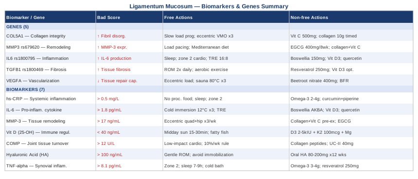
Where to Go From Here
Ligamentum mucosum hypertrophy occupies an awkward clinical space — real enough to cause significant and persistent symptoms, but often overlooked, misdiagnosed, or treated with advice that does not account for the underlying biology. The seven biomarkers covered here — hs-CRP, IL-6, MMP-3, vitamin D, COMP, hyaluronic acid, and TNF-alpha — give you a concrete picture of the inflammatory and tissue-remodeling environment in which your knee is operating. Addressing even two or three of them systematically, with both lifestyle and targeted supplementation strategies, can meaningfully shift the conditions that either perpetuate or resolve the hypertrophy over time.
The genetic layer adds useful depth: understanding whether your tissue remodeling balance, inflammatory responsiveness, or fibrotic healing tendency is constitutionally different from average helps prioritize which interventions carry the highest return for your specific biology. The movement principles from Built to Move translate that understanding into daily practice, and the complementary therapies — photobiomodulation, massage, MBSR, and biofeedback — provide additional tools with real clinical evidence behind them.
The most productive next step is not an overhaul of everything at once. Start with what is most measurable and most impactful: get hs-CRP and 25-OH vitamin D tested, assess your current sleep and loading patterns honestly, and consider bringing this framework to a physiotherapist or sports medicine physician who is open to thinking beyond rest and anti-inflammatories. One well-chosen biomarker result can reframe an entire treatment strategy. That is where meaningful progress usually starts.
Musculoskeletal: Joint Conditions Tendon & Ligament Conditions Sports Injuries
Autoimmune: Inflammatory Conditions Connective Tissue Conditions