This article contains AI generated content.
· UpdatedLimb-Girdle Muscular Dystrophy - 8 Genes And 6 Biomarkers To Track
Introduction
Limb-girdle muscular dystrophy is not a single disease. It is a family of more than 30 genetically distinct conditions that share one outcome: progressive weakness of the shoulder and hip muscles that, over years or decades, erodes independence. For most people living with LGMD, the diagnosis arrives late — after years of being told the fatigue is normal, the weakness is deconditioning, the symptoms are something else entirely. By the time a specific genetic cause is identified, significant muscle loss has often already occurred.
Standard clinical management remains largely reactive. Care focuses on tracking functional decline and responding to complications — cardiac problems, respiratory weakness, contractures — when they appear. That approach is necessary but not enough. It does not tell you why your muscles are breaking down faster during some months than others, which internal signals predict a worsening trend before it becomes obvious, or which biological levers might slow the process.
That is where precision monitoring becomes genuinely useful. Tracking the right biomarkers gives you and your clinical team real-time information about what is happening at the cellular level: how much muscle is being damaged, whether inflammation is amplifying the problem, whether the heart is under subclinical stress. Understanding the specific gene underlying your LGMD subtype opens the door to targeted strategies that work with your specific biology rather than assuming all LGMD is the same.
The goal here is not to promise a cure or suggest genetics can be fully overcome. It is to give you sharper tools. The six biomarkers covered in this article can be tracked with routine blood draws. The eight genes reflect the most common and best-studied LGMD subtypes. The strategies that follow — drawn from muscle physiology research, complementary medicine, and epigenetics — offer layers of support beyond what a quarterly neurology appointment typically provides. Better information, consistently used, leads to better decisions over time.
Summary
This article covers the 6 most actionable biomarkers for monitoring LGMD — starting with the tests your neurologist likely already orders and moving into more sensitive markers that most clinicians have not yet incorporated. For each biomarker, you will find out what it actually reveals, how to get it measured and at what cost, and what to do if the number is unfavorable — both with and without supplements or equipment.
The genetics section examines 8 of the most common LGMD-associated genes, explaining what each does, how its dysfunction leads to muscle breakdown, and which targeted strategies may partially compensate — some backed by human trials, others by strong biological reasoning that has not yet reached the clinic.
Beyond biomarkers and genetics, the article covers 10 muscle physiology insights from recent research that apply directly to managing LGMD, alongside four complementary approaches with meaningful human evidence in this context. Whether you are newly diagnosed or years into managing this condition, this article is designed to help you make more informed decisions about your monitoring, your lifestyle, and your next conversation with a specialist.
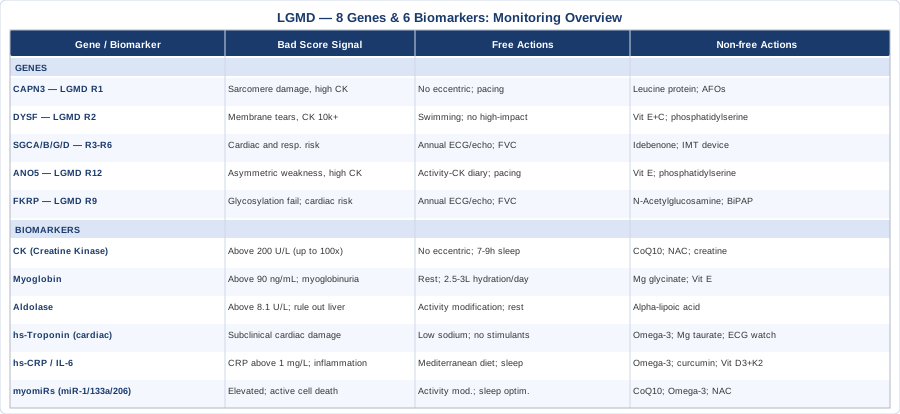
6 Biomarkers That Reveal What Is Actually Happening in Your Muscles
Biomarkers are objective signals. They register changes before symptoms become obvious, and they give you numbers to track over time rather than relying solely on subjective functional assessments. For a condition like LGMD where muscle loss accumulates over years, having reliable measurements is the closest thing available to early-warning infrastructure.
The six biomarkers below are ordered from most established to most emerging. The first three belong in every LGMD monitoring protocol right now. The last two represent the frontier — increasingly available and increasingly informative, but not yet standard of care in most clinics.
Biomarker 1: Creatine Kinase (CK)
Why it matters
Creatine kinase is the most widely used biomarker in neuromuscular disease for good reason. It leaks out of muscle cells when those cells are damaged or dying. In healthy adults, serum CK typically falls below 200 U/L. In many LGMD subtypes — particularly those involving CAPN3 or DYSF mutations — CK can run 10 to 100 times above normal even during periods of apparent clinical stability.
CK is not perfectly specific to LGMD: strenuous exercise, minor trauma, and other muscle diseases also raise it. But as a longitudinal tracking tool in someone with a known LGMD diagnosis, it is invaluable. A sustained upward trend between visits often signals accelerating muscle damage before functional decline becomes clinically apparent. A single high value matters less than the trend.
How to measure it
CK is measured via a standard blood draw and is included in most muscle enzyme panels. Cost: approximately $20–$50 with insurance, $40–$80 out of pocket. Recommended frequency for active LGMD monitoring: every 3–6 months at baseline; more frequently if activity level, treatment, or symptoms change.
If the score is bad, the plan without supplements
Elevated CK reflects ongoing muscle fiber breakdown that is partly driven by the underlying genetic deficit and partly amplified by controllable factors. Eccentric exercise — the lengthening phase of movement, such as walking downhill or lowering a weight — generates far greater mechanical damage than concentric or isometric exercise and should be strictly limited or eliminated. Replace it with low-impact aerobic activity: swimming, cycling, aquatic therapy conducted 3–4 times per week at moderate intensity.
Adequate sleep (7–9 hours per night) is equally critical. Most muscle repair occurs during deep sleep via growth hormone release; chronic sleep restriction measurably worsens inflammatory markers and impairs tissue repair. Pacing — deliberately managing energy output across the day to avoid post-exertional crashes — is one of the most underutilized non-supplement tools for keeping CK from repeatedly spiking.
If the score is bad, the plan with supplements or equipment
Coenzyme Q10 (ubiquinol form): 200–400 mg per day. CoQ10 supports mitochondrial energy production inside muscle cells and has antioxidant properties that reduce oxidative damage to muscle fibers. Cycling: continuous use with no mandatory breaks. Side effects: generally well tolerated; mild gastrointestinal upset possible at higher doses.
N-Acetylcysteine (NAC): 600 mg twice daily. A precursor to glutathione, the body's primary intracellular antioxidant. Oxidative stress is a significant amplifier of muscle fiber breakdown in LGMD. Research published in Free Radical Biology and Medicine has demonstrated that NAC reduces oxidative biomarkers in skeletal muscle disease. Side effects: rare; possible nausea at higher doses.
Creatine monohydrate: 3–5 g per day. A Cochrane systematic review assessing creatine in muscular dystrophies (Kley, Tarnopolsky, and Vorgerd, Cochrane Database of Systematic Reviews) found modest short-term benefits in muscle strength and reduced CK in some subtypes. Evidence specifically for LGMD is limited; discuss with your neurologist before starting, particularly if there is kidney involvement. Side effects: water retention, mild gastrointestinal discomfort.
Aquatic resistance therapy: Exercising against water resistance 2–3 times per week provides meaningful muscular stimulus without the eccentric loading that spikes CK on land. This is one of the most accessible and evidence-consistent exercise interventions in LGMD.
Biomarker 2: Myoglobin
Why it matters
Myoglobin is an oxygen-binding protein concentrated inside muscle fibers. When muscle cells are damaged, myoglobin enters the bloodstream faster than CK — making it a more acutely sensitive marker of muscle injury. Normal serum myoglobin is below 90 ng/mL. In acute exacerbations of LGMD or after unusual exertion, myoglobin can rise sharply and may even enter the urine, producing a brown discoloration called myoglobinuria — a warning sign that requires urgent medical attention due to the risk of acute kidney injury.
Tracking myoglobin alongside CK provides a fuller picture. CK reflects cumulative ongoing damage; myoglobin captures acute spikes. If CK is chronically elevated but myoglobin is normal, the damage pattern is likely chronic and gradual. If both rise together sharply, something has acutely worsened.
How to measure it
Myoglobin is measured via a standard blood draw but is not always included in routine panels and may need to be specifically requested. Cost: $40–$80 out of pocket. Most useful at baseline and during or after symptomatic exacerbations to understand the acute damage pattern.
If the score is bad, the plan without supplements
An acute myoglobin spike is a signal that the muscles are under severe stress. Immediate priority is rest and aggressive hydration — at least 2.5–3 liters of water per day to help clear myoglobin through the kidneys before it causes damage. Identify the trigger: was it unusual physical activity, a viral illness, heat exposure? Documenting triggers directly informs activity planning. If myoglobinuria (brown or cola-colored urine) appears, seek same-day medical evaluation. This is a medical emergency.
If the score is bad, the plan with supplements or equipment
Electrolyte support (magnesium glycinate): 200–400 mg nightly. Magnesium supports cellular membrane integrity and reduces pathological muscle excitability. The glycinate form is better tolerated than oxide. Side effects: loose stools at higher doses.
Vitamin E (mixed tocopherols): 400 IU per day. Vitamin E integrates into cell membranes as a lipid-soluble antioxidant — particularly relevant for DYSF-related LGMD where membrane repair is deficient and membranes are under persistent oxidative attack. Avoid exceeding 1000 IU per day without clinical guidance due to potential anticoagulant effects at high doses.
Biomarker 3: Aldolase
Why it matters
Aldolase is a glycolytic enzyme present in high concentrations in skeletal muscle. Like CK, it enters the bloodstream when muscle fibers break down. Aldolase can be more informative than CK alone in specific LGMD subtypes, and tracking the CK-to-aldolase ratio over time can help characterize disease activity during stable periods. Normal adult range: 1.5–8.1 U/L.
In some LGMD presentations, aldolase rises disproportionately to CK — information that carries diagnostic and prognostic value that a CK-only approach would miss.
How to measure it
Standard blood draw, often ordered alongside CK as part of a muscle enzyme panel. Cost: $40–$80 out of pocket. Recommended testing frequency: every 6–12 months for stable LGMD, or as needed with clinical changes.
If the score is bad, the plan without supplements
High aldolase with elevated CK confirms active muscle breakdown and reinforces the activity modification principles described for CK. A notable exception: high aldolase with normal CK may reflect liver pathology rather than muscle disease, and warrants investigation with liver function tests before attributing it to LGMD progression.
If the score is bad, the plan with supplements or equipment
Alpha-lipoic acid: 300–600 mg per day. A potent antioxidant with both fat- and water-soluble properties that supports mitochondrial function and reduces oxidative stress in skeletal muscle tissue. Works synergistically with CoQ10. Side effects: rare; possible mild hypoglycemia at high doses in sensitive individuals; avoid concurrent use with thyroid medications without guidance.
Biomarker 4: Cardiac Troponin (High-Sensitivity)
Why it matters
This may be the most underprescribed biomarker in LGMD care — and one of the most consequential. Multiple LGMD subtypes carry significant risk of cardiomyopathy: LGMD R1B due to LMNA mutation, LGMD R3–R6 from sarcoglycan defects, and LGMD R9 from FKRP mutation all affect heart muscle progressively and often silently. Because limb weakness limits exercise capacity, the shortness of breath and chest discomfort typically associated with cardiac disease may never develop — leaving cardiomyopathy undetected until it is advanced.
High-sensitivity cardiac troponin I (hs-TnI) or troponin T (hs-TnT) can detect subclinical myocardial damage years before an echocardiogram shows reduced ejection fraction. That is the window where prevention and early pharmacological cardioprotection are most effective.
How to measure it
High-sensitivity troponin requires a blood draw and must be specifically ordered — it is not part of a standard metabolic panel. Cost: $50–$120 out of pocket. Cardiology guidelines for LGMD subtypes with cardiac risk recommend annual or biennial ECG and echocardiography; adding hs-troponin between imaging visits fills the gaps. Interpretation requires a cardiologist familiar with neuromuscular disease, as troponin elevation patterns differ from ischemic heart disease.
If the score is bad, the plan without supplements
Elevated hs-troponin in an LGMD patient should prompt immediate cardiology referral. If cardiomyopathy or arrhythmia is confirmed, pharmacological management — ACE inhibitors, beta-blockers, and in some cases implantable cardiac devices — has strong evidence for delaying progression and reducing mortality in this population. This is not optional.
From a lifestyle standpoint: reduce sodium if there are signs of fluid retention, maintain optimal blood pressure, and avoid all stimulants (high-dose caffeine, decongestants, ephedra-containing products) that increase cardiac preload. Never dismiss fatigue or dyspnea as purely muscular in the context of a known cardiac-risk LGMD subtype.
If the score is bad, the plan with supplements or equipment
Omega-3 fatty acids (EPA + DHA): 2–4 g per day from high-quality fish oil or algae-derived sources. Omega-3s reduce cardiac triglycerides, lower inflammatory burden, improve cardiac electrophysiology, and have broad evidence for cardioprotection. Side effects: mild anticoagulant effect at higher doses; caution if on blood thinners.
Magnesium taurate: 200–400 mg per day. The taurate form has particular affinity for cardiac tissue. Magnesium deficiency is independently associated with arrhythmia risk. Side effects: mild loose stools.
Cardiac wearable monitoring: Devices such as the KardiaMobile single-lead ECG or smartwatches with ECG function allow daily arrhythmia screening between clinical visits. Recommended for all LGMD patients with known cardiac involvement, elevated hs-troponin, or a subtype with established cardiac risk.
Biomarker 5: High-Sensitivity CRP and Interleukin-6
Why it matters
Chronic low-grade inflammation is not simply a downstream consequence of muscle damage in LGMD — it actively accelerates it. When muscle fibers break down, they release damage-associated molecular patterns that trigger an immune response. In healthy tissue, this promotes repair. In LGMD, where the genetic defect impairs repair mechanisms, the inflammatory response becomes destructive rather than restorative — a cycle of damage amplifying further damage.
High-sensitivity CRP (hs-CRP) is a liver-derived marker of systemic inflammation. Optimal for cardiovascular and muscle disease risk: below 1 mg/L. Interleukin-6 (IL-6) is a pro-inflammatory cytokine that drives CRP production and is increasingly measurable through specialty and research-grade labs. Tracking both alongside CK helps distinguish periods of predominantly inflammatory activity from mechanical muscle breakdown — useful information for guiding therapeutic decisions.
How to measure it
hs-CRP is available at virtually any standard lab and is often included in cardiac risk panels. Cost: $20–$50 out of pocket. IL-6 measurement is less standard and requires specialty labs or academic medical center panels. Cost: $80–$200. Recommended testing frequency: every 6 months alongside standard muscle biomarkers.
If the score is bad, the plan without supplements
An anti-inflammatory dietary pattern is the highest-impact non-supplement intervention. The Mediterranean diet — rich in vegetables, olive oil, fatty fish, legumes, and nuts; low in refined carbohydrates and ultra-processed foods — consistently reduces hs-CRP in randomized trials across populations. Reducing vegetable seed oils high in omega-6 (corn, soybean, canola) while increasing omega-3 sources directly improves the inflammatory substrate ratio.
Sleep optimization (7–9 hours nightly) independently lowers IL-6 and CRP. Structured activity that avoids excessive muscle damage — replacing high-impact exercise with swimming or cycling — breaks the damage-inflammation-damage cycle rather than reinforcing it.
If the score is bad, the plan with supplements or equipment
Omega-3 fatty acids: 2–4 g EPA+DHA daily. The most consistently evidence-backed anti-inflammatory supplement across dozens of randomized trials, with effects on both IL-6 and CRP. Cycling: continuous use. Side effects: mild anticoagulant effect at higher doses.
Curcumin with piperine: 500–1000 mg curcuminoids per day, always combined with 5–10 mg piperine (black pepper extract) to overcome poor bioavailability. Anti-inflammatory action via NF-κB pathway inhibition. Avoid at high doses in pregnancy or if on anticoagulants.
Vitamin D3 + K2: Optimize blood 25-OH-D to 50–70 ng/mL, typically requiring 3000–6000 IU D3 daily depending on baseline; pair with K2 (MK-7 form, 100–200 mcg) to direct calcium appropriately. Vitamin D deficiency independently associates with elevated CRP, impaired muscle function, and increased LGMD symptom burden. Monitor blood levels every 6 months when supplementing.
Regular sauna or warm water immersion: 15–20 minutes at 70–80°C, 3–4 times per week. Activates heat shock proteins involved in protein repair and has measurable anti-inflammatory effects in human studies. Start with 10-minute sessions; never use alone, particularly if cardiac involvement is present.
Biomarker 6: Muscle-Specific MicroRNAs (miR-1, miR-133a, miR-206)
Why it matters
This is the frontier of LGMD biomarker science. Muscle-specific microRNAs — called myomiRs — are small RNA molecules produced almost exclusively by skeletal and cardiac muscle. When muscle cells are damaged, these molecules are released into the circulation and can be detected in blood with high sensitivity. Research by Cacchiarelli and colleagues, published in EMBO Molecular Medicine, and subsequent work across multiple neuromuscular disease groups has demonstrated that circulating miR-1, miR-133a, and miR-206 are more sensitive and more specific markers of muscle damage than CK — rising earlier in the disease course, correlating more closely with histological severity, and in some studies distinguishing LGMD subtypes from other neuromuscular conditions.
These markers are not yet standard clinical tests, but they are increasingly available through research programs and some academic medical center genomics laboratories. Establishing your baseline now may prove valuable as this field develops rapidly toward clinical implementation.
How to measure it
Circulating myomiR panels are available through specialized research labs and academic neuromuscular disease programs. They are not typically covered by insurance. Cost: $200–$600 depending on the panel and number of targets measured. If you are enrolled in or eligible for a research program at an academic LGMD center, ask specifically about research-grade biomarker panels — participants often access these at no cost as part of longitudinal cohort studies.
If the score is bad, the plan without supplements
Elevated myomiRs confirm active muscle cell death and reinforce the same management principles as elevated CK: activity modification, structured low-impact exercise, sleep optimization, and anti-inflammatory dietary practices. Currently, myomiRs are most useful as a sensitive tracking tool and for understanding whether an intervention is meaningfully reducing cell damage — rather than as a trigger for any single acute change in management.
If the score is bad, the plan with supplements or equipment
No supplement has yet been demonstrated in a randomized controlled trial to specifically lower circulating myomiRs in LGMD. The rational approach targets the upstream drivers of muscle cell death: CoQ10 for mitochondrial energy support, omega-3 fatty acids for membrane integrity and anti-inflammation, and NAC for oxidative stress reduction. If gene therapy or exon-skipping trials become available for your specific LGMD subtype — an area of active development — baseline myomiR profiles may serve as entry criteria or primary outcome measures, making current baseline data potentially valuable for future eligibility.
More on LGMD genetics and subtypes — MedlinePlus (NIH)
With a monitoring foundation in place, understanding the specific genetic mechanism driving your LGMD subtype opens targeted strategies that biomarkers alone cannot reveal.
The 8 Key Genes Behind Limb-Girdle Muscular Dystrophy
LGMD is genetically heterogeneous — the same clinical picture of proximal weakness can arise from mutations in more than 30 different genes, each disrupting a distinct biological mechanism. The 8 genes covered here represent the most prevalent subtypes and the ones with the most developed evidence for targeted management. Understanding which gene is involved matters because the intervention logic differs significantly depending on what is failing: a membrane repair protein, a structural scaffolding component, an enzymatic regulator, or a glycosylation enzyme.
Gene 1: CAPN3 (Calpain-3) — LGMD R1
What it does
Calpain-3 is a calcium-activated protease — an enzyme that regulates protein turnover within the sarcomere, the contractile unit of muscle. Its primary role is in post-exercise muscle remodeling: cleaving damaged proteins so they can be recycled and replaced. LGMD R1 (formerly LGMD2A) caused by CAPN3 mutations is among the most common LGMD subtypes globally, with particularly high prevalence in the Basque region of Spain and parts of Brazil and Japan.
Without functional calpain-3, damaged proteins accumulate within the sarcomere after each contraction. The muscle fiber progressively becomes dysfunctional and is eventually replaced by fibrous and adipose tissue. Disease onset typically occurs in adolescence or early adulthood with weakness in the hip and shoulder girdles.
If the gene is bad, the plan without supplements
Because CAPN3 dysfunction specifically impairs the post-exercise repair mechanism, exercise selection is the most impactful lifestyle intervention. Eccentric contractions generate the greatest sarcomeric stress and produce microdamage that calpain-3-deficient muscle cannot efficiently process. A structured physical therapy program eliminating or strictly limiting eccentric loading — replacing it with concentric-only or isometric exercises — reduces the accumulation of unrepaired damage. Aquatic therapy with trained neuromuscular physiotherapists is particularly appropriate.
Pacing is essential. The post-exertional worsening that many individuals with LGMD R1 experience represents exceeding the remaining repair capacity of affected muscle. Tracking activity levels with a wearable and keeping a CK response diary helps establish your individual tolerance threshold.
If the gene is bad, the plan with supplements or equipment
Leucine-enriched protein intake: 1.6–2.0 g protein per kg body weight per day, with leucine-rich sources (whey protein isolate, eggs, chicken, fish) prioritized within 30–60 minutes post-exercise. Leucine is the primary activator of mTOR — the key driver of muscle protein synthesis — and may partially compensate for impaired CAPN3-mediated sarcomeric remodeling by upregulating the synthesis side of the equation. Continuous use; no cycling needed.
Assistive mobility devices: For individuals with significant hip girdle weakness, lightweight walking aids or ankle-foot orthoses (AFOs) reduce the mechanical demand on affected muscles during daily activities, decreasing cumulative microdamage accumulation over years of use.
Gene 2: DYSF (Dysferlin) — LGMD R2
What it does
Dysferlin is a calcium-sensitive membrane repair protein. Its primary function is to patch the small tears that naturally occur in the plasma membrane of muscle fibers during contraction — think of it as the muscle cell's emergency sealant. These micro-tears are generated even by normal activity in all muscle; the difference is that healthy muscle repairs them in seconds. Without dysferlin, each tear expands into catastrophic membrane failure and cell death.
LGMD R2 (formerly LGMD2B) typically presents in young adults (late teens to early 30s) with initial calf weakness and CK levels that can reach 10,000–30,000 U/L long before significant functional loss is apparent. The very high CK at early presentation is a characteristic clue.
If the gene is bad, the plan without supplements
Membrane protection is the central principle. High-impact activities — running, jumping, contact sports — dramatically worsen outcomes in dysferlinopathy by generating repeated membrane tears the cell cannot seal. Swimming is the exercise of choice: it provides cardiovascular conditioning with minimal shearing stress on the plasma membrane.
Core temperature management also matters. Dysferlin-mediated membrane repair is calcium-dependent and temperature-sensitive. Maintaining muscle warmth before and during exercise — using warm-up periods, appropriate clothing, and warm water for aquatic exercise — helps maximize residual repair function.
If the gene is bad, the plan with supplements or equipment
Vitamin E (mixed tocopherols) + Vitamin C: E at 400 IU and C at 500 mg daily. Both integrate into or protect cell membranes from oxidative deterioration — amplified when membrane repair machinery is deficient. Use together; the combination has greater antioxidant coverage than either alone.
Phosphatidylserine: 300 mg per day. A structural phospholipid of cell membranes that may support membrane stability in dysferlin-deficient conditions. Human trial evidence specific to DYSF-LGMD is limited; the rationale is mechanistically grounded. Discuss with your specialist before starting.
Serum calcium optimization: Since dysferlin function is calcium-dependent, ensuring serum calcium is in the optimal range (8.6–10.0 mg/dL) and supplementing with calcium citrate (500–1000 mg/day) plus vitamin D3 if dietary intake is suboptimal supports whatever residual repair activity remains.
Genes 3–6: SGCA, SGCB, SGCG, SGCD (The Sarcoglycans) — LGMD R3 through R6
What they do
The four sarcoglycan genes encode proteins that form an interconnected complex within the dystrophin-associated protein complex — the structural scaffolding that links the internal cytoskeleton of the muscle fiber to the surrounding extracellular matrix. This linkage is what allows the muscle to generate and transmit force without tearing itself apart during contraction. When any one of the four sarcoglycans is mutated, the entire complex is destabilized and the membrane is vulnerable to contraction-induced stress.
Sarcoglycanopathies are autosomal recessive and typically present in childhood or adolescence with rapidly progressive proximal weakness. They carry significant risk of both cardiomyopathy and respiratory muscle involvement — particularly SGCD mutations, which have strong cardiac effects.
If the gene is bad, the plan without supplements
The most critical non-supplement intervention is proactive cardiac and respiratory surveillance. All four sarcoglycanopathies warrant annual ECG, echocardiography, and pulmonary function testing — including forced vital capacity (FVC) and maximal inspiratory and expiratory pressure (MIP/MEP). Do not wait for symptoms; cardiac and respiratory involvement in sarcoglycanopathies can progress silently.
For exercise: low-intensity resistance work supervised by a neuromuscular physiotherapist, focused on maintaining functional capacity rather than building strength. Respiratory muscle training using calibrated inspiratory muscle training devices can help preserve breathing mechanics before significant respiratory compromise develops.
If the gene is bad, the plan with supplements or equipment
Idebenone: A synthetic analogue of CoQ10 that penetrates cell membranes more effectively than standard CoQ10. Some cardioprotective evidence exists in Duchenne MD — a related dystrophinopathy — that is biologically applicable to sarcoglycanopathies sharing DAPC disruption. Dose: 150–300 mg three times daily with meals. Side effects: generally well tolerated; nausea possible.
Threshold inspiratory muscle training (IMT) device: These devices (available over the counter, approximately $35–$50) apply calibrated resistance to inspire against. Protocol: 30 breaths per day, 5 days per week, starting at 20–30% of maximum inspiratory pressure and progressing to 40–50% over 6–8 weeks. Evidence in neuromuscular disease populations supports improved inspiratory muscle strength and reduced dyspnea burden.
Early pharmacological cardiac protection: When cardiomyopathy is confirmed — which should be identified by surveillance before symptoms — ACE inhibitors and beta-blockers have strong evidence for delaying cardiac decline in neuromuscular cardiomyopathy. This requires a prescribing cardiologist with neuromuscular disease expertise but is among the most evidence-based interventions available for these subtypes.
Gene 7: ANO5 (Anoctamin-5) — LGMD R12
What it does
ANO5 encodes anoctamin-5, a calcium-activated chloride channel involved in muscle membrane repair and ion homeostasis. Like dysferlin, it participates in sealing membrane injuries after contraction — a functional overlap that explains the clinical similarities between LGMD R12 (formerly LGMD2L) and dysferlinopathy. LGMD R12 is increasingly recognized as one of the most common LGMD subtypes in Northern Europe and North America, yet it remains frequently misdiagnosed for years because it often presents with asymmetric weakness and later onset than other subtypes.
If the gene is bad, the plan without supplements
ANO5-related LGMD often has a milder progression than dysferlinopathy, which creates more time to establish effective management. Exercise tolerance varies significantly between individuals; tracking CK response to different activity types and intensities is particularly useful here. Keeping a structured activity-CK diary — documenting activity type, duration, intensity, and subsequent CK level — helps identify the personal threshold below which CK does not spike significantly. Exercise below that threshold is sustainable long-term.
If the gene is bad, the plan with supplements or equipment
The membrane repair overlap with dysferlin suggests similar supplementation principles: Vitamin E (400 IU daily), phosphatidylserine (300 mg daily), and calcium optimization. Clinical trial data specific to ANO5 supplementation is essentially nonexistent at present; these recommendations are based on shared biological mechanism and general safety profile. Discuss with a specialist before beginning.
Gene 8: FKRP (Fukutin-Related Protein) — LGMD R9
What it does
FKRP encodes an enzyme that glycosylates — adds carbohydrate chains to — alpha-dystroglycan, a protein that serves as the anchor between the muscle fiber and its surrounding extracellular matrix. When glycosylation is impaired, alpha-dystroglycan cannot bind properly to laminin in the extracellular matrix, and the mechanical connection between cell and environment fails under the stress of contraction.
LGMD R9 (formerly LGMD2I) is notable for its unusually wide clinical spectrum. Some individuals carry mutations that result in significant residual FKRP activity and experience relatively mild weakness well into adulthood; others with more severe mutations develop cardiomyopathy and respiratory insufficiency in their 30s. The cardiac and respiratory risks in FKRP-LGMD are often underappreciated and require active monitoring regardless of limb weakness severity.
If the gene is bad, the plan without supplements
Given the cardiac and respiratory risk profile, management parallels the sarcoglycanopathies in requiring regular surveillance. Annual ECG and echocardiography and biennial pulmonary function testing with FVC monitoring are standard. If FVC drops below 50% of predicted, referral for assessment of nocturnal noninvasive ventilatory support (BiPAP) should be initiated without waiting for symptomatic respiratory failure.
For exercise: low-impact activities emphasizing endurance over strength, paced carefully to avoid CK spikes. Respiratory muscle training as described for sarcoglycanopathies applies here equally.
If the gene is bad, the plan with supplements or equipment
N-Acetylglucosamine: 500–1000 mg per day. FKRP participates in a glycosylation pathway, and some researchers have proposed that providing substrate availability through glucosamine derivatives may support residual enzyme activity in individuals with hypomorphic (partially functional) FKRP mutations. Evidence is early-stage — primarily in vitro and animal models. Side effects: generally well tolerated. Discuss with a neuromuscular specialist before starting; this is a biologically plausible but not yet clinically validated approach.
BiPAP/NIV equipment: If respiratory function monitoring shows progressive FVC decline, initiating noninvasive ventilation nocturnally before symptomatic respiratory failure is associated with better long-term survival and quality of life in neuromuscular disease. A pulmonologist with neuromuscular disease experience should guide the timing and setup.
Understanding how muscle works at a cellular level — beyond any one gene or biomarker — opens additional management possibilities that are often overlooked in rare disease care.
What Muscle Physiology Science Reveals About Slowing Functional Decline
The Huberman Lab's extended series on muscle performance science — conducted with exercise physiologist Dr. Andy Galpin — synthesizes decades of human research into principles that, while primarily framed for athletes, have direct and underutilized applications for people managing chronic neuromuscular disease. The following 10 insights are the most applicable to LGMD.
1. Muscle Fiber Type Composition Is Bidirectional and Trainable
Type I (slow-twitch) muscle fibers are more fatigue-resistant, more oxidatively efficient, and more durable under the mechanical stresses common in LGMD. Type II (fast-twitch) fibers generate greater peak force but are more metabolically demanding and typically degenerate faster in LGMD. Sustained low-load, high-repetition endurance training over months to years has been shown in healthy populations to shift functional fiber type composition toward Type I characteristics. In LGMD, this may mean training the more resilient fiber type and allowing it to carry a greater proportion of functional load.
2. Eccentric Loading Is Quantifiably Damaging in Membrane-Repair-Deficient Muscle
Dr. Galpin's review of biomechanics research confirms that eccentric contractions generate 3–4 times greater mechanical stress on the sarcomere than concentric contractions. For LGMD subtypes involving membrane repair defects — DYSF, ANO5, and the sarcoglycanopathies — this translates directly into dramatically higher CK spikes and greater fiber loss following eccentric-dominant exercise. This is not theoretical; it is measurable in CK response within 24–72 hours. The practical takeaway for program design is non-negotiable: eliminate or severely restrict eccentric loading and replace it with concentric-only and isometric variants.
3. Zone 2 Aerobic Training Rebuilds Mitochondrial Density
Zone 2 training — low-intensity aerobic exercise maintained at conversational pace for 30–60 minutes — is the most potent known stimulus for mitochondrial biogenesis (the creation of new mitochondria) in skeletal muscle. Mitochondrial density is reduced in many LGMD subtypes due to progressive muscle cell death. In the surviving fibers, improving mitochondrial density through consistent Zone 2 work — swimming, stationary cycling, rowing at low intensity — improves energy efficiency and resilience to fatigue. This is one of the most achievable and impactful training adaptations available in chronic muscle disease.
4. Sleep Is the Primary Anabolic Window for Muscle Repair
Over 70% of daily growth hormone release occurs during slow-wave sleep. Growth hormone drives muscle protein synthesis and repair — precisely the processes most impaired by LGMD's genetic defects. In a condition where repair mechanisms are already compromised at the molecular level, optimizing the remaining endogenous repair capacity through 7–9 hours of high-quality sleep per night is not optional. Research consistently shows that sleep restriction below 7 hours worsens muscle biomarkers even in healthy individuals; the effect in LGMD is almost certainly amplified.
5. Protein Timing Matters as Much as Total Intake
Distributing protein across 4–5 daily servings of 25–40 g each — rather than concentrating most intake in one or two meals — maximizes the mTOR activation signal throughout the day and sustains muscle protein synthesis for longer. For LGMD patients, this principle supports whatever repair capacity remains. Casein-rich evening protein (Greek yogurt, cottage cheese) has been specifically shown to enhance overnight muscle protein synthesis, making it particularly relevant in a disease where nocturnal repair is critical.
6. Chronic Inflammation Directly Suppresses Muscle Protein Synthesis
Persistently elevated inflammatory cytokines — IL-6, TNF-alpha — inhibit the mTOR pathway, reducing the rate of muscle protein synthesis. In LGMD, this creates a vicious biological cycle: muscle damage triggers inflammation, inflammation suppresses repair, reduced repair leads to greater damage. Every intervention that durably reduces systemic inflammation — omega-3 fatty acids, anti-inflammatory diet, sleep optimization, stress management — also indirectly supports whatever muscle protein synthesis capacity remains.
7. VO2max Is a Proxy for Functional Reserve
VO2max — maximum oxygen uptake capacity — reflects the combined efficiency of cardiovascular and muscular aerobic metabolism. In LGMD, reduced VO2max is common due to both restricted activity and loss of metabolically active muscle mass. In healthy aging populations, VO2max is the single strongest predictor of longevity and functional independence. Even modest improvements in VO2max through regular low-impact aerobic training have been associated with better quality of life and reduced mortality in neuromuscular disease populations.
8. Isometric Training Provides Stimulus Without Membrane Shear
Isometric contractions — pushing or pulling against a fixed resistance without joint movement — generate far less mechanical membrane stress than dynamic contractions while still providing adequate stimulus for motor unit recruitment and neuromuscular connection maintenance. Wall sits, doorframe pushes, and isometric resistance exercises with a physical therapist are particularly appropriate for LGMD and significantly underused in most standard physical therapy protocols. They deserve a central role, not a secondary one.
9. Heat Shock Proteins Support Protein Integrity in Surviving Fibers
Heat shock proteins (HSPs) are molecular chaperones that help misfolded or damaged proteins refold correctly and reduce cellular toxicity from protein aggregation. Regular, controlled heat exposure — whether from sauna, warm water immersion, or heated exercise environments — upregulates HSP expression significantly and consistently in human research. In LGMD, where protein function may be diminished by mutation and surviving fibers are under persistent stress, supporting HSP activity provides a meaningful layer of protection with very low risk if initiated conservatively.
10. Neural Drive Is Preserved Longer Than Muscle Mass
Even in advanced LGMD, the motor nerve pathways that activate muscle contraction often remain intact long after the muscle fibers themselves have degenerated significantly. Neuromuscular electrical stimulation (NMES) devices deliver low-level electrical pulses through surface electrodes to maintain neural activation of muscle fibers that can no longer be voluntarily contracted with adequate force. NMES is commercially available in home-use devices and has evidence in several neuromuscular conditions for slowing atrophy in weakened but not completely denervated muscles. This is a meaningful adjunct for advanced-stage LGMD and deserves wider discussion with physiotherapy teams.
Complementary Approaches with Real Evidence for LGMD
The following four modalities have the strongest applicable human evidence from the approved list for this specific condition. None replaces medical management, but each offers genuine and realistic benefit when properly applied.
Breathing-Based Therapies
Respiratory muscle involvement is one of the most underappreciated complications of LGMD. The diaphragm and intercostal muscles are skeletal muscles subject to the same genetic defects affecting limb muscles. As they weaken, breathing becomes effortful during exertion, sleep quality deteriorates through nocturnal hypoventilation, and the risk of acute respiratory failure during illness increases substantially. Breathing-based therapies — principally calibrated inspiratory muscle training and pursed-lip breathing techniques — directly address this by training respiratory muscles within their functional limits before impairment becomes symptomatic.
A randomized controlled trial in Thorax demonstrated that inspiratory muscle training improved respiratory muscle strength, reduced perceived dyspnea, and improved quality of life in patients with neuromuscular disease including muscular dystrophies. The protocol involved 30 breaths against 30% of maximum inspiratory pressure, 5 days per week for 8 weeks, with incremental resistance increases. Evidence is strongest for preservation of function rather than restoration, which reinforces the importance of beginning before significant respiratory compromise is apparent.
For practical application: the Threshold IMT device (available without prescription, approximately $35–$50) implements this protocol. Start at the lowest resistance setting and increase every 1–2 weeks based on tolerance. Do not practice IMT during acute respiratory illness. Track FVC and MIP/MEP quarterly to assess response. Practice pursed-lip breathing during physical activity — inhaling through the nose for 2 counts, exhaling slowly through pursed lips for 4 counts — to reduce dynamic hyperinflation and reduce respiratory effort throughout the day.
Low-Level Laser Therapy and Photobiomodulation
Photobiomodulation (PBM) involves the application of red (630–700 nm) or near-infrared (780–1100 nm) light to tissue, where it is absorbed by mitochondrial cytochrome c oxidase, the terminal enzyme of the respiratory chain. This absorption increases local ATP production, reduces oxidative stress within the cell, and modulates inflammatory signaling. For LGMD, the relevance is mitochondrial: PBM may enhance energy production in surviving muscle fibers and reduce oxidative damage amplification in membrane-repair-deficient subtypes.
Research published in Lasers in Medical Science found that PBM applied before exercise significantly reduced CK elevation and muscle soreness following exercise-induced muscle damage. Separate research in Duchenne muscular dystrophy — which shares the DAPC disruption mechanism with sarcoglycanopathies — has demonstrated reduction in muscle damage biomarkers with PBM application. Evidence specific to LGMD is limited; biological plausibility is mechanistically grounded and the risk profile is low when used correctly.
For practical application: home-use PBM panels providing 630–850 nm light are available in the $200–$1200 range. Recommended starting protocol: 5–10 minutes per target area (shoulder girdle, hip girdle, quadriceps) 3–5 times per week. Begin with shorter sessions and extend over 2–3 weeks as tolerance is established. Never use PBM directly over the eyes, over areas of known malignancy, or over implanted electronic devices. Consult a physiotherapist or physiatrist experienced in photobiomodulation before beginning, and monitor CK response over the first 4–6 weeks to assess personal benefit.
Mindfulness Meditation and MBSR
Living with a progressive neuromuscular condition carries a psychological burden that standard neurological care rarely addresses adequately. Chronic uncertainty about progression, the experience of progressive functional loss, and managing daily life with a body that is changing unpredictably are measurable stressors that elevate cortisol, worsen inflammatory biomarkers including CRP and IL-6, and impair sleep architecture. These downstream effects, in turn, worsen the biological environment in which muscle repair occurs. Mindfulness-Based Stress Reduction (MBSR) is an 8-week structured program with the strongest evidence base of any psychological intervention in chronic medical illness.
A systematic review in the Journal of Psychosomatic Research found that MBSR significantly reduced anxiety, depression, and perceived pain in patients with chronic medical conditions; parallel evidence from multiple trials documents improvements in inflammatory markers including CRP and IL-6 after sustained mindfulness practice. For LGMD specifically, the body-scan and gentle movement components of MBSR can be adapted to current mobility limitations without requiring positions or movements that stress weakened muscles.
For practical application: the formal 8-week MBSR program is available online through certified providers including the Center for Mindfulness at the University of Massachusetts, where the program originated, as well as various accredited online delivery platforms. Daily sessions of 30–45 minutes are recommended during the program; 15–20 minutes of maintenance practice per day thereafter sustains the benefit. Evidence is particularly strong for sleep improvement, with documented increases in slow-wave sleep duration — directly relevant to the nocturnal muscle repair window discussed throughout this article.
Massage Therapy
For individuals with LGMD, massage therapy addresses several secondary concerns: reducing spasm and tightness in compensatory muscles that are overloaded by the redistribution of mechanical demands, improving local circulation in muscle groups with reduced activity-driven blood flow, decreasing perceived pain, and supporting psychological wellbeing. The critical caveat is that deep-tissue or high-pressure massage is directly contraindicated in LGMD: forceful mechanical pressure on fragile muscle fibers can trigger acute cell damage and measurable CK spikes. The technique must be adapted specifically for neuromuscular fragility.
A systematic review in Complementary Therapies in Medicine found that massage therapy consistently improved pain scores, muscle stiffness, and anxiety in patients with chronic neuromuscular conditions. The primary mechanism in LGMD is circulatory and neurological rather than structural — improving lymphatic flow, reducing perceived tension in overloaded compensatory muscles, and activating parasympathetic responses that support recovery.
For practical application: seek a massage therapist who has specific experience with neuromuscular disease or a physical therapist who incorporates gentle manual techniques. The appropriate approach for LGMD is gentle effleurage and lymphatic drainage — light, rhythmic strokes that move lymphatic fluid and improve local circulation without applying mechanical pressure to muscle tissue. Sessions of 30–45 minutes every 2–4 weeks are appropriate for most individuals. Communicate your diagnosis clearly before every session. Monitor CK levels 48 hours after the first session to assess whether even gentle massage affects your muscle response, then adjust frequency accordingly.
Conclusion
There is no intervention that reverses the genetic defect underlying limb-girdle muscular dystrophy. What does exist — and what remains genuinely underused in most clinical management — is a coherent framework for understanding your biology precisely enough to make better decisions. Tracking the right six biomarkers gives your clinical team early warning of cardiac involvement, inflammatory amplification, and muscle damage rate before these translate into functional decline. Understanding your specific gene clarifies the biological mechanism you are managing and points toward strategies that are tailored rather than generic. The muscle physiology research and complementary approaches add further tools that extend beyond what standard quarterly visits typically address.
The most important next step is ensuring your monitoring is complete: CK, aldolase, high-sensitivity troponin, and hs-CRP should all be part of your regular bloodwork if they are not already. From there, bring your genetics, your biomarker trends, and the targeted strategies in this article into a conversation with a neuromuscular specialist who knows your specific subtype. Better information, applied consistently and collaboratively, is the most realistic path to better long-term outcomes.
Neurological Cardiovascular Respiratory
Musculoskeletal: Muscle Conditions
Autoimmune: Inflammatory Conditions