This article contains AI generated content.
· UpdatedLipoblastoma Genes and Biomarkers — 4 Genes and 6 Biomarkers to Track
Introduction
When a child is diagnosed with lipoblastoma, most families move quickly into surgery mode and leave the hospital with a clean pathology report and a follow-up schedule — but without a clear understanding of what actually drove the tumor to grow. The word "benign" brings enormous relief, yet it does not answer the questions that surface months later: Why did this happen in my child? What does the molecular finding on the report mean? And how tightly should we be watching for recurrence?
Generic information about lipoblastoma being a "rare benign fat cell tumor in infants" is technically accurate. It is also insufficient. The difference between a child whose tumor carried a PLAG1 rearrangement with clean surgical margins and one with an HMGA2 rearrangement and a close margin is not a minor detail — it changes how surveillance should be structured, what molecular retesting is worth requesting, and how to interpret a new finding on an MRI years later.
This article approaches lipoblastoma from a molecular and monitoring standpoint, not a surgical one. The genetics are well-established enough to be clinically useful, and the biomarker panel — while more limited than for adult cancers — has practical value for families navigating the surveillance window. Understanding these layers does not eliminate uncertainty, but it makes the uncertainty legible.
What follows covers four key genes that explain the vast majority of lipoblastoma cases, six biomarkers worth tracking from diagnosis through surveillance, and a set of complementary approaches with genuine evidence for the pediatric caregiving context. The goal throughout is precision, not reassurance — better information genuinely leads to better decisions.
Summary
This article covers four defining genes in lipoblastoma biology — PLAG1, HMGA2, HAS2 as a major fusion driver, and IGF2 as the key downstream effector — explaining what each one does when disrupted and what a confirmed finding means for recurrence risk, surgical planning, and family counseling. You will also find a review of six practical biomarkers: from PLAG1 FISH testing and MRI surveillance to LDH, CRP, AFP, and CBC, each assessed for what it costs, what it reveals, and what to do when a value is off. Beyond the molecular layer, the article includes ten high-impact research insights from the pediatric tumor biology literature, plus a focused look at three complementary approaches — mindfulness for caregivers, music therapy for children during procedures, and post-surgical massage — that have meaningful human evidence in the pediatric oncology context. If the pathology report raised questions that no one has had time to answer in full, this article is the deeper read you have been looking for.
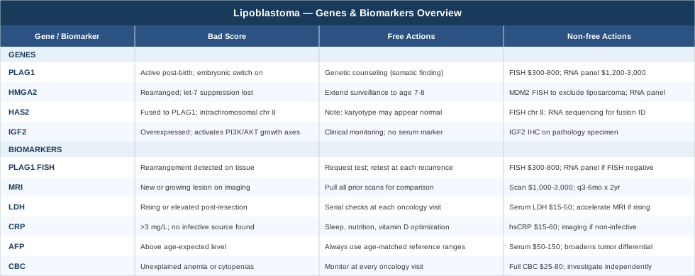
The Four Genes That Define Lipoblastoma Biology
Lipoblastoma is not a random overgrowth of fat cells. Like most tumors, it has a precise molecular signature — a set of genetic alterations that push immature embryonic fat cells called lipoblasts into sustained, uncontrolled proliferation. Research over the past two decades has narrowed this signature down to a small number of recurring genetic events. Understanding these genes does not change the surgical plan, but it refines the surveillance conversation significantly and answers the question families most want answered: why did this grow?
Gene 1 — PLAG1: The Embryonic Switch That Did Not Turn Off
PLAG1 (Pleomorphic Adenoma Gene 1) is a zinc finger transcription factor located on chromosome 8q12. Under normal developmental conditions, it is highly active in the fetus and early infant period, where it drives the expansion of fat cell precursors — exactly what is needed for building adipose tissue during rapid growth. After birth, PLAG1 expression is progressively silenced as the developmental program shifts. In lipoblastoma, a chromosomal rearrangement repositions PLAG1 next to a gene with an active promoter, effectively relocking the switch in the "on" position at a time when it should have gone quiet.
This rearrangement is found in approximately 70 to 80 percent of all lipoblastoma cases, making it the most reliable molecular signature for the diagnosis. The rearrangement works by a promoter-swapping mechanism: PLAG1's own inactive promoter is replaced by the active promoter of a fusion partner, driving high PLAG1 expression from the hybrid gene. The most common partner is HAS2 (Hyaluronan Synthase 2, located on chromosome 8q24) — notably, both PLAG1 and HAS2 sit on chromosome 8, meaning the fusion is an intrachromosomal rearrangement that standard karyotype analysis can miss. Other documented fusion partners include COL1A2 (collagen type 1 alpha 2, chromosome 7q21), COL3A1, and RAD51L1.
The downstream consequences of reactivated PLAG1 are wide-ranging. PLAG1 directly upregulates IGF2 (insulin-like growth factor 2), CRABP2, and ALDH1A2 — genes that collectively amplify cell cycle entry in adipose precursors. This explains why lipoblastomas emerge almost exclusively in very young children: the biological environment (abundant immature lipoblasts, high baseline IGF2 activity, active developmental signaling) is uniquely permissive for PLAG1-driven proliferation.
See this PubMed collection on PLAG1 rearrangements in lipoblastoma for the primary molecular literature.
If PLAG1 rearrangement is confirmed: the plan without supplements or additional tools
A confirmed PLAG1 rearrangement is the expected finding in most lipoblastoma cases. It confirms the diagnosis and carries specific implications for management:
Complete surgical resection remains the primary and curative treatment. Margin status is the strongest predictor of recurrence — achieving an R0 (complete, clear-margin) resection is the single most important modifiable factor in the clinical course. When margins are close or positive, the recurrence rate rises from roughly 5 to 10 percent toward 20 to 25 percent.
Post-surgical surveillance should be structured at intervals agreed with the treating oncology team. MRI or ultrasound (depending on anatomical location) every 3 to 6 months for the first two years is a reasonable standard for intermediate-risk cases. Longer surveillance windows (to age 5 or beyond) are appropriate for retroperitoneal or mediastinal tumors where complete resection is anatomically difficult.
Genetic counseling is a useful but underutilized step. PLAG1 rearrangements in lipoblastoma are somatic — they occur in the tumor cell during development, not in the germline. Most families carry unnecessary anxiety about recurrence risk in siblings or future children. A single genetic counseling consultation can clarify this and significantly reduce long-term distress.
If PLAG1 rearrangement is confirmed: the plan with additional monitoring tools
For families working with teams that offer molecular-guided surveillance:
FISH on any recurrent tissue is recommended rather than imaging-only follow-up of new masses. A recurrent lipoblastoma will typically show the same PLAG1 rearrangement as the primary — confirming this distinguishes true recurrence from a new primary lesion and guides the surgical approach. Cost: $300 to $800 per specimen.
RNA-based fusion panels are increasingly available through academic pediatric centers. These panels detect PLAG1 fusions by sequencing and identify the specific fusion partner — HAS2-PLAG1, COL1A2-PLAG1, and others. The partner gene identity has emerging relevance for understanding local behavior and may eventually guide more individualized surveillance schedules. Cost: $1,200 to $3,000; insurance coverage is improving as molecular diagnostics become standard of care in pediatric sarcoma.
Referral to a pediatric sarcoma specialist is worth requesting if the diagnosis was made at a center that sees lipoblastoma infrequently. High-volume soft tissue tumor programs offer both more experienced molecular interpretation and more granular surveillance protocols. This is not a sign of distrust in the primary surgeon — it is standard practice for rare pediatric tumors.
Note on cycling and side effects: Enhanced molecular surveillance does not carry the procedural risks of repeat imaging but can increase family anxiety. The decision to request additional testing should be made in the context of the clinical picture, not anxiety alone. Discuss the evidence base with the treating team before ordering additional tests independently.
Gene 2 — HMGA2: When Chromatin Architecture Goes Wrong
HMGA2 (High Mobility Group AT-hook 2), located on chromosome 12q14, is the second most commonly rearranged gene in lipoblastoma, found in roughly 10 to 15 percent of cases — predominantly in tumors that do not carry PLAG1 rearrangements. HMGA2 is not itself a transcription factor in the conventional sense. It is a chromatin architectural protein that remodels DNA structure in a way that makes large clusters of growth-promoting genes more accessible to transcriptional machinery.
During normal embryonic development, HMGA2 is highly expressed and critical for adipogenesis. Its expression is suppressed after the embryonic period by the let-7 family of microRNAs, which bind to the 3' untranslated region of the HMGA2 mRNA and trigger its degradation. In lipoblastoma, chromosomal rearrangements that disrupt the 3' end of HMGA2 remove the let-7 binding sites entirely — the suppression never happens, and HMGA2 continues driving fat cell precursor proliferation in postnatal tissue.
Some published series have found that HMGA2-positive lipoblastomas tend to occur in slightly older children within the pediatric age range and may carry a modestly higher recurrence rate after incomplete resection compared to PLAG1-positive tumors. The evidence comes from small retrospective series, so these observations should be treated as hypothesis-generating rather than definitive. The practical implication is that in HMGA2-positive cases with any concern about margin adequacy, a longer and more frequent surveillance schedule is a reasonable precaution.
HMGA2 rearrangements also occur in adult lipoma and, critically, in well-differentiated liposarcoma. This overlap makes molecular differentiation particularly important in atypical presentations — for example, an HMGA2-positive mass in a child older than 5 years warrants very careful pathological review and possibly MDM2 FISH testing to exclude a liposarcoma (MDM2 amplification is not expected in lipoblastoma).
See HMGA2 rearrangement studies in lipoblastoma for the primary research.
If HMGA2 rearrangement is confirmed: the plan without supplements
Management is surgical, as with PLAG1-positive tumors, but with added emphases:
Margin-focused resection planning is even more critical here given the possibly higher recurrence rate. The surgical team should be informed of the HMGA2 finding if it is known preoperatively.
Extended surveillance beyond the standard two-year window is a reasonable precaution — consider maintaining annual imaging to age 7 or 8 in cases with close or positive margins.
MDM2 exclusion is warranted if any histological feature is atypical or if the child is older than the typical lipoblastoma age window. A negative MDM2 FISH result definitively rules out well-differentiated liposarcoma and solidifies the benign prognosis.
If HMGA2 rearrangement is confirmed: the plan with additional tools
RNA fusion sequencing to identify the HMGA2 fusion partner can add diagnostic specificity. Common partners in lipoblastoma include ALDH2 and NFIB — the same fusion partners seen in lipoma — further distinguishing lipoblastoma from malignant liposarcoma variants.
Specialist pathology review at a high-volume soft tissue center is particularly important in HMGA2-positive cases. The diagnostic boundary between lipoblastoma and lipoma in young children can be subtle on histology alone, and HMGA2 positivity in the context of lipoma-like histology benefits from expert interpretation.
Gene 3 — Chromosome 8 Polysomy: Amplification Without a Clear Fusion
A subset of lipoblastoma cases — particularly those in which neither PLAG1 fusion nor HMGA2 rearrangement can be confirmed — shows polysomy of chromosome 8, meaning the tumor cells carry three, four, or more copies of the entire chromosome rather than a focused rearrangement. This is clinically relevant because chromosome 8 carries both PLAG1 at 8q12 and the HAS2 partner gene at 8q24 — extra copies of both effectively amplify PLAG1 activity without producing a classic fusion transcript.
Chromosome 8 polysomy is detectable by conventional karyotype or centromere-specific FISH probes. It is less diagnostically specific than a named fusion but is still supportive of a lipoblastoma diagnosis in the right clinical and histological context. Its presence does not change surgical management, but it is diagnostically useful when the molecular picture is otherwise incomplete.
The recurrence behavior of polysomy-only lipoblastomas is not well-characterized in the literature due to small case numbers. Current clinical practice treats these cases with the same surveillance approach as PLAG1-positive tumors, using margin status as the primary risk stratifier.
If chromosome 8 polysomy is the primary finding: the plan without supplements
Ensure the diagnosis was reviewed by a specialist soft tissue pathologist, as polysomy of chromosome 8 alone can be seen in reactive conditions and in other soft tissue tumors. If PLAG1 FISH was the only molecular test performed, consider requesting RNA fusion sequencing — a subset of polysomy-only cases on FISH will show a cryptic PLAG1 fusion detectable only by RNA-based methods. Standard margin-conscious resection and post-surgical surveillance apply otherwise.
Gene 4 — IGF2: The Growth Signal That Executes the PLAG1 Program
IGF2 (Insulin-like Growth Factor 2) is not rearranged or mutated in lipoblastoma, but it is among the most important direct transcriptional targets of PLAG1 and is central to understanding why the tumor grows. When PLAG1 is reactivated by chromosomal rearrangement, one of its first responses is to massively upregulate IGF2 expression. IGF2 binds to the IGF1 receptor and activates downstream cell cycle machinery — specifically the PI3K/AKT and RAS/MAPK pathways — driving lipoblast proliferation.
This relationship matters beyond lipoblastoma itself. The PLAG1-IGF2 axis is an active therapeutic target across multiple pediatric soft tissue tumors, and research into IGF1R inhibitors in other contexts may eventually produce tools relevant for rare, unresectable lipoblastoma cases. For the moment, IGF2's primary practical role is as a tissue-level biomarker of PLAG1 activity: high IGF2 expression on immunohistochemistry correlates with active PLAG1 signaling and can serve as a surrogate marker when direct FISH is equivocal.
The biological specificity of IGF2 to early childhood also helps explain the age distribution of lipoblastoma: serum IGF2 levels are physiologically elevated in the first years of life as part of normal growth programming, creating a cellular environment that amplifies the consequences of PLAG1 reactivation in exactly the developmental window when most lipoblastomas appear.
See PLAG1-IGF2 signaling in pediatric adipose tumors for the mechanistic research base.
If PLAG1-driven IGF2 overexpression is identified on pathology: practical implications
IGF2 immunohistochemistry (IHC) can be included in the pathology panel when molecular FISH is inconclusive. High IGF2 staining supports the lipoblastoma diagnosis. For standard cases, this is a confirmatory rather than a management-changing finding. In the research context, families treated at academic centers may be offered enrollment in molecular biobanking studies that track IGF2 and related pathway markers over time — this is worth accepting where available, as it contributes to the evidence base for a condition that is rare enough that individual cases meaningfully move the literature forward.
6 Biomarkers Worth Tracking in Lipoblastoma
The biomarker landscape for lipoblastoma is more limited than for adult solid tumors with well-established serum panels. This reflects both the benign nature of the tumor and its rarity — the research investment in surveillance biomarkers is modest. That said, six markers offer practical value across the diagnosis, surgical, and surveillance phases, ranging from low-cost blood tests to molecular pathology assays.
1. PLAG1 FISH Testing — The Molecular Confirmation
What it is: Fluorescence in situ hybridization (FISH) targeting the 8q12 locus to detect PLAG1 rearrangement or chromosome 8 polysomy. This is the gold-standard molecular confirmation for lipoblastoma diagnosis.
Why it matters: A PLAG1 FISH result converts a histological impression into a molecular diagnosis. In cases where histology alone is ambiguous — lipoblastoma versus lipoma or myxoid liposarcoma are not always clearly distinguishable on H&E staining — FISH changes management. It is also valuable when the child is older than the typical lipoblastoma age range, when the tumor is in an unusual location, or when family counseling requires a definitive molecular foundation.
How to measure it: Ordered on FFPE tissue from the surgical specimen or biopsy. Turnaround: 5 to 10 business days. Cost: $300 to $800. Covered by insurance in most health systems when ordered as part of a pediatric sarcoma workup.
If the result is positive: Diagnosis confirmed. Proceed with surveillance plan as described in the genetics section. Retest at each recurrence.
If the result is negative: Does not exclude lipoblastoma. Request RNA fusion sequencing — it is more sensitive than FISH for cryptic rearrangements. A FISH-negative, RNA-negative result in a morphologically typical lipoblastoma in a child under 3 years is still consistent with the diagnosis; molecular findings are supportive, not diagnostic on their own.
2. MRI Surveillance — The Recurrence Detector
What it is: Magnetic resonance imaging of the surgical site and regional anatomy. MRI is the preferred imaging modality for soft tissue tumor surveillance due to its superior contrast resolution compared to CT or ultrasound.
Why it matters: Local recurrence occurs in approximately 20 to 25 percent of cases following incomplete resection. Most recurrences appear within the first two years post-surgery and are detectable on MRI before they become clinically palpable — particularly for deep or retroperitoneal tumors. Early detection of recurrence allows for prompt re-excision, which is curative in the vast majority of cases.
How to measure it: Typically scheduled every 3 to 6 months for the first two post-surgical years, then annually based on risk stratification. Cost: $1,000 to $3,000 per scan with and without contrast. Usually covered by insurance under oncology surveillance protocols.
If a suspicious finding appears: Biopsy is warranted unless the lesion is clearly benign by imaging characteristics. Molecular retesting of recurrent tissue is recommended to confirm the same clone and exclude secondary changes.
Supplement-free optimization: Ensure the interpreting radiologist has access to all prior imaging for comparison — interval change assessment is more sensitive than single-timepoint review. Request the prior scans be pulled actively rather than relying on the radiologist to locate them independently.
3. LDH (Lactate Dehydrogenase) — The Proliferation Signal
What it is: A serum enzyme released by cells undergoing rapid turnover or ischemic stress. LDH is nonspecific but inexpensive and is part of the standard sarcoma monitoring panel at most pediatric oncology centers.
Why it matters: In benign lipoblastoma, LDH should be normal or only mildly elevated at diagnosis and should normalize after complete resection. A rising LDH during surveillance warrants imaging review. More importantly, LDH is part of the differential workup when a child presents with a soft tissue mass — a markedly elevated LDH raises the possibility of a more aggressive process and should accelerate the diagnostic timeline.
How to measure it: Standard serum chemistry, available universally. Cost: $15 to $50. Pediatric normal ranges vary by age; most labs use 140 to 280 U/L for adults, with slightly higher ranges in toddlers. Recheck at each oncology visit.
If LDH is persistently elevated post-resection: Accelerate MRI surveillance. Discuss with the oncology team whether any atypical pathological features from the original specimen warrant review by a specialist. Do not interpret an isolated LDH elevation as a malignancy signal — many causes are benign — but do not ignore it either.
4. C-Reactive Protein — The Inflammation Baseline
What it is: An acute-phase protein synthesized by the liver in response to systemic inflammation. CRP is sensitive but nonspecific — it rises in infection, autoimmune activation, surgical trauma, and, less commonly, tumor-related inflammation.
Why it matters: Tracking CRP serially in the post-surgical period provides a baseline for the child's general inflammatory state. Persistently elevated CRP without a clear infectious source warrants imaging review of the surgical site. It is also a practical general health marker — optimizing a child's inflammatory status through sleep, diet, and micronutrient adequacy supports the healing environment, even if these measures are supportive rather than tumor-directed.
How to measure it: High-sensitivity CRP (hsCRP) on serum. Cost: $15 to $60. Optimal value: below 1 mg/L. Most pediatric labs use 3 mg/L as the upper limit of normal for routine CRP.
If CRP is persistently elevated: Rule out infectious causes first. If none identified, accelerate imaging review. Supportive measures — adequate sleep, age-appropriate nutrition, vitamin D status — are reasonable adjuncts for a child with non-infective persistent inflammation, but these should complement, not replace, clinical evaluation.
5. Alpha-Fetoprotein (AFP) — The Differential Diagnosis Marker
What it is: A glycoprotein normally produced by the fetal liver and yolk sac. AFP levels are physiologically very high in newborns and decline logarithmically through the first year of life. Persistently elevated AFP beyond 12 months can indicate hepatoblastoma, germ cell tumors, or other pediatric neoplasms.
Why it matters for lipoblastoma: AFP is not a marker of lipoblastoma itself, but it is part of the essential differential diagnosis workup for any abdominal or retroperitoneal mass in a young child. An elevated AFP in this context shifts the differential toward germ cell or hepatic tumor before a definitive biopsy is planned. After lipoblastoma is confirmed, AFP should be normal or age-appropriately declining — a rising AFP during lipoblastoma surveillance should prompt expanded differential consideration.
How to measure it: Standard serum test. Cost: $50 to $150. Age-referenced interpretation is essential — a value of 500 ng/mL is alarming in an 18-month-old (normal: under 10) but completely physiologic in a 4-week-old. Always interpret with age-matched reference ranges.
If AFP is elevated at initial presentation: Discuss with the pathology and oncology team before finalizing the diagnostic plan. It does not change the imaging workup but may influence the biopsy approach and the pathological stain panel.
6. Complete Blood Count with Differential — The General Health Baseline
What it is: A full enumeration of red blood cells, white blood cells (with differential), and platelets. The CBC is the most fundamental pediatric health surveillance test.
Why it matters: Lipoblastoma does not cause characteristic CBC changes — it is not a hematologic malignancy and does not typically invade the bone marrow. However, CBC abnormalities in a young child with a known soft tissue mass require explanation and may influence surgical planning (anemia before major surgery, for example). Persistent unexplained leukocytosis, thrombocytopenia, or anemia in a child with lipoblastoma history warrants independent investigation rather than attribution to the tumor.
How to measure it: Standard laboratory panel with any blood draw. Cost: $25 to $80. Typically ordered at each oncology visit as part of the standard surveillance panel.
If the CBC shows persistent abnormalities: Investigate independently of the lipoblastoma management. Most CBC abnormalities in this age group are coincidental — iron deficiency anemia and reactive leukocytosis from concurrent viral infections are far more common than tumor-related cytopenias. Address the underlying cause directly.
What the Research Literature Reveals: 10 High-Impact Insights
The molecular biology of lipoblastoma sits at the intersection of developmental biology, chromosomal rearrangement research, and pediatric soft tissue oncology. No mainstream podcast has yet covered this condition in depth, but the published science contains insights that are underappreciated even by many managing clinicians. The following ten points represent the most practically impactful findings from the research literature — the kind of information that changes the surveillance conversation when families know to ask about it.
1. PLAG1 Is an Embryonic Switch, Not a Broken Gene
PLAG1 reactivation in lipoblastoma is not a catastrophic mutation — it is a developmental timing error. The gene is structurally intact; it is simply active at the wrong postnatal time. This distinction helps explain the tumor's benign behavior: it grows according to a developmental program, not a malignant one, and stops being permissive as the child ages past the lipoblast-rich window of early infancy.
2. The Most Common PLAG1 Fusion Is Invisible on Standard Karyotype
The HAS2-PLAG1 fusion is an intrachromosomal rearrangement on chromosome 8. Because no chromosome-to-chromosome translocation occurs, a standard G-band karyotype may look entirely normal. FISH targeting 8q12 is essential, and RNA sequencing is more sensitive still. A "normal karyotype" report does not exclude lipoblastoma.
3. Age at Diagnosis Is Itself a Molecular Clue
More than 90 percent of lipoblastoma cases occur before age 5, with peak incidence between 1 and 3 years. A soft tissue mass diagnosed as lipoblastoma in a child over 5, or in an adult, requires additional molecular scrutiny — the differential broadens meaningfully outside the peak window, and the probability of lipoma or well-differentiated liposarcoma increases substantially.
4. Recurrence Does Not Mean the Tumor Became Malignant
Local recurrence rates of 20 to 25 percent after incomplete resection are well-documented, and recurrent lipoblastoma remains benign in essentially all reported cases. Re-excision is curative for most recurrences. Families receiving a recurrence diagnosis on surveillance MRI should be explicitly told this — the instinct to equate recurrence with malignancy is understandable but incorrect for lipoblastoma.
5. Retroperitoneal Location Is the Key Anatomical Risk Factor
Lipoblastomas arising in the retroperitoneum or mediastinum carry higher recurrence rates than superficial tumors for a straightforward reason: achieving clear surgical margins is anatomically constrained in these locations. The molecular subtype matters less than the surgical geography in determining recurrence risk.
6. HMGA2 Positive Cases May Warrant Extended Surveillance
Early data from small series suggests a modestly higher recurrence rate in HMGA2-positive tumors. The evidence base is limited to retrospective case series without randomized data. Nevertheless, extending the active surveillance window in HMGA2-positive cases with any margin concern is a reasonable precautionary step given the low cost of continued imaging relative to the benefit of early recurrence detection.
7. Molecular Testing Reclassifies a Meaningful Subset of Lipomas in Young Children
In systematic studies applying FISH and RNA sequencing to soft tissue masses in young children initially classified as lipoma, approximately 10 to 15 percent are reclassified as lipoblastoma based on PLAG1 findings. This has direct management implications: a "lipoma" that is actually a lipoblastoma may warrant more structured surveillance post-resection. Molecular confirmation is not optional for soft tissue masses in the lipoblastoma age group.
8. The IGF2 Pathway Is Being Studied as a Therapeutic Target More Broadly
While lipoblastoma does not require systemic therapy in standard cases, the PLAG1-IGF2 signaling axis is an active area of drug development in other pediatric soft tissue tumors. For the rare case of an unresectable or recurrent lipoblastoma resistant to surgery, this research pipeline may eventually yield targeted options. Families of children with unusual or treatment-resistant presentations can inquire about academic center access to investigational pathways.
9. Second-Opinion Pathology Changes Management in Real Cases
Published reports from high-volume pediatric sarcoma centers document meaningful reclassification rates when cases initially diagnosed at community hospitals are reviewed by specialist soft tissue pathologists. Requesting a second opinion on pathology is standard practice in rare tumor management and should be framed accordingly — not as distrust, but as a quality measure that the published literature supports.
10. Genetic Counseling Is Underutilized but Reliably Reassuring
PLAG1 and HMGA2 rearrangements in lipoblastoma are somatic — they occur in tumor cells during early development and are not heritable. There is no established germline predisposition syndrome for lipoblastoma in the general population. A single genetic counseling appointment can deliver this information clearly and reduce the anxiety about sibling or future-pregnancy risk that many families carry silently for years after the diagnosis.
Complementary Approaches for Families During Treatment and Surveillance
Given that lipoblastoma is primarily managed through surgery in very young children, the complementary care evidence most relevant to this condition centers on the family system and the child's procedural experience. Three modalities from the clinical evidence base are particularly applicable here.
Mindfulness Meditation and MBSR for Caregivers
Navigating a child's tumor diagnosis — even a benign one — generates significant caregiver stress, anxiety, and sleep disruption that can persist well into the surveillance period. Mindfulness-Based Stress Reduction (MBSR) is an eight-week structured program developed at the University of Massachusetts that addresses these impacts through systematic attention training, body awareness, and stress physiology education.
A randomized controlled trial examining MBSR in oncology caregiving contexts (Lengacher et al., published in Psycho-Oncology) found significant reductions in caregiver anxiety, depression, and self-reported physical symptoms following an eight-week MBSR program. A subsequent systematic review of mindfulness interventions specifically for parents of children with cancer documented consistent moderate-effect-size reductions in parental anxiety across multiple trials. The evidence is strongest for adult caregiver outcomes rather than direct child outcomes.
MBSR programs are available through hospital integrative oncology departments, community mindfulness centers, and validated digital platforms including Mindfulness-Based Cancer Recovery. The standard commitment is approximately two hours per week over eight weeks, plus recommended daily home practice of 20 to 30 minutes. Side effects are rare; some participants experience transient emotional activation as suppressed distress surfaces during formal practice. A trained instructor is preferable to unguided apps for caregivers navigating active or recent treatment stress.
Music Therapy for Children During Medical Procedures
Music therapy in pediatric oncology is among the most rigorously studied non-pharmacological interventions for procedure-related anxiety and pain in young children. It involves a board-certified music therapist using live or recorded music interactively to support the child's emotional regulation before and during medical procedures such as IV placement, dressing changes, or MRI preparation.
A meta-analysis in the Journal of Music Therapy found that music therapy significantly reduced pre-procedural anxiety scores in pediatric patients compared to standard care, with consistent results across age groups from infancy through adolescence. A 2020 Cochrane review of music-based interventions in hospitalized children supported meaningful reductions in distress indicators and, in several trials, reduced analgesic requirements during procedures.
For children undergoing post-surgical follow-up visits or surveillance imaging, music therapy can be requested through hospital child life or integrative medicine departments. Sessions are typically 30 to 60 minutes and can be adapted for non-verbal toddlers through live song, rhythm, and sound-based engagement. No adverse effects have been documented in the published literature. The evidence base is particularly strong for reducing needle-related and MRI-related distress in children under 5 — precisely the age group most likely to be in lipoblastoma surveillance.
Massage Therapy for Post-Surgical Recovery
Pediatric massage therapy, calibrated for the pressure and positioning requirements of young children, has evidence for supporting post-operative recovery — specifically in reducing perceived pain intensity and improving sleep quality during the healing period following soft tissue surgery.
A pilot randomized trial in Pediatric Surgery International found that gentle massage therapy in the post-surgical inpatient period reduced caregiver-reported discomfort and reduced analgesic use in children recovering from soft tissue procedures, with a good safety profile. Effect sizes were modest, the evidence base is limited to small trials, and the studies were not specific to lipoblastoma — caution is appropriate in drawing firm conclusions. That said, the safety profile of properly administered pediatric massage is excellent.
In practice, pediatric massage must be performed by a therapist with specific pediatric training and appropriate licensure. The surgical site itself must be avoided until the incision is fully healed and cleared by the surgeon — typically 6 to 8 weeks post-incision. Sessions of 20 to 30 minutes are generally appropriate for toddlers, with technique and pressure substantially gentler than adult therapeutic massage. The likely mechanism is activation of the parasympathetic nervous system rather than any direct effect on wound biology.
Moving Forward with Better Information
Lipoblastoma is a curable diagnosis in the overwhelming majority of children who develop it. But cure and clarity are different things. Families who leave the hospital knowing only that the tumor was benign and the surgery went well are equipped for the best-case scenario. Families who also understand the molecular findings, the recurrence risk profile, and what surveillance is actually detecting are equipped for the realistic range of scenarios — and they tend to navigate the post-treatment years with substantially less ambient anxiety.
The four genes described here — PLAG1, HMGA2, and the chromosome 8 architecture that underlies most cases — give a molecular vocabulary to a diagnosis that otherwise exists primarily in histological language. The six biomarkers offer a practical surveillance framework, most of which is already available through standard oncology follow-up. The complementary approaches are not substitutes for clinical management but genuine supports for the family system that carries the burden of long-term vigilance.
The next practical step for most families is a direct conversation with the managing oncology team about which molecular testing was included in the pathology workup. If PLAG1 FISH was not performed, ask for it — it changes the diagnostic confidence and the family counseling conversation. If the diagnosis was made outside a specialist center, a second pathology opinion from a high-volume soft tissue tumor program is a reasonable and well-supported request. And if the family is carrying unresolved anxiety about recurrence risk in siblings or future children, a single genetic counseling appointment will almost certainly resolve it.
Better information does not eliminate the uncertainty of the surveillance years. It makes that uncertainty navigable — which is what precision medicine, used well, is actually for.