This article contains AI generated content.
· UpdatedLipodermatosclerosis Genes Biomarkers — 5 Genes And 6 Biomarkers To Track
Introduction
Living with lipodermatosclerosis means navigating something that most physicians describe in a few sentences: hardened, discolored skin on the lower legs, typically tied to chronic venous insufficiency, managed with compression stockings. But if you've experienced the burning pain, the skin that seems to tighten and shrink around your leg, the inflammation that doesn't respond to standard advice, you already know that description barely scratches the surface.
What makes LDS particularly frustrating is how personal it is. Two people with the same diagnosis, the same BMI, the same compression prescription — and one improves while the other worsens. Generic management protocols don't account for why your fibrin keeps accumulating, why your inflammation resists basic interventions, or why your skin changes faster than expected. The answers often live deeper, at the level of individual inflammatory biology and metabolic patterns that routine care never examines.
This article takes a different approach. Instead of repeating what you've likely already heard, it maps the specific biological pathways that drive lipodermatosclerosis — the inflammatory and fibrinolytic markers you can measure today, and the genetic variants that may silently shape how your body handles fibrin, vascular integrity, and tissue remodeling. Understanding these doesn't just explain what's happening. It points to specific places to intervene.
The first section covers six targeted biomarkers that reveal how active your inflammatory and fibrotic pathways currently are — with concrete action plans for each result. The second section examines five genetic variants that may predispose you to worse fibrin accumulation, more aggressive inflammation, or impaired tissue repair. A third section distills a framework for long-term metabolic thinking from one of the most rigorous books on preventive medicine available today. A final section covers four complementary approaches with meaningful clinical evidence specifically relevant to venous skin disease. If you've been searching for something more precise than "wear your stockings and lose weight," this is a better starting point.
Summary
Lipodermatosclerosis is driven by a traceable biological chain: venous hypertension → fibrin leakage and cuffing → impaired fibrinolysis → chronic inflammation → fibrosis. Six biomarkers — hsCRP, fibrinogen, D-dimer, PAI-1, homocysteine, and ferritin — each illuminate a different stage of that chain and can be measured affordably. Five genes — SERPINE1, MTHFR, Factor V Leiden, TNF-α, and TGF-β1 — shape how aggressively those pathways operate in your specific body. Each bad result comes with a concrete plan: dietary changes, targeted exercise, supplementation with cycling protocols, dosing frequency, and side effect notes. A section on long-term metabolic thinking drawn from Peter Attia's framework shows how to sustain these interventions over time. Four complementary approaches — photobiomodulation, manual lymphatic drainage, mindfulness-based stress reduction, controlled breathing, and microbiome-directed therapy — round out the picture with protocol-level detail. The goal is not a cure. It is the clearest possible view of your own inflammation biology, so every decision you make is better informed.
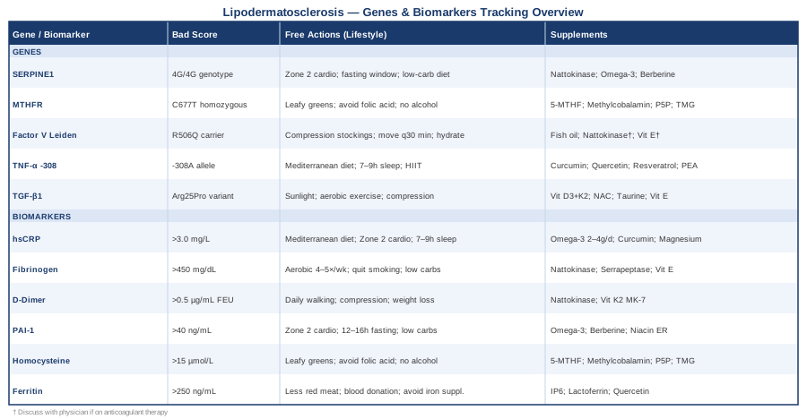
6 Biomarkers to Track if You Have Lipodermatosclerosis
The biology of lipodermatosclerosis is not mysterious — it is traceable. Venous hypertension causes capillary leakage, which deposits fibrin and red blood cells into surrounding tissue. That fibrin should be cleared by the fibrinolytic system, but in many people it isn't — it accumulates, creating the fibrin "cuffs" around capillaries that impair oxygen delivery and trigger the inflammatory and fibrotic cascade that defines LDS. Six biomarkers sit at critical points in this chain, and each one you measure is a window into where the process is specifically breaking down for you.
1. High-Sensitivity C-Reactive Protein (hsCRP)
Why it matters. hsCRP is produced by the liver in response to inflammatory cytokines — mainly IL-6 — and reflects the overall burden of systemic inflammation. In LDS, subcutaneous inflammation is the central process: macrophages infiltrate the tissue, release TNF-α and IL-1β, and drive both ongoing damage and the fibrotic remodeling that characterizes advanced disease. Elevated hsCRP confirms that this systemic inflammatory burden is high, which matters because higher baseline inflammation predicts worse tissue outcomes and slower response to interventions. Research supports the association between elevated CRP and chronic venous disease severity and progression.
How to Measure It
hsCRP is a standard blood test available at essentially every laboratory. Cost range: $20–$50 USD out of pocket, often covered by insurance when physician-ordered. Request specifically "high-sensitivity CRP" — the standard CRP test is not sensitive enough for this purpose. Optimal target: below 1.0 mg/L. Between 1.0 and 3.0 mg/L is moderate. Above 3.0 mg/L reflects active systemic inflammation and warrants investigation. Retest every 3–6 months when tracking changes.
If the Score Is Elevated: Plan Without Supplements
The most powerful lifestyle lever for hsCRP is body composition — visceral fat independently produces IL-6 and TNF-α. Even a 5–10% reduction in visceral mass measurably lowers hsCRP. A Mediterranean-pattern diet (olive oil, oily fish, vegetables, nuts — minimal refined carbohydrates and seed oils) reduces hsCRP by 20–40% in clinical trials. Sleep quality is the second most underrated lever: consistently sleeping fewer than 6–7 hours measurably raises IL-6 and hsCRP within days. Zone 2 aerobic exercise (conversational pace, 45–60 minutes, 3–4 sessions per week) is directly anti-inflammatory through myokine pathways. Frequency: dietary consistency over months matters more than any short-term restriction — this is not an acute intervention.
If the Score Is Elevated: Plan With Supplements
Omega-3 fatty acids (EPA+DHA, 2–4 g/day combined): from fish oil or algae oil. Continuous use is appropriate. At 4 g+, monitor LDL-P (particle count); potential side effects include loose stools, fishy breath, and mild blood thinning — relevant if anticoagulants are prescribed. Curcumin (BCM-95 or liposomal form, 500–1000 mg twice daily with food): cycling of 8–12 weeks on, 2–4 weeks off is reasonable; side effects are rare (occasional GI upset, avoid in gallstone disease). Magnesium glycinate (300–400 mg at night): continuous use; loose stools at higher doses are the main side effect.
2. Fibrinogen
Why it matters. Fibrinogen is the precursor to fibrin — the protein that forms the pericapillary cuffs that are the anatomical signature of LDS. The classic fibrin cuffing hypothesis (established in venous disease literature and extensively documented in subsequent research) proposes that these cuffs act as a physical barrier to oxygen and nutrient diffusion, contributing to tissue hypoxia and necrosis. High fibrinogen doesn't just indicate clotting risk — in the LDS context, it means more substrate for the fibrin deposits that drive the disease process directly. Fibrinogen is also an acute phase reactant, so elevated levels reflect both clotting tendency and inflammatory burden simultaneously. Multiple studies have examined the link between elevated fibrinogen and fibrin deposition in chronic venous insufficiency and lipodermatosclerosis.
How to Measure It
Fibrinogen is measured via standard coagulation panel at most labs. Cost range: $30–$65 USD out of pocket; often included in a comprehensive coagulation workup. Normal laboratory range is 200–400 mg/dL, but for vascular health purposes below 350 mg/dL is the functional target, with below 300 mg/dL being optimal. Levels above 450 mg/dL are significantly elevated and warrant clinical follow-up. Retest every 3–4 months when actively intervening.
If the Score Is Elevated: Plan Without Supplements
Moderate-intensity aerobic exercise (walking, cycling, swimming — 4–5 sessions per week, 40+ minutes) reduces fibrinogen by 10–20% in intervention studies. Smoking cessation is critical: smoking is among the most potent drivers of elevated fibrinogen. Reducing refined carbohydrates and alcohol both independently lower fibrinogen. Reducing processed meat intake is also relevant. Compression therapy is upstream of fibrinogen itself but essential — reducing venous hypertension directly reduces capillary fibrin leakage, the source of the problem.
If the Score Is Elevated: Plan With Supplements
Nattokinase (2000–4000 FU/day, taken in the evening): a fibrinolytic enzyme from fermented soybeans that directly degrades fibrin and has been shown to reduce fibrinogen in small clinical trials. Cycling: 8–12 weeks on, 4 weeks off. Do not combine with anticoagulant therapy (warfarin, apixaban, etc.) without physician supervision — blood thinning is the primary risk. Serrapeptase (40,000–120,000 IU/day on an empty stomach): systemic enzyme with fibrin-degrading properties. Cycling: 8–10 weeks on/off. Side effects: rare nausea; avoid in bleeding disorders. Mixed tocopherol vitamin E (400–800 IU/day, gamma-tocopherol dominant): reduces fibrinogen and improves fibrinolysis. Cycling: 3 months on, 4–6 weeks off. Avoid in combination with anticoagulants.
3. D-Dimer
Why it matters. D-dimer is a fibrin degradation product — it rises when fibrin is being actively formed and broken down. In LDS, ongoing fibrin deposition and attempted clearance mean D-dimer can be persistently elevated at a low-grade level. This is distinct from the emergency setting where D-dimer rules out pulmonary embolism: here, a chronically elevated D-dimer tells you that fibrin turnover in the tissue is ongoing and the fibrinolytic system is struggling to keep pace. It also serves as a sensitive indicator of any underlying thrombophilic tendency — Factor V Leiden, MTHFR variants — that may be quietly feeding the vascular component of LDS. Research connects elevated D-dimer with impaired fibrinolysis and disease activity in chronic venous disease.
How to Measure It
D-dimer is a standard lab test. Cost range: $30–$60 USD out of pocket. In the chronic venous disease context (not acute thromboembolism), mildly elevated D-dimer — above 0.5 µg/mL FEU — is meaningful even when not dramatically elevated. Note that D-dimer rises with age, surgery, pregnancy, and any inflammatory condition — results must be interpreted with clinical context.
If the Score Is Elevated: Plan Without Supplements
Consistent physical activity — even daily 30–45 minute walks — improves fibrinolytic activity and reduces D-dimer over time. Compression therapy is essential: reducing venous hypertension decreases capillary fibrin leakage, which lowers the fibrin turnover rate over weeks and should decrease D-dimer. Weight reduction in LDS patients with obesity is strongly associated with reduced fibrin deposition. Adequate hydration and avoidance of prolonged immobility are foundational habits.
If the Score Is Elevated: Plan With Supplements
Nattokinase (2000–4000 FU, taken at bedtime): directly enhances fibrinolysis. Small clinical trials show reductions in D-dimer with consistent use. Cycling: 8–12 weeks on/off. Contraindicated with anticoagulant therapy; mild blood thinning effect otherwise. Vitamin K2 (MK-7 form, 100–200 mcg/day): supports proper fibrin regulation and reduces vascular calcification. Continuous use is appropriate. K2 (unlike K1) does not meaningfully interfere with warfarin at standard doses, but disclosure to a prescribing physician is always appropriate.
4. PAI-1 (Plasminogen Activator Inhibitor-1)
Why it matters. PAI-1 is the primary brake on fibrinolysis — it blocks tissue plasminogen activator (tPA), which is the enzyme that normally clears fibrin deposits. In LDS, the pericapillary fibrin cuffing exists precisely because fibrin cannot be cleared efficiently. PAI-1 is a central reason why. High PAI-1 means the body's fibrin-clearing machinery is suppressed: fibrin accumulates faster than it is degraded, and the tissue deposits that drive hypoxia and inflammation build up over months and years. PAI-1 is also tightly linked to insulin resistance and visceral adiposity — making it a direct bridge between metabolic health and LDS progression. Studies have documented impaired fibrinolysis and elevated PAI-1 activity in patients with chronic venous insufficiency and lipodermatosclerosis.
How to Measure It
PAI-1 can be measured as plasma activity (functional PAI-1 activity assay) or plasma antigen. Not all standard labs offer it routinely — a specialty or functional medicine lab may be required. Cost range: $50–$120 USD out of pocket. The test is ideally done fasting in the morning (PAI-1 follows a circadian rhythm, peaking in the early morning hours). Target: PAI-1 activity below 20 ng/mL (exact reference ranges vary by laboratory — request the functional activity assay, not just antigen). Levels above 40 ng/mL are significantly elevated.
If the Score Is Elevated: Plan Without Supplements
Aerobic exercise is the most evidence-backed non-supplement intervention for reducing PAI-1. Zone 2 cardio (3–5 sessions per week, 45–60 minutes each) consistently lowers PAI-1 activity over 8–12 weeks of sustained practice. Time-restricted eating (a daily 12–16 hour fasting window) reduces PAI-1 activity, particularly when combined with reduced caloric excess that would otherwise drive insulin elevation. Weight loss — specifically visceral fat reduction — directly suppresses hepatic PAI-1 production. Reducing dietary refined carbohydrates and fructose lowers insulin, which feeds back to suppress PAI-1.
If the Score Is Elevated: Plan With Supplements
Omega-3 fatty acids (EPA+DHA, 3–4 g/day): reduce PAI-1 gene expression independently of other anti-inflammatory effects. Continuous use; same cautions as above. Berberine (500 mg three times daily with meals): improves insulin sensitivity and reduces PAI-1 through insulin-pathway suppression. Cycling: 12 weeks on, 4–6 weeks off — avoid continuous long-term use without monitoring; GI upset is common when starting (start low and titrate); not for use in pregnancy. Extended-release niacin (500–1000 mg/day, titrated slowly): reduces PAI-1 and improves fibrinolysis; note that "flush-free" niacinamide does not lower PAI-1 and should not be substituted. Liver function monitoring is warranted above 1000 mg; flushing is common with immediate-release forms. Evening primrose oil (1000–2000 mg/day): gamma-linolenic acid content may reduce PAI-1 through prostaglandin pathways. Cycling: 8–12 weeks, then reassess.
5. Homocysteine
Why it matters. Homocysteine is an amino acid that accumulates when the methylation cycle is impaired — particularly when B vitamins (B12, B6, folate) are insufficient or when MTHFR variants reduce folate processing efficiency. Elevated homocysteine directly damages endothelial cells through oxidative stress, impairs nitric oxide production, promotes thrombosis, and accelerates vascular inflammation. In LDS — already driven by venous hypertension and capillary dysfunction — elevated homocysteine adds another layer of endothelial fragility that worsens the underlying vascular pathology. Thomas Dayspring has consistently highlighted homocysteine as one of the most underordered yet actionable vascular markers in clinical practice. Research has linked elevated homocysteine to endothelial damage and vascular inflammatory conditions including venous insufficiency.
How to Measure It
Homocysteine is a standard fasting blood test. Cost range: $30–$60 USD out of pocket. Standard labs flag 15 µmol/L as the upper limit of normal, but vascular-focused practitioners use below 10 µmol/L as the functional target, with below 7 µmol/L being optimal. Levels above 15 µmol/L are clinically elevated; above 20 µmol/L is associated with significantly increased vascular risk.
If the Score Is Elevated: Plan Without Supplements
Dietary folate from natural food sources is the most effective non-supplement intervention: dark leafy greens (spinach, kale, romaine), liver (beef or chicken, once weekly), beans, and avocado provide natural folate that partially bypasses MTHFR dependence. Avoid folic acid (the synthetic form in most fortified foods and low-quality supplements) — in MTHFR variant carriers, unmetabolized folic acid competes with active methylfolate and may worsen the situation. Minimize alcohol consumption, which depletes B vitamins and impairs methylation. Regular moderate aerobic exercise mildly improves methylation cycle efficiency.
If the Score Is Elevated: Plan With Supplements
5-MTHF (methylfolate, 400–1000 mcg/day): the pre-activated form of folate that bypasses the MTHFR bottleneck entirely. Start at 400 mcg and increase slowly — some individuals (particularly those sensitive to methylation) experience increased anxiety at higher doses. Continuous use. Methylcobalamin (B12, 1000–5000 mcg/day sublingual or intramuscular): cofactor in the homocysteine remethylation pathway; the methyl form is preferred over cyanocobalamin. Continuous use; extremely safe at these doses. P5P (pyridoxal-5-phosphate, active B6, 25–50 mg/day): required for the transsulfuration pathway that backs up when remethylation is impaired. Warning: regular pyridoxine (B6) above 100 mg/day can cause peripheral neuropathy over time — use P5P and stay within range. Cycle: 3–4 months, then reassess. Betaine/TMG (trimethylglycine, 500–1500 mg/day with food): provides a MTHFR-independent remethylation route through the BHMT enzyme. An effective add-on when homocysteine remains elevated despite B vitamin optimization.
6. Ferritin and Iron Panel
Why it matters. The brownish-rust skin discoloration characteristic of LDS is caused by hemosiderin — iron-containing deposits from red blood cells that leak from capillaries under venous pressure. These deposits are not merely cosmetic: hemosiderin triggers macrophage activation, generates reactive oxygen species through Fenton chemistry (iron-catalyzed oxidation), and sustains the local inflammatory cycle that drives tissue damage. Ferritin — the body's iron storage protein — reflects both iron burden and inflammatory status (it is also an acute phase reactant). High ferritin in the context of LDS may signal excessive iron available for oxidative damage in affected tissue. A full iron panel (ferritin, serum iron, TIBC, transferrin saturation) gives a more complete picture than ferritin alone. Studies document hemosiderin-driven macrophage activation and oxidative inflammation as a pathological feature of lipodermatosclerosis.
How to Measure It
Ferritin is a standard blood test. Cost range: $20–$45 USD out of pocket; a full iron panel adds $30–$60. Target for ferritin: 30–150 ng/mL for women; 30–200 ng/mL for men. Above 250 ng/mL warrants investigation to distinguish iron overload (hereditary hemochromatosis, excess supplementation) from inflammatory elevation. Transferrin saturation above 45% suggests true iron excess rather than an inflammatory ferritin rise.
If the Score Is Elevated: Plan Without Supplements
If ferritin is elevated due to iron excess (transferrin saturation also elevated), reduce dietary heme iron sources: red meat, organ meats, shellfish. Pair iron-containing foods with calcium or polyphenol-rich foods (coffee, tea, turmeric) to inhibit absorption. Regular blood donation (1–2 donations per year for eligible individuals) is the most efficient method of reducing iron stores — measurable ferritin reduction occurs within 12–18 months. Addressing the underlying venous disease to reduce capillary leakage is the most direct approach to reducing ongoing hemosiderin deposition.
If the Score Is Elevated: Plan With Supplements
IP6 (inositol hexaphosphate, 1000–2000 mg twice daily on an empty stomach): a natural iron chelator that binds free iron and reduces iron-catalyzed oxidative stress. Cycling: 3–6 months, then reassess ferritin. Take separately from other supplements and meals by at least 2 hours, as it also chelates other minerals. Lactoferrin (200–300 mg/day): a milk-derived protein that modulates iron availability and carries anti-inflammatory properties in iron-loaded states. Continuous use; minimal side effects; caution in dairy protein allergy. Quercetin (500–1000 mg/day with food): polyphenol with iron-chelating and anti-inflammatory properties relevant to macrophage activation in LDS tissue. Cycling: 8–12 weeks on, 2–4 weeks off.
The six biomarkers above provide a traceable map of where your LDS biology is currently most active. Each is measurable, each is actionable, and each points to a different stage of the disease pathway. The next question is: why do some people's pathways run harder than others? That is where genetic variants become worth examining.
What Your Genes May Be Telling You About Lipodermatosclerosis
Genetic variants don't cause lipodermatosclerosis on their own — but they can tilt the playing field significantly. Five variants are particularly relevant to the pathways that drive LDS: fibrinolysis capacity, thrombotic tendency, homocysteine metabolism, inflammatory cytokine production, and fibrotic remodeling. If you have had genetic testing through a consumer genomics service or a clinical panel ordered by a functional medicine physician, these are the variants worth examining. Even without testing, the protocols below apply broadly to people whose biomarkers suggest impairment in these specific pathways.
SERPINE1 — The PAI-1 Gene
What it does. The SERPINE1 gene encodes PAI-1, the principal inhibitor of fibrinolysis. The 4G/5G polymorphism in the promoter region is the most studied variant: the 4G allele is associated with significantly higher PAI-1 transcription, meaning more PAI-1 protein, less fibrin clearance, and more fibrin accumulation in tissues. Homozygous 4G/4G individuals produce substantially more PAI-1 under inflammatory conditions. This is directly relevant to the fibrin cuffing mechanism at the heart of LDS — the same mechanism that fibrinogen and D-dimer biomarkers reflect. The SERPINE1 4G/5G variant has been studied in venous disease, thrombophilia, and impaired fibrinolysis contexts.
If the Gene Is Problematic: Plan Without Supplements
Consistent aerobic exercise (zone 2, 3–5 sessions per week) is the most powerful lifestyle modifier of PAI-1 expression. Time-restricted eating (16-hour daily fasting window, practiced most days of the week) reduces PAI-1 activity, especially combined with visceral fat reduction. Reducing dietary refined carbohydrates and fructose is essential — insulin resistance directly upregulates PAI-1 through the 4G allele pathway. Morning exercise specifically appears to reduce the early-morning PAI-1 peak that is most relevant to overnight fibrin accumulation.
If the Gene Is Problematic: Plan With Supplements
Nattokinase (2000–4000 FU, taken at bedtime to target the morning PAI-1 peak): provides direct fibrinolytic activity independent of the tPA/PAI-1 system. Cycling: 8–12 weeks on, 4 weeks off. Contraindicated with prescription anticoagulants; mild blood thinning effect at standard doses. Omega-3 EPA+DHA (3–4 g/day): reduces SERPINE1 gene expression. Continuous use; standard cautions apply. Berberine (500 mg × 3/day with meals): improves insulin sensitivity, reducing the metabolic trigger for PAI-1 upregulation in 4G/4G or 4G/5G carriers. Cycle 12 weeks on, 6 weeks off.
MTHFR — The Methylation Gene
What it does. The MTHFR gene encodes the enzyme that converts dietary folate into its active form (5-MTHF). The C677T variant reduces enzyme activity by 30–65% depending on whether you carry one or two copies. The A1298C variant also impairs function. In LDS, impaired MTHFR leads to elevated homocysteine — which damages the endothelium, promotes thrombosis, and worsens the vascular fragility underlying venous hypertension. Gary Brecka has brought significant public attention to MTHFR variants and their downstream vascular and inflammatory consequences. The MTHFR C677T variant's connection to elevated homocysteine and endothelial dysfunction is among the most replicated findings in vascular genetics.
If the Gene Is Problematic: Plan Without Supplements
Maximize dietary folate from natural food sources: dark leafy greens, liver, beans, and avocado provide natural folate that partly bypasses MTHFR dependence. Avoid folic acid in processed foods and low-quality supplements — unmetabolized folic acid can compete with active methylfolate in variant carriers. Regular moderate exercise supports SAM (S-adenosylmethionine) production, the body's primary methyl donor. Minimize alcohol, which depletes B vitamins and impairs methylation at multiple steps.
If the Gene Is Problematic: Plan With Supplements
5-MTHF (methylfolate, 400–800 mcg/day, up to 1 mg for confirmed homozygous C677T): bypasses the MTHFR enzymatic step entirely. Start low and increase slowly — some individuals experience anxiety or irritability at higher doses; reduce if this occurs. Continuous use. Methylcobalamin (B12, 1000–2000 mcg sublingual or intramuscular): cofactor for the MTHFR-dependent remethylation reaction; the methyl form is required. Continuous; extremely safe. P5P (active B6, 25–50 mg/day): supports the transsulfuration pathway that backs up when remethylation is impaired. Cycle 3–4 months, reassess. Do not exceed 100 mg/day of any B6 form to avoid peripheral neuropathy risk. TMG/betaine (1000–2000 mg/day with food): MTHFR-independent remethylation via the BHMT enzyme — particularly useful for confirmed compound heterozygotes or persistently elevated homocysteine.
Factor V Leiden (F5 Gene, R506Q Variant)
What it does. Factor V Leiden is a point mutation in the Factor V clotting gene that makes Factor Va protein resistant to inactivation by activated protein C. This creates a prothrombotic state — venous clots form more easily and persist longer. The connection to LDS is direct: a history of DVT (deep vein thrombosis) is a major risk factor for developing LDS, and Factor V Leiden is the most common inherited thrombophilia in people of European descent (approximately 5% prevalence). Even without clinical DVT, subclinical microthrombosis in the venous system may worsen venous hypertension and capillary leakage over years. Factor V Leiden is the most extensively studied thrombophilic variant in venous thromboembolism research.
If the Gene Is Problematic: Plan Without Supplements
Compression therapy is especially critical for Factor V Leiden carriers with LDS or venous insufficiency — medical-grade compression (20–30 mmHg minimum) should be consistent, not optional. Prolonged immobility — long flights, extended desk work — is particularly high-risk; movement breaks every 30–60 minutes are essential. Weight management is a clinical priority: obesity multiplies thrombotic risk exponentially in Factor V Leiden carriers. Adequate daily hydration (2+ liters) reduces blood concentration and clotting factor density.
If the Gene Is Problematic: Plan With Supplements
Important: all supplement choices in Factor V Leiden carriers must be discussed with a physician, particularly if prescription anticoagulant therapy is already prescribed or under consideration. Fish oil (EPA+DHA, 2–3 g/day): mild antiplatelet effect, generally safe in the absence of anticoagulant therapy. Nattokinase (2000 FU/day): enhances endogenous fibrinolysis — do not combine with warfarin, apixaban, or similar agents without physician supervision. Vitamin E (mixed tocopherols, 400 IU/day): mild anticoagulant properties. Cycle 3 months on/1 month off. Discontinue 1–2 weeks before any surgery.
TNF-α (Tumor Necrosis Factor Alpha, -308 G>A Polymorphism)
What it does. TNF-α is a master inflammatory cytokine produced primarily by macrophages. The -308 G>A promoter polymorphism increases TNF-α transcription — more inflammatory signal in response to any trigger. In LDS, macrophages infiltrate the subcutaneous tissue and produce TNF-α and IL-1β, driving both ongoing inflammation and the fibrotic remodeling of the chronic phase. Carriers of the -308A allele may experience more aggressive inflammation, faster progression to the sclerotic phase, and greater tissue damage for an equivalent degree of venous hypertension compared to non-carriers. The TNF-α -308 variant has been studied in chronic inflammatory and dermatological conditions with inflammatory components.
If the Gene Is Problematic: Plan Without Supplements
A Mediterranean-style anti-inflammatory diet reduces TNF-α activity significantly through high polyphenol intake (olive oil, berries, cruciferous vegetables). Visceral fat reduction is the highest-leverage lifestyle intervention: visceral adipocytes are primary producers of TNF-α, so even 5–8% visceral mass reduction measurably lowers circulating levels. Sleep quality (7–9 hours, consistent schedule) suppresses inflammatory cytokine production nightly. High-intensity interval training (HIIT, 2 brief sessions per week) has specific TNF-α-suppressing effects through IL-6 and irisin signaling pathways, complementing the steady state zone 2 work.
If the Gene Is Problematic: Plan With Supplements
Curcumin (BCM-95 or liposomal/phytosomal form, 500–1000 mg twice daily with food): NF-κB inhibitor that directly reduces TNF-α transcription. Cycling: 8–12 weeks on, 4 weeks off. Rare GI irritation; avoid in gallstone disease. Quercetin (500–1000 mg/day): inhibits TNF-α-mediated signaling and mast cell degranulation — synergistic with curcumin. Resveratrol (250–500 mg/day with a fat-containing meal): SIRT1 activator with TNF-α-suppressing properties. Cycling: 3 months on, 4–6 weeks off. PEA (palmitoylethanolamide, 600 mg twice daily): endogenous lipid mediator with strong local anti-inflammatory effects specifically studied in subcutaneous and peripheral inflammatory contexts. Cycling: 12 weeks on, reassess. Well-tolerated with no significant side effects documented in current literature.
TGF-β1 (Transforming Growth Factor Beta 1, Codon 25 Polymorphism)
What it does. TGF-β1 is the master regulator of fibrosis — it activates myofibroblasts, drives collagen deposition, and is the molecular explanation for why LDS skin becomes progressively hardened and leathery in its chronic phase. The codon 25 (Arg25Pro) polymorphism is associated with altered TGF-β1 expression and downstream signaling. In LDS, dysregulated TGF-β1 activity means more aggressive fibrotic remodeling once the inflammatory phase begins — faster progression to the indurated, "inverted wine bottle" appearance characteristic of advanced disease. TGF-β1 has been documented as a central driver of fibrosis in lipodermatosclerosis and chronic venous skin changes.
If the Gene Is Problematic: Plan Without Supplements
Vitamin D3 from sunlight (20–30 minutes of midday sun on exposed skin when possible) has TGF-β1 modulating properties — vitamin D3 signaling competes with and partially suppresses TGF-β1-driven fibrotic pathways. Aerobic exercise reduces fibrotic remodeling generally by improving tissue oxygenation and reducing hypoxia — which is the primary trigger for TGF-β1 upregulation in LDS-affected tissue. Consistent compression therapy that reduces venous hypertension is the most direct lifestyle anti-fibrotic approach, as it removes the upstream trigger for TGF-β1 release in the affected tissue.
If the Gene Is Problematic: Plan With Supplements
Vitamin D3 + K2 (4000–8000 IU D3 with 100–200 mcg K2-MK7, daily with a fat-containing meal): D3 at these doses requires K2 for proper calcium routing and to prevent soft tissue calcification. Monitor serum 25(OH)D quarterly — target 50–80 ng/mL. Reduce dose if levels exceed 100 ng/mL. N-acetylcysteine (NAC, 600–1200 mg/day): glutathione precursor that reduces oxidative stress driving TGF-β1 expression in hypoxic tissue. Cycling: 3 months on, 4–6 weeks off. Occasional mild GI effects. Taurine (2–3 g/day): amino acid with documented anti-fibrotic properties; downregulates TGF-β1-mediated collagen synthesis in preclinical and early human data. Continuous use; excellent safety profile. Gamma-tocopherol-dominant vitamin E (400–800 IU/day): the gamma-tocopherol isoform specifically has anti-fibrotic properties and reduces TGF-β1 activity in tissue. Cycle 3 months on/4 weeks off. Avoid combination with anticoagulant therapy.
Between the biomarkers and the genetic variants, you now have both a real-time measurement of how active these pathways are and a structural understanding of why they may run harder in your particular biology. What remains is a framework for organizing all of this into a coherent long-term practice — which is where the next section becomes genuinely useful.
A Framework for Long-Term Thinking: Key Lessons From Outlive
Peter Attia's Outlive: The Science and Art of Longevity is not a book about lipodermatosclerosis — but it may be the most practically useful framework available for managing it. Attia's core argument is that modern medicine is effective at treating acute disease and poorly structured to prevent the chronic, slow-moving pathologies driven by inflammation, metabolic dysfunction, and vascular damage over decades. Lipodermatosclerosis fits that description precisely: it moves slowly, compounds over time, and responds poorly to reactive management.
1. Track Biomarkers Before Symptoms Worsen
Attia argues for routinely measuring hsCRP, homocysteine, fibrinogen, and inflammatory markers — not just when clinical deterioration is obvious. The same logic applies directly to LDS: waiting for visible skin changes to worsen before testing means losing the window for intervention during a more reversible phase. The six biomarkers described earlier are exactly the early warning system Attia advocates for.
2. Zone 2 Exercise Is the Closest Thing to an Anti-Inflammatory Drug
Attia calls zone 2 cardio "the single most powerful tool available for longevity." For LDS specifically: sustained moderate aerobic effort (conversational pace, 45–90 minutes, 3–5 times per week) improves mitochondrial efficiency, reduces PAI-1 activity, lowers hsCRP and fibrinogen, and improves venous return from the lower extremities. Lower-impact zone 2 options (cycling, pool walking, swimming) protect affected areas while delivering full systemic benefit.
3. Visceral Fat Is a Cytokine Factory
Attia consistently identifies visceral adiposity — not BMI — as the metabolic variable most predictive of inflammation severity. Every kilogram of visceral fat continuously produces IL-6, TNF-α, and PAI-1. In a condition where macrophage-driven inflammation is already active, high visceral fat reinforces the inflammatory signal around the clock. Targeting visceral fat — measured by waist circumference or DEXA scan — is more informative and actionable than watching the scale.
4. Homocysteine Is One of the Most Underordered Markers in Medicine
Both Attia and Thomas Dayspring note that homocysteine is systematically undertested in standard care despite being cheap, fully reversible with B vitamins, and directly connected to endothelial damage. For LDS patients whose disease involves vascular fragility, this is a readily correctable missed opportunity. A target below 7 µmol/L is what Attia works toward with patients.
5. Sleep Is Not Optional for Inflammation Control
A single week of sleeping below 6 hours measurably raises IL-6, TNF-α, and hsCRP. Attia documents this relationship extensively and treats poor sleep as the most efficient way to undermine every other intervention simultaneously. For LDS patients managing chronic pain that disrupts sleep, addressing sleep quality is a clinical priority — not a lifestyle preference to get around to eventually.
6. Insulin Resistance Governs PAI-1 More Than Most Realize
Attia's framework returns repeatedly to insulin sensitivity as an upstream regulator of vascular health. Chronically elevated insulin drives PAI-1 production through the liver, and elevated PAI-1 suppresses fibrinolysis. Reducing carbohydrate load, implementing a time-restricted eating window, and improving insulin sensitivity through exercise and resistance training all feed directly into the fibrin cuffing mechanism that drives LDS progression.
7. The Omega-3 Index Is a Better Target Than Dose
Attia tracks the omega-3 index (EPA+DHA as percentage of red blood cell fatty acids) rather than prescribing a fixed dose. An index below 4% is pro-inflammatory; above 8% is cardioprotective and anti-inflammatory. For LDS patients, testing the omega-3 index (~$100 specialty test) tells you whether your current omega-3 intake is sufficient for your body's actual needs — and prevents both under-dosing and unnecessary over-supplementation.
8. Strength Training Complements Cardio in the Inflammation Picture
Resistance training preserves muscle mass, which functions as a metabolic buffer against inflammation and insulin resistance. Attia recommends 2–3 resistance training sessions per week alongside zone 2 cardio. For LDS patients, lower-extremity exercises should be adapted to avoid trauma to affected areas during flares — seated resistance, pool-based resistance training, or upper body focus are practical alternatives.
9. Targeted Supplementation Follows Lab Work, Not the Other Way Around
Attia explicitly argues against generic supplement stacks — he prescribes based on individual biomarker results. Omega-3s, magnesium, vitamin D, and methylated B vitamins are in his standard toolkit, but only when labs indicate deficiency or suboptimal levels. This aligns exactly with the biomarker-first approach of this article: test first, supplement specifically based on results, reassess at 3–6 months.
10. Reframe the Question From Treating LDS to Optimizing Its Drivers
Attia's most useful reframe is this: the question is not "how do I treat this skin condition" but "what does optimal inflammatory, metabolic, and vascular health look like for my specific biology?" When you optimize for those upstream variables — tracked through the biomarkers above, shaped by your genetic variants — LDS is often one of several conditions that improves as a downstream consequence. It reorients the work from reactive to proactive and from condition-specific to biology-wide.
With this long-term framework in mind, the following section looks at specific complementary approaches that have meaningful clinical evidence for the symptoms and pathways directly relevant to LDS.
Complementary Approaches With Clinical Evidence
Low-Level Laser Therapy / Photobiomodulation
Photobiomodulation (PBM) uses specific wavelengths of red and near-infrared light (typically 630–850 nm) to stimulate mitochondrial function, reduce local inflammation, improve microcirculation, and promote tissue healing. In LDS and chronic venous ulceration — conditions characterized by tissue hypoxia and impaired local blood flow — the rationale is direct: improved microcirculation addresses one of the core physiological deficits, and reduced local inflammation may slow fibrotic progression. Randomized controlled trials and systematic reviews have evaluated photobiomodulation as an adjunct in chronic venous leg ulcers, with several showing improvements in healing rates compared to standard care alone.
A typical protocol in clinical settings uses devices delivering 630–850 nm light at 4–10 J/cm² per session, 3 sessions per week for 8–12 weeks, applied directly over the affected lower leg area. FDA-cleared home devices are available in the $200–$800 range, though clinical devices provide more precise dosimetry. The evidence is stronger for established venous ulcer healing than for the pre-ulceration LDS stage specifically — results are additive to compression therapy, not a replacement for it.
For practical application: begin with 3 sessions per week, 10–20 minutes per session applied to the affected lower leg. Avoid applying PBM over active infection or openly macerated skin without medical supervision. The most realistic benefit for LDS is reduction in local inflammation and improved skin oxygenation over a consistent 8–12 week course. Discuss with your dermatologist or wound care specialist before starting if you have active LDS.
Manual Lymphatic Drainage
Manual lymphatic drainage (MLD) is a specialized massage technique designed to improve lymphatic and venous drainage from the extremities using very light, rhythmic pressure applied in the direction of lymphatic flow. In lipodermatosclerosis, venous hypertension and secondary lymphatic dysfunction contribute to fluid stagnation, tissue edema, and ongoing local inflammation. Unlike deep tissue massage — contraindicated over acutely inflamed LDS skin — MLD uses gentle superficial pressure appropriate for sensitive, fibrotic tissue. Clinical studies support manual lymphatic drainage as an effective adjunct to compression therapy in chronic venous insufficiency and associated skin changes.
The established clinical protocol for venous-related skin disease is complete decongestive therapy (CDT) — a combination of MLD, multilayer compression bandaging, therapeutic exercise, and skin care. MLD is performed by a certified lymphedema therapist (CLT), 3–5 sessions per week during the intensive phase, then 1–2 per week for maintenance. Session duration: 45–60 minutes. Cost: $80–$150 per session; insurance coverage exists in many countries when prescribed. Evidence from multiple RCTs supports CDT as superior to compression bandaging alone for chronic venous skin changes.
For practical application: request a referral to a certified lymphedema therapist from your dermatologist or vascular specialist. If regular professional sessions are not accessible, validated self-MLD techniques for the lower extremities are teachable and maintain meaningful benefit between professional sessions. MLD without subsequent compression loses most of its effect — the two must be combined.
Mindfulness-Based Stress Reduction (MBSR)
The standardized 8-week MBSR program has documented anti-inflammatory effects — including measurable reductions in hsCRP, IL-6, and TNF-α — in multiple randomized controlled trials. In the context of LDS, which involves chronic pain, sleep disruption, and the psychological burden of a visible and disfiguring condition, MBSR addresses a genuine clinical gap that conventional management does not. The mechanism is physiologically direct: chronic cortisol elevation and sympathetic nervous system activation drive inflammatory cytokine production; reducing the stress response measurably lowers this inflammatory load over time. Meta-analyses of mindfulness and MBSR programs document reductions in CRP and inflammatory cytokines in participants with chronic inflammatory conditions.
The standard MBSR protocol involves 8 weeks of structured practice: a 2.5-hour weekly group session, a full-day retreat at week 6, and 45 minutes of daily home practice. In clinical trials, this format produced measurable reductions in hsCRP and improvements in sleep quality and pain tolerance in chronic pain populations. For LDS, the most relevant outcomes are reduced systemic inflammatory load, improved sleep, and reduced pain-driven avoidance of movement. Digital and app-based structured programs (Headspace Clinical, Calm structured 8-week programs, Insight Timer) offer accessible entry points, though the evidence base is stronger for the full in-person format.
Commit to the complete 8-week structured format at least once. Brief daily practice (10–20 minutes) maintains the benefit afterward. MBSR works best as one component of a comprehensive strategy — it is synergistic with the lifestyle and supplement interventions described in this article, not a standalone answer.
Breathing-Based Therapies
Controlled breathing practices — including diaphragmatic breathing, slow breathing at approximately 6 breaths per minute, and paced resonance frequency breathing — have documented effects on heart rate variability (HRV), autonomic tone, inflammatory cytokine levels, and venous return dynamics. The venous connection is physiologically direct: the diaphragm acts as a pump during inhalation, creating pressure gradients that draw venous blood from the lower extremities toward the heart. In LDS, where venous return is already compromised by structural venous disease, deliberate breathing practice offers a self-administered tool that works on both the autonomic and mechanical level simultaneously. Studies document that slow-paced resonance breathing improves heart rate variability and reduces inflammatory markers in chronic disease contexts.
The most studied protocol is resonance frequency breathing (coherence breathing): 6 full cycles per minute (5-second inhale, 5-second exhale) for 20 minutes, ideally twice daily. This specific rhythm maximizes HRV, improves vagal tone, and reduces inflammatory cytokine levels over 4–8 weeks of consistent practice. A more accessible entry point: 4-7-8 breathing (inhale 4 seconds, hold 7, exhale 8) for 10–15 minutes before sleep. Apps like Breathwrk, Elite HRV, and Othership provide guided paced protocols with heart rate feedback.
For LDS-specific application: practice slow breathing in an elevated leg position (legs above heart level at 30–45 degrees, supported by pillows) to simultaneously benefit from venous drainage and autonomic modulation. Duration: 15–20 minutes per session, ideally morning and before sleep. Side effects are minimal; individuals with CO2 sensitivity should start with shorter sessions (5–10 minutes) and build up gradually.
Microbiome-Directed Therapies
Emerging research connects gut microbiome composition to systemic inflammatory tone, including modulation of TNF-α, IL-6, and gut permeability that allows inflammatory lipopolysaccharide (LPS) to enter circulation and amplify the body-wide inflammatory background. In LDS — a condition of chronic subcutaneous inflammation — reducing systemic inflammatory burden through gut health is a physiologically plausible adjunct, even if direct LDS-specific microbiome studies are currently limited. The pathway runs through the gut-immune axis: gut dysbiosis is associated with elevated hsCRP, impaired barrier integrity, and higher circulating LPS, all of which add to the macrophage activation driving LDS tissue damage. Multiple studies have linked gut dysbiosis to elevated systemic inflammatory markers including CRP in chronic inflammatory conditions.
Practical microbiome interventions with clinical evidence include: multi-strain probiotic supplementation — specifically formulas including Lactobacillus rhamnosus GG, Bifidobacterium longum, and Lactobacillus acidophilus at ≥10 billion CFU/day — which have shown reductions in hsCRP and IL-6 in RCTs. Prebiotic fiber intake (inulin, FOS, beta-glucan from oats) feeds beneficial bacteria that produce short-chain fatty acids — butyrate specifically reduces NF-κB signaling and lowers TNF-α production in colonic epithelium and systemically. Practical dose: 20–30 grams of diverse prebiotic fiber daily from whole plant foods.
The highest-leverage intervention is dietary: eliminate ultra-processed foods, increase plant species diversity (a target of 30+ different plant foods per week has been associated with measurable microbiome diversity improvements in the American Gut Project data), and include fermented foods (yogurt, kefir, kimchi, sauerkraut) regularly. If supplementing probiotics, choose formulas with clinical strain identification and dose transparency, and take consistently for 8–12 weeks before assessing effect. The evidence in LDS specifically is indirect — the expected benefit is systemic inflammatory reduction rather than direct treatment of the skin condition.
Conclusion
Lipodermatosclerosis is a complex condition, but it is not opaque. Its biology is traceable — from venous hypertension to fibrin cuffing, from impaired fibrinolysis to macrophage-driven inflammation, from cytokine activity to progressive fibrotic remodeling. Every stage of that chain has a measurable marker or a genetic variable that reflects it. Tracking hsCRP, fibrinogen, D-dimer, PAI-1, homocysteine, and ferritin gives you a real-time window into where your biology is currently most stressed. Understanding SERPINE1, MTHFR, Factor V Leiden, TNF-α, and TGF-β1 helps explain why your specific body runs these pathways harder than someone else's with the same diagnosis.
Neither piece of information cures LDS. But together they change what you do with it — and what you ask for when you sit across from a physician. The next step is simply to start: request a baseline panel of the six biomarkers at your next appointment, ask about genetic testing through a functional medicine provider or consumer genomics service, and map what you find against the protocols described here. The science of lipodermatosclerosis has advanced considerably beyond compression alone. The way you manage it should reflect that.
Cardiovascular: Blood Vessel Conditions Vascular Conditions
Skin: Inflammatory Skin Conditions
Autoimmune: Inflammatory Conditions