This article contains AI generated content.
· UpdatedLipohemarthrosis: 6 Biomarkers And 5 Genes To Track
Introduction
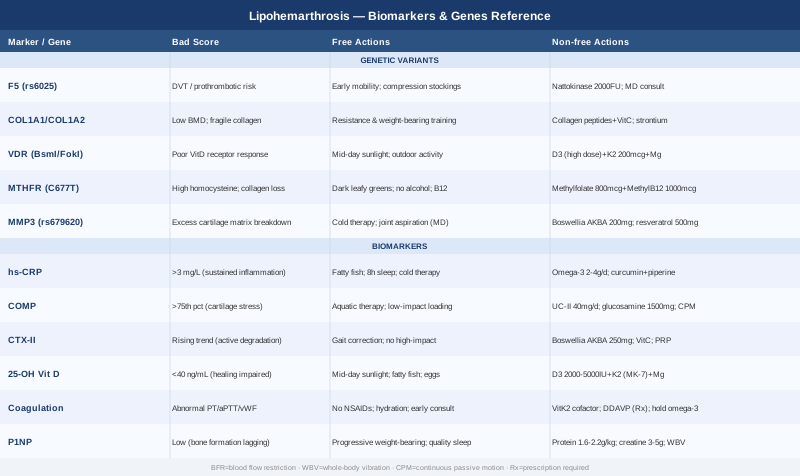
Lipohemarthrosis arrives suddenly — a swollen, painful joint, most often the knee, following a fall or forceful impact. When fat from bone marrow leaks into the joint space alongside blood, it is a reliable radiological signal that an intra-articular fracture has occurred. The imaging confirms it. The management protocol begins. And then, for many people, the conversation essentially ends: immobilize, rehabilitate, wait. What rarely gets discussed is why some individuals recover cleanly while others face months of persistent inflammation, progressive cartilage loss, or a second episode that happens faster and heals slower than the first.
The answer lies in individual biology. Coagulation capacity, baseline inflammatory tone, cartilage matrix quality, bone formation dynamics, and genetic predispositions all determine how a joint responds to this kind of internal injury — and how well it recovers afterward. Joint blood is not benign; iron from hemoglobin generates oxidative stress and activates proteolytic enzymes that damage cartilage matrix. The longer the joint environment remains inflamed and unresolved, the worse the downstream consequences. Standard protocols address the mechanics of recovery but rarely account for these biological variables.
This is exactly where the approach shifts. Rather than generic recommendations, the tools available now — blood tests, urine biomarkers, and genetic panels — can reveal what is happening inside the joint at a molecular level. Some of these tests cost less than a copay. Others are more specialized but increasingly accessible through functional medicine or sports medicine clinicians. Together, they replace assumptions with data.
Better information leads to better decisions, and in the context of joint health that distinction compounds over time. The sections that follow cover two complementary strategies: tracking the six most informative biomarkers that reflect real-time joint and systemic status after lipohemarthrosis, and understanding the five genetic variants that may explain underlying vulnerabilities in coagulation, bone quality, cartilage regulation, and inflammation. Both approaches are oriented toward action, not just awareness. Each bad result points toward a concrete plan.
6 Biomarkers to Track After Lipohemarthrosis
Biomarkers are measurable signals — from blood or urine — that reflect biological processes the body is running in real time. For lipohemarthrosis, the most informative cluster around four domains: inflammation, cartilage integrity, bone turnover, and coagulation. Tracking them at the right intervals creates a living picture of recovery rather than a one-time snapshot.
1. hs-CRP (High-Sensitivity C-Reactive Protein)
Why it matters: CRP is synthesized by the liver in response to inflammatory signals, including those originating from joint injury. The high-sensitivity version (hs-CRP) is precise enough to detect the low-grade but sustained inflammation that characterizes impaired joint recovery. After lipohemarthrosis, a persistently elevated hs-CRP in the weeks following injury suggests the inflammatory cascade is not resolving — a pattern that drives synovial membrane thickening, cartilage-degrading enzyme activation, and ultimately accelerates post-traumatic osteoarthritis. It is also one of the most actionable markers, since the interventions that lower it are broad and well-documented.
How to measure it: Standard blood draw, widely available. Cost: $10–$40 depending on location and coverage. Optimal is below 1 mg/L; 1–3 mg/L indicates moderate sustained inflammation; above 3 mg/L warrants direct attention. Test at baseline once the acute phase begins to resolve (around 3–4 weeks post-injury), then retest every 4 weeks during recovery to track trajectory.
If the score is bad, the plan without supplements: Eliminate the pro-inflammatory dietary inputs that chronically elevate CRP: refined seed oils, ultra-processed foods, high-glycemic carbohydrates. Increase consumption of fatty fish (salmon, sardines, mackerel) three to four times per week. Prioritize 7–9 hours of consistent sleep — cortisol dysregulation from poor sleep is a significant independent driver of CRP. Apply localized cold therapy to the affected joint (10–15 minutes, two to three times daily) during the subacute phase to reduce local inflammatory signaling without systemic side effects. Introduce low-impact movement as early as cleared — joint stasis prolongs inflammation.
If the score is bad, the plan with supplements or equipment: Omega-3 fatty acids (combined EPA+DHA, 2–4 g/day) have well-documented anti-inflammatory effects and are among the most consistently supported interventions for reducing hs-CRP. Long-term continuous use is safe and appropriate; retest at 8–12 weeks to confirm response. At high doses, omega-3s have a mild antiplatelet effect — relevant during the acute hemarthrosis phase, so introduce at a reduced dose in the first two weeks and increase thereafter. Curcumin with piperine (500–1000 mg twice daily) targets NF-kB inflammatory pathways and has clinical evidence for reducing CRP in joint-related inflammation; cycle three months on, one month off, retest. Blood flow restriction (BFR) training during rehabilitation maintains muscle mass around the joint at very low mechanical load, preserves metabolic fitness, and indirectly reduces systemic inflammatory tone — access through a physical therapist or with a guided protocol.
2. COMP (Cartilage Oligomeric Matrix Protein)
Why it matters: COMP is a structural glycoprotein embedded in articular cartilage, tendons, and the meniscus. When cartilage is mechanically stressed or damaged — as occurs during an intra-articular fracture and the joint bleeding that follows — COMP is released from the matrix into synovial fluid and from there into the bloodstream. Serum COMP is one of the most studied early-warning biomarkers for cartilage damage, and its post-injury elevation correlates with the severity of joint stress and the risk of developing post-traumatic osteoarthritis over time. It captures cartilage damage that MRI may not yet visualize. Sports medicine researchers use it specifically because it responds to change faster than imaging findings.
How to measure it: Serum blood test, available through rheumatology and functional medicine labs. Cost: $80–$200. Reference ranges are age-adjusted; values consistently above the 75th percentile in the context of recent joint injury represent meaningful cartilage stress. Baseline at 4–6 weeks post-injury (once acute swelling is resolving), then retest at 3 months to determine whether cartilage stress is increasing, plateauing, or declining.
If the score is bad, the plan without supplements: Cartilage has no direct blood supply; it receives nutrients through mechanical compression and decompression of the synovial fluid. Prolonged immobilization starves it. The core non-supplement strategy is progressive low-impact loading: aquatic therapy (walking in water eliminates impact while maintaining movement), stationary cycling at low resistance, and carefully graded weight-bearing through a physical therapist's guidance. The goal is to restore the rhythmic mechanical stimulus that feeds cartilage without overloading the healing fracture site.
If the score is bad, the plan with supplements or equipment: Undenatured type II collagen (UC-II, 40 mg/day) preserves the native three-dimensional structure of collagen, stimulating an oral tolerance immune mechanism that may reduce cartilage-directed inflammation. Human trials have shown improvements in joint function and pain with this specific form — note that it differs from standard hydrolyzed collagen in mechanism and dose. Use daily for 3 months, retest COMP, then assess continuation. Side effects are minimal. Glucosamine sulfate (1500 mg/day) has mixed long-term evidence for cartilage support and is more useful as a sustained maintenance strategy than an acute rescue. Continuous passive motion (CPM) devices, which provide gentle cyclical joint movement without active muscle effort, promote cartilage fluid exchange and are sometimes prescribed post-surgically; they require a medical prescription but can be rented for home use.
3. CTX-II (C-Terminal Crosslinking Telopeptide of Type II Collagen)
Why it matters: Where COMP reflects acute stress on the cartilage matrix, CTX-II reflects actual degradation of type II collagen fibers — the structural backbone of articular cartilage. It is measured in urine and serves as a downstream marker of ongoing cartilage breakdown. In the months following lipohemarthrosis, elevated urinary CTX-II signals that the joint environment is still actively degrading cartilage rather than recovering — often because inflammation is persisting, mechanical loading is asymmetrical, or underlying biological vulnerabilities are accelerating the damage. Post-traumatic osteoarthritis is partly defined by exactly this pattern of unresolved cartilage breakdown following joint injury, making CTX-II a critical early-warning signal.
How to measure it: First-morning urine sample, normalized to creatinine. Available through musculoskeletal and functional medicine labs. Cost: $100–$180. Values are age- and sex-adjusted. The trend over time matters more than a single measurement: rising CTX-II over 3 months is a more concerning signal than a moderately elevated stable value. Test at 6 weeks post-injury and again at 3 months.
If the score is bad, the plan without supplements: The most impactful non-supplement intervention is addressing joint mechanics. Abnormal loading patterns — limping, avoidance behaviors, muscle weakness creating joint instability — chronically stress cartilage in asymmetrical ways that elevate CTX-II. Gait analysis by a physical therapist can identify these patterns and correct them through targeted strengthening and movement retraining. Simultaneously, eliminating high-impact loading (running, jumping, heavy axial leg compression) during the active cartilage recovery window (typically 3–6 months post-injury) is non-negotiable. Restoring proprioception — the joint's positional awareness — through balance training reduces microstress to cartilage during daily activities.
If the score is bad, the plan with supplements or equipment: Boswellia serrata (standardized AKBA extract, 100–250 mg twice daily) inhibits the 5-lipoxygenase enzyme, which drives cartilage-damaging inflammation and contributes to MMP (matrix metalloproteinase) upregulation. Clinical trials in knee osteoarthritis have shown reductions in pain, stiffness, and cartilage-breakdown markers. Cycle: 3 months on, 4 weeks off; mild GI side effects occasionally occur. Vitamin C (500–1000 mg/day) is a necessary cofactor for collagen cross-link formation and may slow ongoing type II collagen breakdown when dietary intake is borderline. Platelet-rich plasma (PRP) injections — delivered by a sports medicine physician — concentrate growth factors including TGF-beta and PDGF that directly promote cartilage matrix repair; this is an evidence-supported adjunct for post-traumatic cartilage damage and has the strongest evidence base at 2–3 injections over 6 weeks.
4. 25-OH Vitamin D (Calcidiol)
Why it matters: Vitamin D insufficiency is pervasive in general populations and has a disproportionate impact on the specific processes most critical after lipohemarthrosis: fracture healing, bone remodeling, immune modulation, muscle function, and synovial health. When vitamin D is insufficient, the enzymatic machinery for mineralizing new bone at the fracture site functions suboptimally, muscle weakness persists longer, and the immune regulation of joint inflammation is impaired. Clinicians including Peter Attia, who advocates for vitamin D targets considerably above conventional minimums, argue that the standard clinical definition of "normal" (above 20 ng/mL) is too low to support optimal tissue repair and musculoskeletal performance.
How to measure it: 25-OH vitamin D blood test — one of the most commonly ordered preventive panels. Cost: $30–$80. Optimal for musculoskeletal recovery: 40–60 ng/mL (100–150 nmol/L). Below 30 ng/mL: insufficient. Below 20 ng/mL: deficient, and a genuine impediment to fracture healing. Test at baseline; retest at 8–12 weeks after initiating supplementation to confirm adequate response and adjust dose.
If the score is bad, the plan without supplements: Mid-day sunlight exposure (20–30 minutes on significant skin surface area: arms and legs, not just face and hands) can meaningfully raise vitamin D in lighter-skinned individuals during summer months. Dietary sources — fatty fish, egg yolks, fortified dairy — contribute modestly. In practice, for someone recovering from an intra-articular fracture with an active demand for bone healing, food and sunlight alone are rarely sufficient to correct a meaningful deficit quickly enough to matter clinically.
If the score is bad, the plan with supplements or equipment: Vitamin D3 (cholecalciferol), 2000–5000 IU/day, is the starting dose for correcting insufficiency. Always co-administer with vitamin K2 (MK-7 form, 100–200 mcg/day), which directs calcium into bone matrix rather than arterial walls — an essential cofactor that is often omitted. Magnesium (glycinate or malate form, 200–400 mg/day at bedtime) is required for vitamin D conversion and activation; it is frequently co-depleted. Continuous use of this trio is safe and appropriate long-term; toxicity with D3 below 10,000 IU/day is rare but do not exceed this without medical monitoring. Retest at 12 weeks and adjust to achieve the 40–60 ng/mL target.
5. Coagulation Panel (PT, aPTT, Factor VIII, von Willebrand Factor)
Why it matters: Lipohemarthrosis involves bleeding into the joint — and how much blood enters the joint, and how quickly it is cleared, is partly determined by coagulation biology. An undetected mild bleeding tendency — subclinical Factor VIII deficiency, mild von Willebrand disease, or platelet function impairment — can explain heavier-than-expected hemarthrosis, slower joint clearing, and greater secondary cartilage damage from prolonged blood exposure. Joint blood is directly toxic to cartilage: iron from hemoglobin drives oxidative damage to proteoglycans, and macrophages responding to blood debris release matrix metalloproteinases that degrade the cartilage matrix. People with unrecognized mild coagulopathies may experience this toxic exposure for longer, with correspondingly worse cartilage outcomes. This panel becomes especially important in anyone with recurrent hemarthrosis or unexpectedly heavy joint bleeding from low-energy trauma.
How to measure it: Basic PT/aPTT: $30–$60. Extended factor panel including Factor VIII, Factor IX, von Willebrand antigen, and von Willebrand activity: $100–$300 depending on number of factors tested. Platelet count is included in standard CBC and adds negligible cost. A hematologist should interpret results in context; mild factor deficiencies are commonly missed on initial screening because aPTT can be near-normal even with partial factor deficiency.
If the score is bad, the plan without supplements: If a coagulation disorder is confirmed, the plan is fundamentally clinical and must be managed with a hematologist. Lifestyle-level precautions include strictly avoiding medications that impair platelet function (NSAIDs, aspirin at analgesic doses) unless medically necessary, and eliminating high-impact activities during the acute recovery window to reduce re-bleed risk. Ensuring adequate hydration — which affects blood viscosity and synovial fluid circulation — is a practical daily baseline.
If the score is bad, the plan with supplements or equipment: Vitamin K2 (100–200 mcg/day) supports proper coagulation factor function as a cofactor in the carboxylation of clotting proteins; this is a general supportive measure rather than a treatment for diagnosed deficiency. Desmopressin (DDAVP) is a medically prescribed option used by hematologists in von Willebrand disease and mild Factor VIII deficiency to transiently increase factor levels before surgery or trauma — not a supplement, but worth knowing about for planned medical procedures. Importantly: hold omega-3 supplementation above 2 g/day during the acute hemarthrosis phase, as higher doses have a mild antiplatelet effect that could prolong bleeding; reintroduce gradually at 2–3 weeks post-injury once the acute phase has resolved.
6. P1NP (Procollagen Type 1 N-Terminal Propeptide)
Why it matters: P1NP is the most sensitive serum marker of bone formation activity, reflecting how actively osteoblasts are synthesizing new bone matrix. For lipohemarthrosis — which occurs because of a fracture — tracking bone formation alongside bone resorption (measured by serum CTX-I) gives a real-time picture of the bone repair balance. The ideal recovery trajectory shows P1NP appropriately elevated (active new bone formation) while CTX-I normalizes toward baseline (decreasing resorption relative to formation). A pattern where resorption remains chronically elevated and P1NP is low signals that the fracture site and surrounding bone may not be healing efficiently — and it may indicate underlying bone quality issues worth addressing proactively before a future fracture occurs.
How to measure it: Blood draw, morning fasting preferred for consistency. Cost: $60–$120. Best interpreted alongside serum CTX-I (bone resorption marker, $50–$100) for a complete turnover picture. The P1NP:CTX-I ratio gives the most clinically useful signal: a ratio shifting favorably toward formation over time indicates healthy bone healing. Test at 4–6 weeks post-injury and again at 3 months.
If the score is bad, the plan without supplements: Mechanical loading is the most powerful physiological stimulator of osteoblast activity — compressive force through the bone directly signals the Wnt/beta-catenin pathway to increase bone formation. Guided progressive weight-bearing rehabilitation, cleared by your orthopedist at each stage, is not just physical therapy; it is a direct biological intervention on P1NP. Sleep quality is equally critical: growth hormone released during slow-wave sleep is a primary anabolic signal for osteoblasts, and sleep restriction consistently lowers bone formation markers. Prioritize sleep hygiene as aggressively as exercise during recovery.
If the score is bad, the plan with supplements or equipment: Dietary protein at 1.6–2.2 g/kg body weight per day provides the amino acid substrate — particularly glycine, proline, and lysine — for collagen synthesis in new bone matrix. This is one of the most impactful and underutilized nutritional interventions in fracture recovery. Creatine monohydrate (3–5 g/day, no loading phase required) has emerging evidence for supporting bone mineral density and muscle retention during recovery; it can be used continuously without cycling, and side effects are minimal. Calcium (500 mg twice daily from calcium citrate if dietary intake is below 1000 mg/day) combined with vitamin D3 and K2 provides the mineralization substrate for new bone matrix. Whole-body vibration (WBV) devices (25–40 Hz, 10–20 minutes per session, 3–5 times per week) deliver a bone-stimulating mechanical signal supported by clinical trials in fracture recovery populations — begin only after your orthopedist confirms adequate fracture consolidation.
With the biomarker picture established, it is worth stepping back to consider what lies beneath these numbers — the genetic architecture that predisposes some individuals to more difficult recoveries in the first place.
The Genetic Layer: 5 Variants That Shape Your Risk and Recovery
Biomarkers tell you what is happening now. Genetics reveal why some of these tendencies exist in the first place — and what long-term vulnerabilities may need ongoing monitoring rather than one-time correction. For lipohemarthrosis, five genetic loci stand out as particularly relevant to coagulation dynamics, bone quality, cartilage regulation, and inflammation control.
F5 — Factor V Leiden (rs6025)
What it affects: The Factor V Leiden variant results in a form of coagulation Factor V resistant to inactivation by activated protein C. This creates a prothrombotic tendency — a bias toward clot persistence rather than clot breakdown. In the context of lipohemarthrosis, this has nuanced implications: hemarthrosis resolution depends partly on fibrinolytic activity in the joint, and dysregulated coagulation dynamics can affect how synovial blood is cleared. More practically, Factor V Leiden carriers have significantly elevated deep vein thrombosis risk during post-injury immobilization — a reality that must inform rehabilitation planning. The mutation is present in approximately 5% of Northern European ancestry populations and is the most common inherited thrombophilia in those groups. Genetic testing through a panel (23andMe or clinical hereditary thrombophilia panel) identifies it reliably.
If the gene is bad, the plan without supplements: Early mobilization within safe limits is critical — prolonged bed rest or immobilization in Factor V Leiden carriers carries elevated DVT risk that must be actively managed. Compression garments (graduated compression stockings, 15–20 mmHg) on the unaffected and recovering limb during periods of reduced mobility are appropriate. Any surgical procedure (arthroscopy, fracture fixation) requires the surgical team to be explicitly aware of the mutation so that perioperative anticoagulation planning can be adjusted. Physical therapy prescription should emphasize early mobility even if the fracture timeline is conservative.
If the score is bad, the plan with supplements or equipment: Nattokinase (2000 FU/day) is a serine protease extracted from fermented soybeans with fibrinolytic activity, sometimes used in functional medicine to support healthy clot resolution. Evidence for this specific application in Factor V Leiden carriers is limited to mechanistic plausibility rather than controlled trials. Cycle: 3 months on, assess with follow-up coagulation markers, 4 weeks off. Do not combine with prescribed anticoagulants without explicit physician guidance — the interaction potential is real and the stack is not well-studied. This is an adjunct at best; the primary management of Factor V Leiden remains clinical.
COL1A1 / COL1A2 — Type I Collagen Genes
What they affect: COL1A1 and COL1A2 encode the two alpha chains of type I collagen — the structural protein that makes up roughly 90% of bone matrix and is also present in cartilage, ligaments, and tendons. Variants in COL1A1 (particularly the Sp1 site polymorphism in the promoter region) are associated with lower bone mineral density, altered collagen fibril geometry, and increased fracture risk. For lipohemarthrosis, this is directly relevant on two levels: the fracture that caused the hemarthrosis may reflect underlying bone quality limitations, and the speed of bone healing at the fracture site is partially influenced by the collagen scaffold quality. COL1A1 variants are well-studied in the osteoporosis literature, where the association with fracture susceptibility is consistent across multiple large cohorts. Testing is available through clinical bone health panels or hereditary skeletal disorders panels.
If the gene is bad, the plan without supplements: Progressive resistance training — particularly weight-bearing exercise with progressive overload — is the most robustly evidence-supported strategy for improving bone mineral density regardless of genetic starting point. The osteogenic signal comes from mechanical strain on bone tissue: walking, resistance training, stair climbing, and (when fully healed) impact-loading activities like jogging all contribute. The evidence is clear enough that resistance training should be considered a biological intervention on bone metabolism, not merely a fitness activity. For someone with COL1A1 variants, this is not optional maintenance — it is the primary countermeasure.
If the score is bad, the plan with supplements or equipment: Hydrolyzed collagen peptides (10 g/day taken with 50 mg vitamin C) have been studied in bone and joint health trials, providing the glycine, proline, and hydroxyproline precursors that the body uses to synthesize new collagen matrix. The vitamin C co-dosing is essential for hydroxylation steps in collagen assembly. Use continuously during the recovery period; side effects are negligible. Strontium (strontium citrate, 340–680 mg/day elemental strontium, taken separately from calcium) has bone-building evidence from osteoporosis trials — it stimulates osteoblast activity and inhibits osteoclast function, with effects on bone microarchitecture beyond density measurement. Do not take within 2 hours of calcium, as they compete for absorption; cycle 6 months on, 4 weeks off, monitor kidney function if using long-term. Whole-body vibration therapy at 25–40 Hz specifically stimulates osteoblast lineage cells and may partially compensate for reduced type I collagen transcription in COL1A1-variant individuals.
VDR — Vitamin D Receptor (BsmI, FokI, TaqI Polymorphisms)
What it affects: Your VDR genotype determines how effectively cellular machinery transduces vitamin D signals into biological response — bone mineralization, immune regulation, muscle function, anti-inflammatory gene expression. The FokI polymorphism (rs10735810) is particularly well-studied: the ff genotype produces a shorter VDR protein with reduced transcriptional activity, meaning carriers effectively need higher circulating vitamin D levels to achieve the same biological effect as FF carriers. This explains a clinically common and puzzling pattern: patients with apparently normal 25-OH vitamin D levels who still present with poor bone density, impaired healing, and elevated inflammatory markers. BsmI and TaqI variants add further variation in vitamin D receptor expression and stability. Testing is available through consumer genetic panels.
If the gene is bad, the plan without supplements: Maximize natural vitamin D activation through morning sunlight exposure for circadian entrainment, mid-day exposure for vitamin D synthesis, and regular outdoor activity. Prioritize dietary sources more aggressively than a VDR wild-type individual would need to. Resistance training activates some of the same osteogenic downstream pathways as vitamin D signaling (Wnt/beta-catenin, IGF-1), providing partial functional compensation when vitamin D receptor efficiency is reduced.
If the score is bad, the plan with supplements or equipment: VDR polymorphism carriers should target the higher end of optimal vitamin D range (50–70 ng/mL rather than the standard 40 ng/mL), requiring individualized D3 dosing adjusted to serum response rather than following fixed-dose charts. Vitamin K2 (MK-7, 200 mcg/day) amplifies VDR-mediated effects on osteocalcin activation in bone. Magnesium is non-negotiable as a cofactor for vitamin D conversion to active 1,25-OH form. For VDR-variant individuals, the threshold for initiating supplementation should be lower — treat a serum level of 35 ng/mL as functionally insufficient rather than borderline adequate, and adjust accordingly. Retest every 12 weeks during dose titration.
MTHFR — Methylenetetrahydrofolate Reductase (C677T, A1298C)
What it affects: MTHFR encodes an enzyme central to folate metabolism, specifically the conversion of 5,10-methylenetetrahydrofolate to 5-methyltetrahydrofolate — the active form that drives the methylation cycle and the remethylation of homocysteine to methionine. Reduced MTHFR activity (70% reduction in the homozygous C677T TT genotype) causes homocysteine accumulation. Elevated homocysteine is directly relevant to lipohemarthrosis recovery because it impairs lysyl oxidase, the enzyme responsible for collagen and elastin cross-link formation in both bone and cartilage. Structural collagen integrity is compromised when lysyl oxidase function is inhibited. Additionally, homocysteine drives endothelial dysfunction and chronic low-grade inflammation that raises CRP and IL-6. This is not a rare variant — approximately 10–12% of the general population carries the homozygous TT genotype.
If the gene is bad, the plan without supplements: A diet naturally rich in folate (dark leafy greens like spinach and romaine, legumes, asparagus, eggs) supports the methylation cycle by increasing folate substrate availability, partially compensating for reduced enzyme efficiency. Eliminating alcohol is particularly important, as alcohol depletes folate and directly inhibits MTHFR activity. Adequate dietary B12 (from meat, fish, eggs, and dairy) supports the downstream methionine synthase step. Reducing metabolic burden — adequate sleep, stress management, limiting processed foods — decreases the overall methylation demand and reduces homocysteine accumulation.
If the score is bad, the plan with supplements or equipment: Methylfolate (5-MTHF, 400–800 mcg/day) is the active, already-converted form of folate that bypasses the impaired MTHFR enzyme entirely, directly entering the methylation cycle. This is the specific form required for MTHFR variants — folic acid (the synthetic form in many multivitamins) requires MTHFR conversion and may accumulate unmetabolized in variant carriers. Methylcobalamin (B12 in active methyl form, 1000 mcg/day sublingual for best absorption) works synergistically by supporting methionine synthase. Homocysteine should be tested (cost: $30–$60) and ideally maintained below 8 µmol/L. Long-term continuous use of methylfolate and methylB12 is appropriate and safe; monitor homocysteine every 3–6 months and adjust dose to response.
MMP3 — Matrix Metalloproteinase 3 (rs679620)
What it affects: MMP3 (stromelysin-1) is a zinc-dependent endopeptidase that degrades extracellular matrix components including fibronectin, laminin, collagen types II, III, IV, IX, and X — collectively, the structural architecture of articular cartilage. When functioning normally, MMP3 participates in healthy tissue remodeling. When overexpressed — as occurs in carriers of the rs679620 A allele, which is associated with higher MMP3 promoter activity — it drives accelerated cartilage matrix breakdown, particularly in the presence of inflammatory triggers. Joint blood is one of those triggers: hemarthrosis strongly upregulates MMP3 through iron-mediated oxidative stress and cytokine signaling. In a genetic context where MMP3 expression is already elevated at baseline, the cartilage-damaging consequences of hemarthrosis may be amplified and persist longer after the acute event. This association has been reported in rheumatoid arthritis and osteoarthritis research populations.
If the gene is bad, the plan without supplements: The most impactful non-supplement intervention is rapid resolution of joint hemarthrosis — specifically, aspiration of the joint by a qualified clinician if significant effusion is present. Removing the blood removes the primary MMP3-triggering stimulus. Cold application during the acute phase (10–15 minutes, multiple times daily) limits the local inflammatory cascade that activates MMP3 transcription. Avoiding prolonged NSAID use paradoxically matters here: while NSAIDs reduce inflammation, some studies suggest they may impair the healthy resolution phase of inflammation necessary for tissue repair. Discuss the specific NSAID strategy with your orthopedist.
If the score is bad, the plan with supplements or equipment: Boswellia serrata (AKBA extract, 100–250 mg twice daily) specifically inhibits 5-lipoxygenase and has been shown in joint-specific trials to reduce pain, improve function, and decrease cartilage-degrading enzyme activity, including MMP-related pathways. This is arguably the most condition-relevant supplement option for elevated MMP3 expression. Cycle: 3 months on, 4 weeks off. Resveratrol (500 mg/day with a fatty meal) activates SIRT1, which in turn downregulates NF-kB and has been shown in laboratory studies to reduce MMP3 expression in synovial cells; human trial evidence is more limited but the safety profile is good. A high-polyphenol dietary baseline — berries, green tea, dark chocolate in moderation, olive oil — provides broad MMP-inhibitory effects through multiple pathways and is a sustainable long-term foundation that does not require cycling.
The genetic and biomarker picture together forms a more complete understanding of vulnerability and recovery capacity. But numbers and gene reports are only useful when translated into habit and context — which brings us to a framework from one of the most rigorous voices in this space.
What Peter Attia's Outlive Gets Right About Joint and Bone Recovery
Outlive: The Science and Art of Longevity by Peter Attia (with Bill Gifford, 2023) is structured around what Attia calls "Medicine 3.0" — a proactive, data-driven approach to health that prioritizes prevention and early detection over late-stage disease management. While not written specifically about lipohemarthrosis, its principles are directly applicable to understanding what shapes fracture risk, joint recovery quality, and long-term musculoskeletal resilience. Several of Attia's core arguments actively challenge the standard clinical approach to post-injury care.
1. Bone Density Is a Leading Indicator, Not a Lagging One
Attia argues that by the time someone sustains a fracture, the underlying bone density problem has typically existed for years — often decades. DXA scanning, FRAX scores, and bone turnover markers are not tools reserved for elderly patients. He advocates tracking bone density from the fourth decade of life onward so that intervention can happen while the bone deficit is still reversible. For lipohemarthrosis patients, this reframes the event: the fracture is not random — it may reflect a bone quality trajectory that warranted attention earlier.
2. Muscle Mass Is the Most Underrecognized Protective Factor
Lean muscle mass protects against fractures through two mechanisms: it absorbs impact force in falls, and it exerts continuous osteogenic stimulus on adjacent bone through mechanical tension. Attia emphasizes that people with higher muscle mass at the time of trauma fracture less, recover faster, and rebuild more completely. Preserving muscle mass during lipohemarthrosis rehabilitation — through BFR training, upper-body work, and protein optimization — is fracture recurrence prevention, not merely rehabilitation comfort.
3. Zone 2 Cardio Has Genuine Anti-Inflammatory Systemic Effects
Low-intensity aerobic training at the conversational threshold (Zone 2) drives mitochondrial efficiency and consistently reduces systemic inflammatory markers including hs-CRP over time. Attia prescribes approximately 3–4 hours weekly. During lipohemarthrosis recovery, modified Zone 2 (pool walking, stationary cycling at minimal resistance) can begin relatively early and maintains anti-inflammatory metabolic tone during the period when the joint itself cannot tolerate more demanding loading.
4. Sleep Is When Joint Repair Happens
Growth hormone — the body's primary anabolic repair signal — is secreted predominantly during deep slow-wave sleep. Attia argues that chronic sleep deprivation is one of the most pervasive and underappreciated causes of poor recovery outcomes. For someone healing from an intra-articular fracture with joint bleeding, seven to nine hours of consistent quality sleep (darkness, cool temperature, fixed schedule) is not optional comfort — it is the hormonal environment in which collagen synthesis, osteoblast activation, and cartilage repair occur.
5. Protein Intake Must Be Deliberate, Not Assumed
Attia targets 1.6–2.2 g of protein per kilogram of body weight per day — a range significantly above what most people consuming a typical Western diet actually achieve. This protein intake provides glycine, proline, and hydroxyproline for collagen synthesis, as well as branched-chain amino acids for muscle protein synthesis. During recovery, protein adequacy is one of the few dietary variables with direct evidence for improving tissue repair outcomes.
6. Omega-3 Index Is Chronically Underestimated
Attia uses the Omega-3 Index (EPA+DHA as a percentage of red blood cell fatty acids) as a clinical biomarker, targeting above 8%. Most people test between 4–5%. The index responds to supplementation over 8–12 weeks and provides a more stable, personalized measure of omega-3 status than dietary recall. Optimizing it to the 8%+ range reduces systemic inflammatory load — one of the most consistently replicated effects in nutritional epidemiology.
7. Blood Sugar Stability Directly Affects Collagen and Cartilage
Glycation — the non-enzymatic attachment of glucose to proteins — stiffens collagen fibers and accelerates cartilage matrix degradation through advanced glycation end-product (AGE) formation. Attia argues that glycemic variability, even in non-diabetic ranges, causes meaningful long-term tissue damage. During recovery, post-meal walking, protein-first meal structure, and limiting refined carbohydrates reduce the glycemic burden on healing joint collagen.
8. Resistance Training Frequency Beats Volume
Two to three resistance sessions per week with progressive overload drives the anabolic signaling — mTOR, IGF-1, osteocalcin — that supports bone and muscle health long-term. During active lipohemarthrosis recovery, training the uninjured limbs and upper body maintains systemic anabolic signaling while the affected joint heals. This is not working around the injury — it is active biological maintenance of the entire recovery environment.
9. Balance and Proprioception Training Belong in Every Healthy Adult's Routine
Attia frames fall prevention not as an elderly concern but as a training priority for anyone in their 30s and beyond. Single-leg balance work, reactive neuromuscular drills, and stability training reduce the probability of the falls and unexpected mechanical loads that cause intra-articular fractures in the first place. For someone who has experienced lipohemarthrosis, restoring and then exceeding baseline proprioception is both rehabilitation and recurrence prevention.
10. Regular Testing Beats Annual Guessing
Attia's "Medicine 3.0" approach is built on frequent, purposeful biomarker measurement rather than annual blood panels that detect problems only after they have become clinically obvious. For post-lipohemarthrosis management, this translates to quarterly hs-CRP checks, a bone turnover panel at 6 and 12 weeks post-injury, and Omega-3 Index testing before and after supplementation. The information created by regular testing enables interventions when they are still effective — not after damage is done.
Complementary Modalities Worth Considering
Standard care — fracture management, aspiration when indicated, rehabilitation — is the foundation. The following modalities do not replace it, but each has meaningful supporting evidence relevant to the inflammation, pain, and tissue recovery aspects of lipohemarthrosis.
Low-Level Laser Therapy (Photobiomodulation)
Photobiomodulation (PBM) uses red (630–700 nm) and near-infrared (780–1100 nm) light to stimulate mitochondrial cytochrome c oxidase, increasing ATP production, reducing intracellular oxidative stress, and promoting cellular repair signaling. It is particularly relevant for lipohemarthrosis because it may accelerate synovial inflammation resolution, support collagen remodeling in periarticular soft tissues, and reduce pain without pharmacological side effects. Unlike ultrasound or thermal therapies, PBM operates through photochemical rather than thermal mechanisms, making it safe to apply over healing tissue.
A systematic review and meta-analysis published in Lasers in Medical Science evaluated PBM for musculoskeletal pain and inflammation and found consistent improvements in pain intensity and functional scores across multiple joint conditions. While direct lipohemarthrosis trials are not available (the condition is not studied in isolation frequently enough), the synovial and cartilage mechanisms are directly applicable to the post-hemarthrosis joint environment.
In practice, PBM can be delivered through physiotherapy clinics using medical-grade devices, or at home with consumer-grade red/near-infrared panels ($200–$600). For joint recovery, apply over the affected joint for 10–20 minutes per session, three to five times per week. Begin after the acute bleeding phase has resolved — typically 3–4 weeks post-injury. Consumer devices are lower-powered than clinical units and may require longer treatment periods to achieve comparable effects. Use as an adjunct within a supervised rehabilitation program.
Mindfulness Meditation (MBSR)
Mindfulness-Based Stress Reduction (MBSR), the 8-week program developed by Jon Kabat-Zinn at the University of Massachusetts, trains present-moment attention through body scan, seated meditation, and mindful movement. Its relevance to lipohemarthrosis recovery operates on two pathways. First, it directly modulates pain processing: mindfulness alters prefrontal-limbic connectivity in ways that reduce the perceived intensity of musculoskeletal pain without altering the underlying injury. Second, it reduces chronic psychological stress — which, when sustained, elevates cortisol and inflammatory cytokines including IL-6 and TNF-alpha, both of which impair synovial healing and cartilage repair.
A randomized controlled trial published in Psychosomatic Medicine demonstrated reductions in serum IL-6 and CRP following MBSR in a population with chronic pain conditions — indicating a biological anti-inflammatory effect, not merely subjective pain relief. Pain catastrophizing — the cognitive pattern of interpreting pain as worse and less controllable than it is — is a well-established predictor of poor musculoskeletal recovery outcomes, and MBSR directly and measurably reduces it.
The MBSR program is accessible online through structured platforms (the UMass CFM offers digital versions; apps like Waking Up and Insight Timer include guided programs). Twenty to thirty minutes of daily practice for the full 8-week protocol produces the clearest evidence-based effects. Begin 2–3 weeks into recovery once the acute phase is managed. Cost is low, risk is negligible, and the psychological component of recovery — often entirely ignored in orthopedic rehabilitation — deserves the same deliberate attention as physical rehabilitation.
Massage Therapy
Manual massage applied to the periarticular musculature surrounding an injured joint — the quadriceps, hamstrings, iliotibial band, and gastrocnemius in the case of knee lipohemarthrosis — reduces muscle guarding (a reflex protective tension response that, when sustained, creates abnormal joint loading patterns), promotes local circulation in soft tissues, and supports lymphatic clearance of inflammatory mediators from the injured area. This is not deep tissue work directly over the injured joint — that is contraindicated during the acute phase — but skilled work on the surrounding musculature has direct mechanical and circulatory relevance to joint recovery.
A systematic review published in PLoS ONE (2015) found that massage therapy produced significant reductions in perceived pain and disability scores across musculoskeletal conditions, with evidence of modest anti-inflammatory effects measurable in biomarkers. The mechanism for surrounding muscle work is primarily through reducing the chronic mechanical stress patterns that impaired muscle tone imposes on the joint during daily activity.
Two to three sessions per week with a licensed massage therapist during the subacute phase (weeks 2–8 post-injury), focusing on periarticular muscles rather than the joint itself, is a practical protocol. Self-massage tools — foam rollers, percussion devices — can extend the benefit between sessions at minimal cost and give the recovering individual agency over their own tissue quality. Evidence specific to hemarthrosis is limited; use it as a supported adjunct to physical therapy rather than a primary intervention.
Conclusion
Lipohemarthrosis marks a moment where individual biology — coagulation dynamics, bone quality, cartilage resilience, inflammatory regulation — becomes impossible to ignore. The injury itself is acute, but the consequences unfold over months, and the quality of that recovery is shaped by factors most standard protocols do not measure or address.
The most practical next step is to bring data into the conversation: request a baseline hs-CRP and vitamin D panel, ask for a bone turnover panel (P1NP and CTX-I) at your next follow-up, and — if the hemarthrosis was heavier than expected or you have a history of unexplained bleeding — ask for a coagulation evaluation including von Willebrand factor. If you have access to genetic testing, the five variants discussed here can identify upstream vulnerabilities worth managing before the next injury rather than after it. Discuss the COMP and CTX-II options with a sports medicine physician or functional medicine clinician who will interpret them in context.
No single test changes everything. But collectively, moving from assumption to measurement — and from generic advice to targeted action — is the most reliable route to a joint that recovers well and stays functional long-term. That is a goal worth pursuing with the same rigor you would apply to any other health priority.
Musculoskeletal: Bone Conditions Joint Conditions Sports Injuries
Autoimmune: Inflammatory Conditions Connective Tissue Conditions