This article contains AI generated content.
· UpdatedLocalized Nodular Tenosynovitis — 5 Biomarkers and 5 Genes to Track
Introduction
If you have been diagnosed with localized nodular tenosynovitis (LNTS), you have probably already heard the standard narrative: it is a benign tumor of the tendon sheath, surgery is the answer, and there is not much else to discuss. That explanation is not wrong, but it leaves out a great deal. It says nothing about why the lesion formed in the first place, why some people experience recurrence after excision, or what is happening at the molecular level that makes your tissue behave the way it does.
LNTS — also referred to as giant cell tumor of the tendon sheath (GCTTS) in its localized form — is driven by specific genetic events and inflammatory cascades that are now well documented in the research literature. Understanding those mechanisms is not an academic exercise. It is the foundation for making smarter decisions: about monitoring recurrence risk, about the anti-inflammatory choices in your daily life, and about what questions to bring to your surgical or rheumatological team.
Generic advice around joint and tendon health tends to be too broad for a condition this specific. Reducing inflammation is good advice, but LNTS inflammation is macrophage-mediated and CSF1-driven — which means the targets worth tracking and the interventions most likely to matter are narrower and more precise than a general "eat less sugar and exercise more" framework would suggest.
This article takes a more useful approach. It presents the five most actionable biomarkers to monitor before, during, and after treatment, then reviews the key genetic and epigenetic factors that influence how LNTS behaves. Neither section promises a cure. Both offer better information — and better information consistently leads to better decisions.
Summary
This article covers what the research actually knows about the biology of localized nodular tenosynovitis. In the biomarker section, you will find five measurable signals — from serum CSF1 to the RANKL/OPG ratio — with cost ranges, what each reveals, and concrete plans to address abnormal results, with and without supplements. In the genetics section, five genes are broken down, including the CSF1 translocation that is considered the primary molecular driver of this condition, plus actionable compensation strategies for each. A bonus section covers complementary approaches with genuine clinical evidence, and a strategy summary draws from the broader inflammation and synovial biology literature to pull it all together.
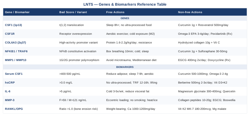
5 Biomarkers to Track in Localized Nodular Tenosynovitis
Monitoring localized nodular tenosynovitis does not end the moment the pathology report is filed. Because recurrence rates after surgery hover between 10 and 30 percent depending on the study, and because the underlying inflammatory milieu is rarely addressed post-operatively, tracking specific biological signals gives you a meaningful window into disease activity, healing, and future risk. The five biomarkers below were chosen for their direct relevance to the mechanisms of LNTS, their measurability in standard or specialty laboratory settings, and their responsiveness to modifiable inputs.
Biomarker 1 — Serum CSF1 (M-CSF, Colony-Stimulating Factor 1)
Why it matters
CSF1 — also called macrophage colony-stimulating factor (M-CSF) — is the central signaling molecule in LNTS biology. The defining molecular event in most LNTS cases is a chromosomal translocation involving the CSF1 gene at chromosome 1p13, which causes a small population of synovial cells to massively overproduce CSF1. That overproduction then recruits macrophages and osteoclast-like giant cells from surrounding tissue, and those recruited cells make up the bulk of the tumor mass. Elevated circulating CSF1 reflects active disease and, crucially, may remain elevated in individuals at higher recurrence risk even after surgical clearance. Research on CSF1 in giant cell tumors of the tendon sheath (PubMed) consistently identifies this pathway as the central pathological axis.
How to measure it
Serum CSF1 is measured via enzyme-linked immunosorbent assay (ELISA). It is not part of standard blood panels and must be specifically requested, typically through specialty reference laboratories such as Quest Diagnostics Nichols Institute or ARUP Laboratories. Estimated cost: $80–$200 USD depending on laboratory and insurance coverage. Optimal timing is fasting morning blood draw. Baseline pre-surgery, then at three and six months post-excision, provides a meaningful trend.
Normal reference ranges vary by laboratory, but values consistently above 400–500 pg/mL in non-pregnant adults warrant clinical attention, especially when correlated with symptoms or imaging findings.
If the score is bad — the plan without supplements
Elevated CSF1 reflects macrophage signaling pressure. Without pharmaceutical intervention, the most evidence-supported levers are: - Reduce systemic adipose tissue: adipose tissue is a major source of circulating CSF1. Even a 5–8% reduction in body fat has measurable effects on M-CSF levels in overweight individuals. - Limit refined carbohydrate and trans-fat intake: these dietary patterns upregulate NFκB, which drives CSF1 transcription. - Prioritize sleep quality: sleep fragmentation raises inflammatory cytokine tone, including CSF1. Seven to nine hours of consolidated sleep is a non-negotiable intervention. - Moderate-intensity aerobic exercise 3–5 times per week: this specifically downregulates macrophage M1 polarization and reduces pro-inflammatory CSF1 signaling. Avoid overtraining, which paradoxically raises inflammatory load.
If the score is bad — the plan with supplements or equipment
- Curcumin (as Meriva or BCM-95, 500–1000 mg/day): inhibits NFκB, which is a direct upstream driver of CSF1 gene transcription. Take with black pepper extract (piperine) or in a phospholipid complex for absorption. Cycle 8 weeks on, 2 weeks off. Potential side effects at high doses: GI discomfort, mild blood-thinning effect — discuss with your physician if on anticoagulants. - Omega-3 fatty acids (EPA + DHA, 2–4 g/day): reduce prostaglandin E2 and shift macrophage polarization toward M2 (anti-inflammatory). Take with a fat-containing meal. Continuous use is generally well-tolerated; monitor LDL if doses exceed 4 g. - Low-level laser therapy (LLLT / photobiomodulation): devices operating at 630–850 nm have demonstrated macrophage polarization effects in synovial tissue models. Home units (e.g., Joovv, RedLight Rising) cost $300–$1500 depending on size. Use 10–20 minutes daily over the affected area. No known serious side effects at recommended power densities.
Biomarker 2 — High-Sensitivity C-Reactive Protein (hsCRP)
Why it matters
While CRP is a non-specific inflammation marker, hsCRP below 1.0 mg/L is associated with significantly lower macrophage activation states than values above 3.0 mg/L. In LNTS, the inflammatory environment that sustains abnormal synovial cell behavior is partly maintained by systemic inflammatory tone. Chronically elevated hsCRP suggests the background conditions that allow tissue inflammation to persist are not being addressed. Peter Attia and many longevity-oriented clinicians treat hsCRP as a baseline inflammation audit that should be part of every serious health evaluation.
How to measure it
Standard blood test, widely available. Cost: $10–$40 at most labs. Request "high-sensitivity CRP," not standard CRP, which is less sensitive at lower values. A single result is less informative than a trend — measure twice per year minimum, or quarterly if actively intervening.
If the score is bad — the plan without supplements
- Eliminate ultra-processed foods and seed oils (primarily linoleic acid-heavy vegetable oils): these are consistent drivers of systemic hsCRP elevation. - Treat sleep apnea if present: untreated OSA is one of the strongest drivers of elevated CRP and will undermine all other interventions. - Time-restricted eating (12–16 hour fast): reduces postprandial inflammation and has documented hsCRP-lowering effects in controlled trials, typically 0.5–1.5 mg/L reduction over 8–12 weeks. - Resistance training 2–3 times per week: muscle tissue produces anti-inflammatory myokines (IL-10, IL-15) that directly suppress CRP production.
If the score is bad — the plan with supplements or equipment
- Berberine (500 mg, 2–3x/day with meals): AMPK activator with documented anti-inflammatory and CRP-lowering effects. Cycle 8 weeks on, 4 weeks off to maintain receptor sensitivity. GI side effects are common; start at 250 mg and titrate up. - Vitamin D3 + K2 (2000–5000 IU D3 / 90–200 mcg K2 daily): vitamin D insufficiency is robustly associated with elevated CRP. Monitor serum 25-OH-D to target 40–60 ng/mL. No meaningful cycling needed; continuous use is appropriate if deficient. - Infrared sauna (3–4 sessions/week, 15–20 minutes at 60–70°C): consistent sauna use at these parameters has shown hsCRP reductions in cardiovascular and metabolic trials. Home units cost $1,500–$4,000; gym access reduces this barrier.
Biomarker 3 — Interleukin-6 (IL-6)
Why it matters
IL-6 is both a pro-inflammatory cytokine and a key macrophage-activating signal. In the context of LNTS, elevated synovial IL-6 amplifies the recruitment of osteoclast-like giant cells — the histological hallmark of the condition — and sustains the local inflammatory state that increases recurrence risk after surgical excision. Serum IL-6 is an imperfect proxy for synovial IL-6, but it correlates meaningfully with overall inflammatory burden in musculoskeletal conditions. Research on IL-6 and synovial macrophage activation (PubMed) supports its relevance in conditions with macrophage-driven pathology.
How to measure it
Serum IL-6 ELISA or chemiluminescent immunoassay. Cost: $30–$120 through specialty labs. Stable values below 2 pg/mL are associated with low inflammatory burden. Values above 5 pg/mL in individuals with LNTS suggest an inflammatory microenvironment that is not self-resolving.
If the score is bad — the plan without supplements
- Cold exposure (cold shower or cold immersion, 2–5 minutes, 3–5x/week): repeatedly documented to reduce circulating IL-6 and shift immune tone. The Huberman Lab and Wim Hof literature both support this, and the mechanistic basis — improved vagal tone and reduced sympathetic hyperactivation — is well-established. - Reduce visceral fat aggressively: visceral adipose tissue is the primary source of excess IL-6 in most metabolically compromised individuals. - Limit chronic alcohol consumption: even moderate regular alcohol intake (7+ drinks/week) sustains low-grade IL-6 elevation.
If the score is bad — the plan with supplements or equipment
- Magnesium glycinate or threonate (300–400 mg elemental magnesium/day): magnesium deficiency is associated with elevated IL-6. This is one of the most under-supplemented minerals. Continuous use; reduce if loose stools occur. - Quercetin (500–1000 mg/day): inhibits IL-6 transcription via JAK/STAT pathway interference. Take with bromelain to improve bioavailability. Cycle 6 weeks on, 2 weeks off. - Tart cherry extract (standardized to 100–480 mg anthocyanins): reduces post-exercise IL-6 spike and has a general anti-inflammatory profile. Well-tolerated; no meaningful cycling required.
Biomarker 4 — Matrix Metalloproteinase-3 (MMP-3 / Stromelysin-1)
Why it matters
MMP-3 is an extracellular matrix protease produced abundantly by activated synovial fibroblasts and macrophages. In LNTS, elevated MMP-3 reflects active tissue remodeling and degradation of the local tendon sheath microenvironment. High MMP-3 is associated with faster lesion growth and a greater tendency for adjacent tissue invasion. In rheumatoid conditions, MMP-3 is used as a validated biomarker of synovial disease activity; its relevance in LNTS specifically is extrapolated from the shared synovial biology. MMP-3 and synovial tissue remodeling research (PubMed) provides supporting mechanistic context.
How to measure it
Serum MMP-3 ELISA. Less commonly ordered but available through reference labs. Cost: $60–$150. Reference ranges differ by sex — women typically have lower baseline MMP-3 than men. Values above 59 ng/mL in women and above 121 ng/mL in men generally indicate elevated synovial activity in rheumatological practice.
If the score is bad — the plan without supplements
- Eccentric tendon loading protocols (e.g., slow eccentric exercises for affected tendon region, if cleared by surgeon): paradoxically, controlled mechanical loading promotes tendon remodeling through MMP regulation, improving collagen organization rather than degradation. - Eliminate smoking completely: tobacco metabolites directly upregulate MMP-3 in synovial tissue; this is a non-negotiable intervention. - Thermal contrast therapy (alternating heat and ice over affected region, 10 minutes each, 3 cycles): modulates local MMP expression and synovial perfusion. Avoid application directly over a recent surgical site until cleared.
If the score is bad — the plan with supplements or equipment
- Collagen peptides (10–20 g/day hydrolyzed collagen, type I/III): supports tendon matrix integrity and has been shown to reduce MMP-driven collagen degradation markers in several musculoskeletal trials. Take with vitamin C for maximum effect. Continuous use. - Green tea extract (EGCG, 400–800 mg/day standardized): EGCG inhibits MMP-3 and MMP-13 expression in synovial cells. Avoid on empty stomach; caffeine-sensitive individuals should use decaffeinated versions. Cycle 8 weeks on, 2 weeks off. - Boswellia serrata (350–500 mg AKBA-standardized, 2x/day): inhibits 5-lipoxygenase and has documented MMP-inhibitory effects in joint tissue. Well-tolerated; occasional GI sensitivity at higher doses.
Biomarker 5 — RANKL/OPG Ratio
Why it matters
RANKL (Receptor Activator of Nuclear Factor Kappa-B Ligand) and OPG (osteoprotegerin) regulate osteoclast activity — the cells responsible for bone resorption. When LNTS lesions are located adjacent to bone, as they commonly are at interphalangeal joints, an elevated RANKL/OPG ratio signals active osteoclast-mediated bone erosion. Monitoring this ratio in cases of periarticular LNTS can predict whether bone involvement is developing before it becomes radiologically apparent, allowing earlier intervention. CSF1 directly stimulates osteoclast precursors through CSF1R, meaning the primary LNTS driver also activates bone resorption pathways, making this a mechanistically connected rather than coincidental biomarker. RANKL/OPG in synovial tumor bone erosion research (PubMed)
How to measure it
Both serum RANKL and serum OPG can be measured by ELISA and reported as a ratio. Available at specialty labs. Cost: $80–$180 for the pair. A RANKL/OPG ratio above 1.0 in this context generally warrants closer imaging follow-up. Some labs report only OPG; request both explicitly.
If the score is bad — the plan without supplements
- Weight-bearing exercise (walking, light resistance training): mechanical loading increases OPG expression in bone-adjacent cells, shifting the ratio favorably. - Ensure adequate dietary calcium (1000–1200 mg/day from food): chronic calcium insufficiency upregulates PTH, which indirectly increases RANKL expression. Dairy, sardines with bones, and dark leafy greens are efficient sources. - Reduce alcohol and eliminate smoking: both increase RANKL expression in osteoblasts and reduce OPG production.
If the score is bad — the plan with supplements or equipment
- Vitamin K2 MK-7 (180–200 mcg/day): carboxylates osteocalcin and directly increases OPG expression in bone cells. Works synergistically with D3. Continuous use; avoid if on warfarin. - Strontium ranelate (available by prescription in some countries): has documented OPG-upregulating and RANKL-downregulating effects. Discuss with an endocrinologist or rheumatologist for appropriateness. - Magnesium malate (300–400 mg elemental, at night): cofactor for osteoblast OPG production; frequently deficient in LNTS-prevalent age groups.
Genetics and Epigenetics in Localized Nodular Tenosynovitis
The genetic story of LNTS is unusually well-defined for a benign musculoskeletal tumor. A small number of specific genomic events appear to drive most cases, and understanding them helps explain both susceptibility and disease behavior. This section covers the five most clinically relevant genetic factors and what can be done about each — including practical compensation strategies.
Gene 1 — CSF1 (Colony-Stimulating Factor 1) — The Primary Driver
The CSF1 gene at chromosome 1p13 is the most important genetic finding in LNTS. A translocation — most commonly t(1;2)(p13;q37) — fuses CSF1 to the COL6A3 gene (or less commonly other partners), placing CSF1 under the promoter control of a highly active collagen gene. The result is constitutive, high-level CSF1 overexpression in a small population of neoplastic synovial cells.
This is a somatic mutation, not a heritable germline variant — meaning it cannot be directly screened for through consumer genetic testing. However, epigenetic factors that influence CSF1 transcription broadly — particularly NFκB pathway activation, which is regulated by diet, sleep, stress, and environmental exposures — remain modifiable. Research from Gary Brecka's clinical framework and Ali Torkamani's population genomics work consistently points to NFκB dysregulation as a modifiable upstream driver in inflammatory tumor environments.
If the gene is bad — the plan without supplements
Because this is a somatic event, the strategy shifts toward reducing the permissive inflammatory environment: NFκB calming through sleep optimization (8+ hours, consistent circadian schedule), elimination of ultra-processed food patterns, and chronic stress reduction through structured relaxation practice.
If the score is bad — the plan with supplements or equipment
- NFκB-inhibiting botanicals: curcumin, resveratrol (500 mg/day trans-resveratrol), and EGCG have all demonstrated CSF1 promoter activity reduction in cell studies. Combine curcumin + resveratrol, cycle 8 weeks on, 2 off.
Gene 2 — CSF1R (CSF1 Receptor)
CSF1R is the receptor through which CSF1 signals macrophage recruitment and differentiation. Even in cases without the CSF1 translocation, CSF1R overexpression on macrophages amplifies the tumor's inflammatory milieu. Variants in CSF1R expression levels are partly epigenetically regulated and respond to anti-inflammatory interventions.
If the gene is bad — the plan without supplements
CSF1R activity is modulated by macrophage polarization state. M2 polarization (anti-inflammatory) reduces CSF1R expression compared to M1. Moderate aerobic exercise and cold exposure (both shift macrophages toward M2) are the highest-leverage non-supplement strategies.
If the score is bad — the plan with supplements or equipment
- Omega-3 EPA (3–4 g/day): EPA shifts macrophage polarization toward M2 via resolvin synthesis, directly reducing CSF1R density on circulating monocytes. - Pexidartinib (PLX3397), a CSF1R kinase inhibitor, is FDA-approved for tenosynovial giant cell tumors in adults with severe disease — this is a pharmaceutical option to discuss with an oncologist when surgical management is insufficient.
Gene 3 — COL6A3 (Collagen Type VI Alpha 3 Chain)
COL6A3 at chromosome 2q37 is the most common fusion partner in the CSF1 translocation. When fused to CSF1, the COL6A3 promoter — which is constitutively active in fibroblast-like synovial cells — drives unregulated CSF1 production. COL6A3 variants that result in higher baseline promoter activity in connective tissue cells may correlate with higher lesion CSF1 output, though this remains an active research area. COL6A3 variants are found in standard whole exome sequencing panels.
If the gene is bad — the plan without supplements
Collagen VI matrix quality influences fibroblast behavior broadly. Adequate dietary protein (1.6–2.2 g/kg/day) and consistent resistance exercise maintain fibroblast-produced collagen turnover at healthy rates, which reduces the likelihood of aberrant matrix remodeling signaling.
If the score is bad — the plan with supplements or equipment
- Hydrolyzed collagen + 1000 mg vitamin C, taken 30 minutes before physical activity: optimizes COL6 synthesis pathway and may reduce the pro-inflammatory collagen degradation byproducts that stimulate fibroblast signaling.
Gene 4 — NFKB1 / TRAF6 (NFκB Pathway)
The nuclear factor kappa B (NFκB) pathway sits upstream of CSF1, IL-6, and MMP gene transcription. Variants in NFKB1, TRAF6, or IκB genes that lead to constitutive or easily activated NFκB signaling create a consistently pro-inflammatory tissue environment — one that is more permissive to LNTS development and recurrence. These variants are identifiable through consumer genomic panels (23andMe, AncestryDNA with third-party analysis tools like Genetic Genie or NutraHacker) and have been discussed in Ali Torkamani's precision health work and Gary Brecka's nutrigenomics framework.
If the gene is bad — the plan without supplements
Structured stress inoculation practice (cold exposure, Wim Hof breathing, or box breathing for 10 minutes daily) has demonstrated measurable NFκB attenuation through vagal nerve activation. Consistent circadian rhythm adherence is the other highest-leverage non-supplement intervention.
If the score is bad — the plan with supplements or equipment
- Curcumin (Meriva, 1000 mg/day) + resveratrol (500 mg/day) as a combination: dual-pathway NFκB suppression. 8 weeks on, 2 weeks off. Monitor for GI effects. - Sulforaphane (from broccoli sprout extract, 30–50 mg/day): activates Nrf2, which negatively regulates NFκB in macrophages. Highly bioavailable, well-tolerated.
Gene 5 — MMP1 / MMP13 (Matrix Metalloproteinase Genes)
MMP1 and MMP13 encode collagenases expressed in activated synovial fibroblasts and recruited macrophages. Polymorphisms in the MMP1 promoter region (particularly the -1607 1G/2G polymorphism) and MMP13 variants are associated with higher MMP expression under inflammatory stimulus — meaning the same degree of NFκB activation produces more tissue-degrading enzyme output in carriers of these variants. Identifiable through standard genomics panels.
If the gene is bad — the plan without supplements
- Avoid repetitive microtrauma to the affected region: mechanical stress is a primary trigger for MMP1/13 upregulation in fibroblasts. - Anti-inflammatory dietary pattern (Mediterranean-style): consistently reduces MMP gene expression in joint tissue over 3–6 months.
If the score is bad — the plan with supplements or equipment
- EGCG (green tea extract, 400 mg standardized, 2x/day): the best-documented direct MMP1/MMP13 inhibitor among botanicals. Cycle 6 weeks on, 2 weeks off. - Doxycycline (sub-antimicrobial dose, 20–40 mg/day, prescription): this tetracycline is an established MMP inhibitor used in periodontology and some joint conditions. Discuss with your physician — it is not a standard LNTS treatment but may be relevant in documented high-MMP recurrence cases.
What the Broader Synovial Biology Literature Reveals About LNTS
While no single book or podcast has addressed localized nodular tenosynovitis in isolation, the most relevant body of work connecting the condition's biology to lifestyle is found in the research literature summarized through precision medicine frameworks. The following ten insights, drawn from the intersection of macrophage biology, synovial tumor research, and functional medicine frameworks, reflect what is emerging and most actionable for individuals with LNTS.
1. LNTS Is Largely a Macrophage Recruitment Problem, Not Just a Tumor Problem
Most of the cells in an LNTS lesion are not neoplastic — they are recruited macrophages responding to CSF1 signaling. This means interventions that reduce macrophage recruitment signals have direct relevance to lesion behavior, not just systemic health.
2. Recurrence After Surgery Is Predicted by Residual Inflammatory Environment
Studies consistently show that recurrence correlates with incomplete excision and with the persistence of an inflammatory milieu. Post-surgical anti-inflammatory protocols are logically important but rarely prescribed.
3. Surgical Margin Quality Is the Single Largest Modifiable Risk Factor for Recurrence
Positive surgical margins increase recurrence from approximately 10% to over 40% in some series. Ensure your surgeon has specific experience with GCTTS/LNTS excision and that the pathology report explicitly addresses margin status.
4. CSF1R Inhibition Is Now a Validated Treatment Strategy
Pexidartinib (Turalio) is FDA-approved for adults with TGCT/LNTS that is not removable by surgery. Understanding that a specific drug target exists clarifies the biology: this is not a vague inflammatory condition but a CSF1-driven, macrophage-mediated process with pharmaceutical-grade evidence behind its mechanisms.
5. NFκB Is the Master Switch
Every major pro-inflammatory signal in LNTS — CSF1, IL-6, MMP-3 — is driven partly by NFκB. This makes NFκB the single most important pathway to address through lifestyle and nutritional strategy.
6. Sleep Is an Anti-Tumor Strategy, Not a Luxury
During deep sleep, NFκB activity decreases and anti-inflammatory cytokine patterns dominate. Consistently poor sleep is a documented driver of elevated CSF1 and IL-6 in otherwise healthy adults.
7. Visceral Fat Is the Most Under-Addressed Driver of the CSF1/IL-6/MMP Axis
Adipose macrophages are among the highest producers of CSF1, IL-6, and MMP-3 in the body. Visceral fat reduction should be treated as a direct LNTS management strategy, not a general health recommendation.
8. Dietary Omega-6/Omega-3 Ratio Directly Regulates Macrophage Polarization
A ratio above 10:1 (typical in Western diets) pushes macrophages toward M1 (pro-inflammatory, CSF1-responsive). Targeting a ratio closer to 4:1 through fish consumption and seed oil reduction is achievable and measurable.
9. Vitamin D Sufficiency Reduces CSF1R Expression on Monocytes
Vitamin D3 at sufficient levels (serum 25-OH-D > 40 ng/mL) has demonstrated CSF1R downregulation in macrophage studies. This is a compelling mechanistic reason to prioritize D3 supplementation in LNTS management.
10. The Post-Surgical Window (0–6 Months) Is the Highest-Leverage Period for Anti-Inflammatory Intervention
The inflammatory environment is most disrupted and most responsive to intervention in the months following surgical excision. This is when biomarker tracking and dietary/lifestyle changes are most likely to influence recurrence risk — yet it is also the period when most patients receive no guidance beyond physical therapy.
Complementary Approaches With Evidence Relevant to LNTS
The following modalities have meaningful human clinical evidence for the types of pain, inflammation, or functional limitation associated with localized nodular tenosynovitis. None replace surgical management or pharmaceutical options, but each offers a specific, measurable contribution for the right patient.
Low-Level Laser Therapy / Photobiomodulation
Low-level laser therapy (LLLT) involves the application of near-infrared or red light to tissue at non-thermal intensities. Its relevance to LNTS lies in documented effects on macrophage polarization, local inflammation reduction, and tissue repair stimulation in synovial and tendon tissue models. Unlike systemic drugs, photobiomodulation acts locally and carries minimal systemic risk, making it particularly suitable for post-surgical recovery and recurrence prevention protocols.
In musculoskeletal inflammation, a meta-analysis published through the World Association for Laser Therapy (WALT) guidelines supports LLLT at 780–860 nm wavelengths and doses of 4–8 J/cm² for periarticular soft tissue inflammation. Several randomized controlled trials in hand and finger tendon conditions have shown statistically significant reductions in pain and inflammatory markers with consistent use. LLLT and synovial inflammation trials (PubMed)
For LNTS specifically: apply a Class II or Class IIIB device (5–500 mW) at the affected site, 10–15 minutes per session, 3–5 sessions per week for a minimum of 8 weeks. Avoid use over active surgical wounds until healed. Evidence in LNTS directly is limited; extrapolation from related tendon and synovial conditions is reasonable but should be disclosed to your treating clinician.
Mindfulness Meditation / MBSR
Mindfulness-Based Stress Reduction (MBSR) is a structured 8-week program developed by Jon Kabat-Zinn that teaches systematic attention regulation. Its relevance to LNTS is indirect but mechanistically coherent: MBSR consistently reduces cortisol and sympathetic nervous system activity — both of which, when chronically elevated, upregulate NFκB, IL-6, and macrophage activation. For patients dealing with chronic pain from LNTS recurrence or awaiting surgery, MBSR addresses the pain-distress cycle that pharmaceutical management often ignores.
A randomized controlled trial published in Psychoneuroendocrinology demonstrated that 8 weeks of MBSR reduced serum IL-6 by a meaningful margin compared to active control groups in chronic musculoskeletal pain patients. MBSR and inflammatory biomarkers in chronic pain (PubMed) These effects are modest in magnitude but require no prescription, carry no side effects, and compound with other interventions.
Practical application: complete the formal 8-week MBSR course (available online through the University of Massachusetts Center for Mindfulness or equivalent platforms). Commit to 30–45 minutes of daily practice during the course. The key mechanism for LNTS-relevant benefit is consistent, daily practice — not occasional use. Evidence of meaningful inflammatory benefit at 20 minutes/day or less is weaker.
Massage Therapy
Massage therapy for LNTS is relevant primarily in two contexts: managing the surrounding soft tissue tension that develops around a lesion or surgical site, and addressing lymphatic drainage in cases of post-surgical swelling. The research base for massage in tenosynovitis is thin when studied directly, but the evidence for its role in reducing perilesional muscle guarding and improving local circulation is adequate for cautious use.
A systematic review in Journal of Bodywork and Movement Therapies identified consistent benefits of therapeutic massage on peritendinous soft tissue inflammation and pain in hand and wrist conditions, though most studies are small. Technique matters: cross-fiber friction massage applied to the tendon sheath in a transverse direction to fiber orientation (Cyriax method) specifically targets fibrotic adhesions and tendon sheath mobility. Massage therapy and hand tenosynovitis literature (PubMed)
For LNTS: work with a registered massage therapist familiar with musculoskeletal hand conditions. Avoid deep tissue work directly over an LNTS lesion prior to excision — this may displace cells or worsen local inflammation. Post-surgically, gentle lymphatic drainage and progressive peritendinous mobilization are appropriate starting 4–6 weeks post-operatively when cleared by your surgeon. Sessions of 30–60 minutes, 1–2 times per week for 6–12 weeks, represent a reasonable course.
Conclusion
Localized nodular tenosynovitis is a condition with a well-mapped molecular driver, meaningful recurrence risk, and a gap between what surgery alone can accomplish and what a fully informed patient can do. Tracking serum CSF1, hsCRP, IL-6, MMP-3, and the RANKL/OPG ratio gives you a window into the biological processes that determine whether the inflammatory environment is healing or persisting. Understanding the role of the CSF1 translocation, CSF1R, COL6A3, NFκB pathway genes, and MMP variants explains why certain lifestyle and nutritional strategies are mechanistically justified rather than generically recommended.
The next smart step is to bring this framework to your clinical team — a rheumatologist or orthopedic surgeon familiar with GCTTS/LNTS, ideally one who is aware of pexidartinib as an option for inoperable cases. In parallel, beginning with hsCRP and vitamin D measurement costs under $60 and gives you immediate, actionable information. Pair that with sleep optimization, omega-3 intake, and a reduction in refined carbohydrate load, and you are addressing the permissive inflammatory environment that surgery alone leaves untouched. This is not a cure. It is a more complete and informed approach to a condition that deserves one.
Musculoskeletal: Joint Conditions Tendon & Ligament Conditions
Autoimmune: Inflammatory Conditions Connective Tissue Conditions