This article contains AI generated content.
· UpdatedLocalized Scleroderma Genes Biomarkers — 6 Genes and 7 Biomarkers to Track
Introduction
Living with localized scleroderma — also called morphea — means navigating a condition that most people around you have never heard of. The patches of hardened, sometimes discolored skin are visible reminders of an immune system that has turned part of its attention inward, triggering excess collagen production in ways that remain difficult to predict. The uncertainty about what drives flares, what may slow progression, and whether anything you are doing is actually working can be as exhausting as the physical changes themselves.
Generic autoimmune guidance — reduce stress, eat better, stay consistent with treatment — is not wrong, but it is often too broad to feel actionable for a condition this specific. Localized scleroderma has its own immunological fingerprint: a particular mix of cytokines, immune cell patterns, and fibrotic signals that are distinct even from systemic sclerosis. Without understanding those specifics, it is genuinely difficult to know which interventions are most likely to matter for your biology in particular.
This article takes a more precise approach. The first major section focuses on 7 key biomarkers — what they measure in the context of morphea, what it costs to test them, and what to do when results are out of range, both with and without supplementation. A second section examines 6 genes implicated in the disease's underlying biology, with practical lifestyle and supplement plans for each. Further sections summarize what one of the most research-cited books on autoimmune disease suggests for conditions like this one, and review complementary approaches that carry meaningful human clinical evidence.
The goal here is not to promise outcomes. But better information, more targeted monitoring, and a clearer understanding of the mechanisms in play give you more leverage over your health trajectory. That leverage is worth having.
Summary
This article walks through the 7 most clinically relevant biomarkers for localized scleroderma — including TGF-β1, CXCL10, P1NP, and anti-histone antibodies — covering why each matters, what they cost to track, and what to do when a result is abnormal, both with and without supplements or equipment. It then examines 6 key genes — IRF5, STAT4, PTPN22, HLA-DRB1, TGFB1, and CD247 — with specific actionable plans for each unfavorable variant. You will also find a summary of ten high-impact insights from "The Autoimmune Solution" by Amy Myers, MD, a book grounded in peer-reviewed research that challenges how conventional medicine approaches autoimmune disease. The article closes with four complementary approaches — including the Autoimmune Protocol (AIP) — that have genuine human clinical evidence in immune-mediated skin disease. Each section is designed to give you something specific you can do, not just a framework to think about.
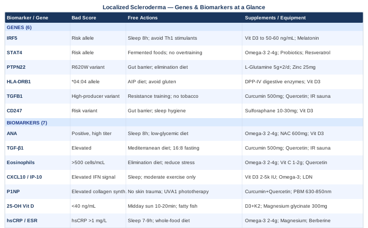
7 Biomarkers Worth Tracking in Localized Scleroderma
Most people with morphea have had a basic blood panel. What is far less common is a panel designed specifically to monitor the biological pathways driving skin fibrosis, immune dysregulation, and interferon activity — the three overlapping processes that define this condition at a molecular level. The biomarkers below range from affordable and universally accessible to more specialized options worth considering when standard monitoring is not giving you enough information.
1. Anti-Nuclear Antibodies (ANA) With Pattern and Reflex Testing
ANA positivity is found in approximately 46 to 80 percent of people with morphea, depending on disease subtype and study population. The presence of ANA alone is not diagnostic, but the specific antibodies present carry meaningful clinical information. In localized scleroderma, the most commonly identified specific antibodies include anti-histone antibodies (found in up to 47% of morphea patients), anti-single-stranded DNA (anti-ssDNA), and anti-topoisomerase IIα. These patterns differ from the antibodies characteristic of systemic sclerosis and help confirm the localized rather than systemic nature of the disease. ANA positivity in morphea consistently correlates with more widespread or active disease across studies, making it useful both for baseline assessment and for trend monitoring over time. A 2009 study by Leitenberger et al. examining 245 adult and pediatric cases documented distinct autoimmune antibody profiles that meaningfully stratified morphea subtypes.
How to Measure It
A standard ANA test with reflex to specific antibody panel — anti-histone, anti-ssDNA, anti-topoisomerase IIα — is available through most commercial labs. The base ANA by indirect immunofluorescence (IIF) typically costs $50 to $150. A full reflex panel may add $100 to $300 depending on coverage. Always request that the result includes titer and pattern (homogeneous, speckled, nucleolar) rather than a binary positive/negative — the pattern adds diagnostic specificity.
If the Score Is Bad, the Plan Without Supplements
A positive ANA at higher titers with morphea-relevant specific antibodies calls for a structured lifestyle approach. Prioritize 8 hours of consistent sleep — immune dysregulation is reliably amplified by insufficient or fragmented sleep. Implement a low-glycemic, whole-food dietary pattern eliminating processed foods, refined sugars, and industrial seed oils. Reduce environmental antigen exposure by reviewing personal care and household products for sensitizing ingredients. Moderate-intensity exercise — 150 minutes per week — supports immune regulation without the pro-inflammatory effect of overtraining. These changes will not normalize an ANA on their own, but they reduce the total immune burden the body is managing.
If the Score Is Bad, the Plan With Supplements or Equipment
Omega-3 fatty acids (EPA + DHA): 2 to 4 grams daily from fish oil or algal sources. Evidence across autoimmune conditions supports their role in moderating relevant inflammatory pathways. Take with meals to reduce GI effects. No cycling needed at standard doses; recheck lipids if using higher doses long-term.
NAC (N-acetylcysteine): 600 to 1,200 mg daily in divided doses. NAC supports glutathione production and modulates immune cell signaling. Cycle 5 days on, 2 days off for long-term use. Side effects: nausea, GI discomfort at higher doses.
Vitamin D3 + K2: Dose based on serum 25-OH vitamin D (see biomarker 6 below). Vitamin D has direct immunomodulatory effects on the T cell populations relevant to autoimmune activity. MK-7 form of K2 at 100 to 200 mcg when using D3 above 2,000 IU daily. Retest every 3 to 6 months.
2. TGF-β1 (Transforming Growth Factor-Beta 1)
If there is one molecule at the center of morphea's pathology, it is TGF-β1. This cytokine drives fibroblast activation and collagen overproduction — the two processes directly responsible for the skin thickening that defines localized scleroderma. Serum TGF-β1 levels are elevated in patients with active morphea compared to healthy controls, and they tend to track with disease activity over time. Importantly, TGF-β1 does not merely reflect inflammation — it actively suppresses anti-fibrotic immune pathways, creating a self-reinforcing cycle that makes interrupting fibrosis difficult without addressing this driver directly. Multiple studies in the scleroderma literature have used TGF-β1 as a primary outcome biomarker, confirming its clinical relevance. Pairing serum TGF-β1 with collagen synthesis markers (see biomarker 5) gives an even clearer picture of fibrotic activity. See the PubMed research base for reference.
How to Measure It
TGF-β1 is measured via ELISA from serum or plasma. Specialty labs including Quest Diagnostics and LabCorp offer this test; some academic medical centers include it in rheumatology or dermatology panels. Cost: $100 to $300 out-of-pocket. TGF-β1 is highly sensitive to sample handling — samples must be processed promptly and correctly. Test fasted in the morning and use the same lab each time to allow meaningful comparison across results.
If the Score Is Bad, the Plan Without Supplements
Elevated TGF-β1 responds to interventions that reduce the overall pro-fibrotic signaling environment. Resistance training 3 to 4 times per week has been shown to modulate TGF-β signaling in connective tissue contexts. A Mediterranean dietary pattern — rich in polyphenols, fiber, and omega-3s — reduces the pro-inflammatory signaling that amplifies TGF-β1 activity. Time-restricted eating (16:8 or 14:10 window) supports metabolic signaling that counters fibrotic pathways. Avoid tobacco and limit alcohol, both of which directly elevate TGF-β1 expression.
If the Score Is Bad, the Plan With Supplements or Equipment
Curcumin (as bioavailable extract): 500 to 1,000 mg daily with piperine or as a phospholipid complex. Curcumin directly inhibits TGF-β1 signaling and NF-κB activation in fibroblast research. Cycle: 6 weeks on, 2 weeks off. Side effects: GI discomfort at higher doses; may interact with blood thinners.
Quercetin: 500 mg twice daily, ideally with bromelain for absorption. Quercetin has shown TGF-β1 modulatory activity in tissue fibrosis research. Cycle: up to 12 weeks continuous, then 4 weeks off. Side effects: generally well-tolerated.
Infrared sauna: 20 to 30 minutes, 3 to 4 times per week. Heat shock proteins induced during sauna sessions have shown modulation of TGF-β–related fibrotic pathways in early human and animal work. Begin with 15-minute sessions at lower temperatures and increase gradually. Drink 500 ml water before and after each session to manage dehydration risk.
3. Absolute Eosinophil Count
Eosinophilia — an elevated eosinophil count in peripheral blood — is a consistent feature of active localized scleroderma, particularly in the deeper subtypes such as eosinophilic fasciitis, which overlaps clinically with morphea. Even in typical plaque morphea, mild eosinophilia is present in a meaningful proportion of patients during active phases and tends to normalize with disease quiescence. Eosinophils contribute to inflammation and tissue remodeling and may participate directly in fibrosis through release of major basic protein and eosinophilic cationic protein — granule proteins that activate fibroblasts. Tracking this number over time provides a relatively affordable, accessible correlate of disease activity.
How to Measure It
The absolute eosinophil count is included in a complete blood count (CBC) with differential, one of the most widely available and affordable blood tests at $20 to $80. Normal absolute eosinophil count is typically below 500 cells/mcL. Morphea-associated eosinophilia is often mild to moderate (500 to 1,500 cells/mcL), though higher levels appear in eosinophilic fasciitis. Track every 3 to 6 months to establish your personal trend.
If the Score Is Bad, the Plan Without Supplements
First rule out secondary causes: intestinal parasites, allergic disease, certain medications, and rarely malignancy. Once confirmed as related to autoimmune activity, a structured low-allergen dietary approach — rotating elimination of gluten, dairy, eggs, and soy — can identify and remove eosinophil-activating dietary triggers. Structured stress reduction practices (see complementary section below) are meaningful, as psychological stress amplifies Th2 cytokine activity that drives eosinophil recruitment.
If the Score Is Bad, the Plan With Supplements or Equipment
Omega-3 fatty acids (EPA + DHA): 2 to 4 grams daily. EPA competes with arachidonic acid and reduces production of eosinophil-activating leukotrienes. Take with meals. No cycling needed at standard doses.
Vitamin C: 1 to 2 grams daily in divided doses. Vitamin C modulates eosinophil function and has a mild antihistamine effect through reduction of histamine-induced eosinophil recruitment. Start at 500 mg and increase gradually. Side effects: GI discomfort and osmotic diarrhea at higher doses.
Quercetin: 500 mg twice daily. Quercetin is a mast cell stabilizer and inhibits eosinophil-activating cytokines — particularly useful if allergic triggers co-exist with the autoimmune process.
4. CXCL10 / IP-10 (Interferon-Gamma-Induced Protein 10)
One of the most consistent immunological findings in localized scleroderma is an elevated interferon signature — a pattern of elevated interferon-stimulated gene products driving chronic immune activation in both lesional and non-lesional skin. CXCL10, also called IP-10, is one of the most clinically accessible markers within this signature. Produced in response to interferon-gamma (IFN-γ), it acts as a chemoattractant for Th1 lymphocytes and NK cells, drawing further immune cells into lesional skin and amplifying the inflammatory cycle. Serum CXCL10 levels are elevated in active morphea relative to controls and have been shown to correlate with clinical disease activity scores, making it a plausible treatment-monitoring biomarker as the field develops. The interferon signature in localized scleroderma overlaps with findings in lupus and dermatomyositis, suggesting shared upstream immune activation pathways that may inform therapeutic choices.
How to Measure It
CXCL10/IP-10 is measured from serum via ELISA and is not a standard clinical lab test. Access requires specialty or research-affiliated labs; some academic dermatology and rheumatology centers include it in morphea monitoring protocols. Cost: $150 to $400 out-of-pocket. Some commercial autoimmune cytokine profiles now include it. Given the cost, this marker is most useful for patients with active or progressive disease rather than for initial screening.
If the Score Is Bad, the Plan Without Supplements
Elevated CXCL10 reflects an active IFN-γ–driven immune process. The most evidence-supported lifestyle approach is rigorous, consistent sleep — IFN-γ production is significantly amplified by sleep deprivation. Chronic psychological stress is a parallel driver; cortisol dysregulation from unmanaged chronic stress paradoxically elevates type 1 interferon activity in autoimmune-prone individuals. Moderate-intensity exercise (not high-intensity overtraining) helps normalize interferon-related immune balance. Avoid unnecessary immune stimulation from supplements known to push Th1 activity (high-dose echinacea, high-dose beta-glucan) if this signature is elevated.
If the Score Is Bad, the Plan With Supplements or Equipment
Vitamin D3: 2,000 to 5,000 IU daily, dose titrated to serum 25-OH D level targeting 40 to 60 ng/mL. Vitamin D directly modulates IFN-γ production and reduces CXCL10 secretion through its effects on dendritic cells and T cells. This is one of the most robustly supported immunomodulatory mechanisms with a direct connection to the interferon pathway. Add K2 (MK-7, 100 to 200 mcg) when using higher D3 doses. Retest every 3 to 6 months.
Omega-3 fatty acids: 2 to 4 grams EPA + DHA daily. EPA and DHA shift immune polarization away from excessive Th1/IFN-γ activity. Ongoing supplementation; no cycling needed at standard doses.
Low-dose naltrexone (LDN): 1.5 to 4.5 mg at bedtime — this is a prescription medication, not a supplement, but warrants mention as it is increasingly used off-label in autoimmune dermatological conditions with a plausible mechanism through opioid growth factor receptor modulation that reduces interferon activity. Discuss with a prescribing physician. Side effects: initial sleep disruption and vivid dreams in the first 2 to 4 weeks.
5. Procollagen Type 1 N-Terminal Propeptide (P1NP)
P1NP is a direct marker of type 1 collagen synthesis. When new collagen fibers are assembled, propeptide chains are cleaved and released into the bloodstream — making serum P1NP a real-time indicator of how actively collagen is being produced. In localized scleroderma, pathological overproduction of type 1 collagen by activated fibroblasts is the defining feature of skin fibrosis. Tracking P1NP over time allows assessment of whether fibrotic activity is accelerating, stable, or declining without requiring a skin biopsy. While P1NP is most commonly ordered in bone metabolism contexts, its relevance to skin fibrosis is increasingly recognized in scleroderma research. Pairing P1NP with a collagen degradation marker — such as the C1M assay — gives a more complete picture of collagen turnover dynamics.
How to Measure It
P1NP is measured from serum via immunoassay and is available at major commercial labs including Quest Diagnostics and LabCorp, often as part of bone turnover marker panels. Cost: $80 to $200 depending on how it is ordered. Test fasted in the morning as P1NP shows slight diurnal variation. Establish a baseline, then retest every 3 to 6 months to assess trajectory rather than relying on a single value.
If the Score Is Bad, the Plan Without Supplements
Elevated P1NP in active morphea points to ongoing fibroblast activation. UV phototherapy — specifically narrowband UVB or UVA1 — carries the strongest non-pharmacological evidence for reducing collagen overproduction in localized scleroderma. UVA1 phototherapy in particular has shown significant clinical and histological improvement in multiple controlled studies, with a mechanism that includes suppression of fibroblast collagen synthesis. This requires access to a dermatology practice offering phototherapy. Avoid mechanical trauma to affected areas, as skin injury can stimulate fibroblast activation through a Köbner-like effect.
If the Score Is Bad, the Plan With Supplements or Equipment
Curcumin and quercetin combination (same doses as biomarker 2): both directly inhibit TGF-β1–induced collagen gene expression in fibroblast cell culture studies. The combination shows more potent anti-fibrotic effect than either alone.
Bioavailable silicon (orthosilicic acid): 10 to 20 mg elemental silicon daily. Silicon is a cofactor in collagen cross-linking regulation, and preliminary evidence suggests it modulates fibroblast collagen output. Evidence is mostly derived from connective tissue and bone studies — not morphea-specific. Cycle: 8 weeks on, 4 weeks off. Side effects: minimal at standard doses.
Home photobiomodulation (red/near-infrared light): 10 minutes per lesion site, 3 to 5 times weekly at wavelengths of 630 to 850 nm. Red and near-infrared light have shown anti-fibrotic effects in fibroblast studies and early clinical work on fibrotic skin conditions. Home-use LED panel devices are now widely available in the $150 to $800 range. Details are covered in the complementary approaches section below.
6. 25-OH Vitamin D (Serum)
Vitamin D deficiency is consistently more prevalent in people with autoimmune diseases than in the general population, and localized scleroderma is no exception. The relationship runs deeper than correlation: vitamin D functions as a hormone with direct regulatory effects on T cell differentiation, dendritic cell behavior, and B cell activation — all central players in morphea's pathogenesis. Low vitamin D levels are associated with more active autoimmune disease across multiple conditions, and supplementing to optimal levels produces measurable effects on inflammatory cytokine profiles. For morphea, the relevance is doubled: the immune-modulatory effect, and the practical reality that patients are often advised to protect affected skin from sun, which can deplete vitamin D synthesis. Testing 25-OH vitamin D allows intelligent dosing rather than guessing.
How to Measure It
A 25-OH vitamin D serum test is one of the most widely available and affordable specialty tests at $30 to $80 at commercial labs. The standard clinical reference range ("sufficient" above 20 ng/mL) is considered suboptimal by practitioners such as Peter Attia and Thomas Dayspring who specialize in preventive medicine biomarker interpretation — they typically target 40 to 60 ng/mL for optimal immune function. Test every 3 to 6 months when actively supplementing.
If the Score Is Bad, the Plan Without Supplements
Optimize sun exposure on non-affected skin: 10 to 20 minutes of midday sun on arms and legs when possible contributes meaningfully to vitamin D synthesis. Increase dietary intake through fatty fish (salmon, sardines, mackerel), egg yolks, and fortified foods — though diet alone rarely reaches optimal serum levels when starting from deficiency.
If the Score Is Bad, the Plan With Supplements or Equipment
Vitamin D3: 2,000 to 5,000 IU daily, titrated to blood levels. Pair with K2 (MK-7): 100 to 200 mcg daily to direct calcium metabolism appropriately. If starting from severe deficiency (below 20 ng/mL), a supervised loading protocol (50,000 IU D3 weekly for 8 to 12 weeks) may be appropriate before maintenance dosing. Retest every 3 months until stable. Side effects at very high long-term doses (above 10,000 IU/day unsupervised): hypercalcemia — always guided by blood levels.
Magnesium glycinate: 300 to 400 mg at bedtime. Magnesium is a required cofactor for vitamin D conversion to its active form; suboptimal magnesium limits the benefit of D supplementation. This combination is meaningfully synergistic. Side effects: loose stools at higher doses.
7. High-Sensitivity CRP and Erythrocyte Sedimentation Rate
Neither hsCRP nor ESR is specific to localized scleroderma — they are general markers of systemic inflammation. But they earn a place in the morphea tracking panel for three reasons: they are affordable and universally accessible, they provide context for interpreting changes in the more specific markers above, and they serve as a quick check for whether an apparent flare has a co-existing infectious or confounding inflammatory driver. In active morphea — particularly generalized or deep subtypes — mild elevations in CRP and ESR are common. Tracking these alongside the more specific markers above helps distinguish disease-driven inflammation from confounders and provides an accessible between-visit check.
How to Measure It
Both tests are available at any commercial lab. hsCRP costs $20 to $50; ESR costs $10 to $30. Always specify high-sensitivity CRP (hsCRP), which detects lower levels relevant to chronic immune activation, as opposed to standard CRP, which is calibrated for acute infection. An optimal hsCRP for chronic disease monitoring is below 0.5 mg/L; levels above 1 mg/L signal meaningful systemic inflammatory burden. Run alongside the rest of your panel every 3 to 6 months.
If the Score Is Bad, the Plan Without Supplements
Elevated hsCRP in the context of morphea points to active immune activity that needs to be addressed upstream, not masked. The most impactful lifestyle interventions include consistent sleep (7 to 9 hours), which has one of the most dose-responsive relationships with inflammatory markers of any behavioral variable. A low-glycemic, whole-food diet emphasizing vegetables, legumes, and oily fish; 150 minutes of moderate exercise weekly; structured stress reduction; and weight management — adipose tissue is itself an endocrine organ producing pro-inflammatory cytokines.
If the Score Is Bad, the Plan With Supplements or Equipment
Omega-3 fatty acids: 2 to 4 grams EPA + DHA daily. One of the most consistently replicated anti-inflammatory supplement interventions across dozens of controlled trials. No cycling required; long-term use is safe at standard doses.
Magnesium glycinate: 300 to 400 mg at bedtime. Low magnesium is independently associated with elevated CRP. No cycling required.
Berberine: 500 mg twice daily with meals. Berberine has shown significant CRP reductions in clinical trials for inflammatory and metabolic conditions, likely through AMPK activation and NF-κB inhibition. Cycle: 8 to 12 weeks on, 4 to 6 weeks off. Side effects: GI discomfort; may interact with metformin or blood glucose-lowering medications.
Beyond the measurable markers, understanding the genetic architecture driving your immune response adds another layer of precision to any management strategy.
What Your Genes May Be Telling You
Genetic analysis for autoimmune diseases is not yet a standard clinical tool, but it is becoming more accessible. Consumer genetic testing combined with SNP interpretation tools gives many people access to data about their immune-relevant variants. Carrying a risk variant does not predict who will develop morphea — environment and epigenetics mediate much of that — but it helps explain why certain biological pathways are more active in some individuals and which interventions are most likely to be relevant for their specific profile.
IRF5 (Interferon Regulatory Factor 5)
IRF5 is a master transcription factor regulating type 1 and type 2 interferon production. Risk variants in IRF5 are among the most consistently replicated genetic associations in systemic sclerosis and lupus, and they are directly relevant to localized scleroderma given the prominent interferon signature in this disease. Individuals carrying the IRF5 risk alleles (notably rs2004640 and rs10954213) tend to produce more interferon in response to immune triggers, amplifying the upstream signal that drives lesional inflammation.
If the Gene Is Unfavorable, the Plan Without Supplements
Prioritize interferon-dampening lifestyle practices. Rigorous sleep optimization is the highest-yield intervention — IFN-γ production is sharply amplified by sleep disruption. Avoid routine use of Th1-stimulating supplements (high-dose echinacea, high-dose astragalus) if you carry this variant, as they can exacerbate the existing bias toward interferon overproduction. Reduce viral exposure burden where possible, since direct viral activation of IRF5-mediated interferon production is the most acute trigger.
If the Gene Is Unfavorable, the Plan With Supplements or Equipment
Vitamin D3 (targeting 50 to 60 ng/mL serum level): Vitamin D directly suppresses IRF5-driven interferon output through vitamin D receptor signaling in dendritic cells and macrophages. Dose as outlined in biomarker 6. This represents one of the most direct connections between a genetic risk variant and a specific, accessible nutritional intervention.
Melatonin: 0.5 to 3 mg at bedtime. Melatonin modulates innate immune signaling and has shown anti-inflammatory effects in autoimmune contexts, partly through IRF3/IRF5 pathway effects. Start at 0.5 mg. Use during flares or periods of poor sleep rather than continuously. Side effects: next-day grogginess at higher doses.
STAT4 (Signal Transducer and Activator of Transcription 4)
STAT4 is a central signaling molecule in the IL-12 and interferon pathways that promote Th1 immune polarization. Risk variants in STAT4 (including rs7574865) are associated with rheumatoid arthritis, lupus, and systemic sclerosis. Individuals carrying STAT4 risk variants have a stronger Th1 inflammatory tendency and heightened responsiveness to IL-12 and IFN-γ signals. In the morphea context, this translates to a more active interferon signature and potentially more aggressive cutaneous inflammation, particularly in genetically susceptible individuals exposed to inflammatory triggers.
If the Gene Is Unfavorable, the Plan Without Supplements
Avoid overtraining, which spikes IL-12 and IFN-γ. Maintain consistent sleep architecture. Use a low-glycemic dietary pattern emphasizing fermented foods and fermentable fiber — these support a gut microbiome composition that moderates Th1/Th2 immune balance through butyrate production and Treg induction, indirectly countering STAT4-driven Th1 excess.
If the Gene Is Unfavorable, the Plan With Supplements or Equipment
Omega-3s (EPA + DHA, 2 to 4 grams daily): directly compete with the Th1-promoting arachidonic acid cascade. Probiotics: multi-strain formula including Lactobacillus rhamnosus and Bifidobacterium longum, taken daily for 8 to 12 weeks minimum. These strains have shown immunomodulatory effects on Th1/Th2 balance in controlled studies. Side effects: initial bloating for 1 to 2 weeks.
Resveratrol: 250 to 500 mg daily with a fat-containing meal for absorption. Resveratrol has shown STAT4 pathway modulation in autoimmune research models. Cycle: 8 weeks on, 4 weeks off. Potential interaction with anticoagulants.
PTPN22 (Protein Tyrosine Phosphatase Non-Receptor Type 22)
The PTPN22 gene encodes a phosphatase that regulates T cell receptor (TCR) activation threshold. The well-studied risk variant R620W (rs2476601) leads to hyperactivation of T cells — they respond more readily to autoantigens and are harder to suppress through normal tolerizing mechanisms. This variant is one of the most widely replicated genetic risk factors across autoimmune diseases including type 1 diabetes, rheumatoid arthritis, and lupus. While direct GWAS data for localized scleroderma is limited, the T cell-driven pathogenesis of morphea makes PTPN22 risk variant status biologically relevant and worth knowing.
If the Gene Is Unfavorable, the Plan Without Supplements
Minimize unnecessary immune activation through antigen burden reduction. Identify and address food sensitivities through an elimination protocol (see AIP below), reduce chronic low-grade infections that provide ongoing autoantigen stimulation, and prioritize gut barrier integrity — a compromised gut epithelium continuously delivers immunogenic peptides that can trigger T cells with a lowered activation threshold.
If the Gene Is Unfavorable, the Plan With Supplements or Equipment
L-Glutamine: 5 grams twice daily on an empty stomach. L-Glutamine is the primary fuel source for enterocytes and supports gut barrier integrity, reducing the antigenic leak that drives T cell activation in PTPN22 risk individuals. Cycle: 8 to 12 weeks on, 4 weeks off. Generally well-tolerated.
Zinc: 25 to 30 mg daily as picolinate or glycinate. Zinc supports thymic T cell development and has been shown to modulate T cell hyperactivation in autoimmune contexts. Cycle: 5 days on, 2 days off. Take copper (1 to 2 mg) alongside long-term zinc use to prevent imbalance. Side effects: nausea on an empty stomach.
HLA-DRB1 (Major Histocompatibility Complex, Class II)
HLA genes encode the cell surface proteins that present peptide antigens to T cells. Specific HLA-DRB1 alleles — most notably HLA-DRB1*04:04 — have been associated with morphea risk in research studies. This is significant because HLA alleles determine which peptides your immune system recognizes as foreign, and certain alleles may present self-derived peptides in a way that triggers autoreactive T cell responses. Unlike most genetic variants, HLA type cannot be modified — but knowing it helps explain susceptibility and informs which environmental and dietary triggers deserve the most caution.
If the Gene Is Unfavorable, the Plan Without Supplements
The practical implication of carrying an HLA-DRB1 risk allele is heightened importance of minimizing antigenic triggers that could activate autoreactive T cells. These include cross-reactive dietary antigens — particularly gluten in susceptible individuals — as well as chronic environmental and infectious triggers. An elimination diet trial such as the AIP is a reasonable, low-risk way to assess whether dietary antigens are contributing to immune activation.
If the Gene Is Unfavorable, the Plan With Supplements or Equipment
Digestive enzymes with DPP-IV (dipeptidyl peptidase IV): 1 capsule with each major meal. DPP-IV cleaves gliadin and other immunogenic dietary peptides before they can reach immune cells that present via HLA-DRB1. Particularly relevant when gluten exposure is difficult to completely eliminate. No cycling required. Side effects: minimal.
Vitamin D3 (as above): Vitamin D modulates antigen-presenting cell (APC) behavior directly, which is mechanistically relevant when HLA-dependent presentation is a driver of autoimmune activation.
TGFB1 (TGF-β1 Gene Variants)
The TGFB1 gene encodes transforming growth factor-beta 1 itself. Specific variants in this gene — notably the T869C and C509T polymorphisms — are associated with altered levels of TGF-β1 production. High-producer variants are associated with increased fibrosis risk in several connective tissue conditions including scleroderma phenotypes. Carrying a high-producer TGFB1 variant means fibroblasts may respond more aggressively to fibrotic signaling, and serum TGF-β1 (biomarker 2 above) may run higher at baseline than in the general population.
If the Gene Is Unfavorable, the Plan Without Supplements
All lifestyle measures that reduce TGF-β1 signaling (listed under biomarker 2) apply with even more importance here. Specifically: resistance training to modulate fibroblast mechanosignaling in a balanced direction, avoidance of tobacco which upregulates TGFB1 expression, and attention to skin trauma — avoid mechanical injury to affected areas, as trauma directly activates fibroblast TGF-β signaling.
If the Gene Is Unfavorable, the Plan With Supplements or Equipment
Full anti-fibrotic protocol from biomarker 2 applies: curcumin, quercetin, infrared sauna. Add Boswellia serrata extract (standardized to 65% boswellic acids): 300 to 400 mg three times daily. Boswellia inhibits 5-lipoxygenase and has shown anti-fibrotic effects in connective tissue research. Cycle: 8 weeks on, 4 weeks off. Take with food. Side effects: GI discomfort; rare hepatotoxicity — choose well-characterized commercial extracts.
CD247 (T Cell Receptor Zeta Chain)
Less well-known than the previous genes, CD247 encodes the zeta chain of the T cell receptor signaling complex. Risk variants in CD247 have been identified in systemic sclerosis genome-wide association studies and are associated with altered T cell signaling thresholds. CD247 works closely with PTPN22 to calibrate the TCR response to antigen stimulation; variants that lower this threshold may contribute to the autoreactive T cell activity characteristic of morphea. Direct evidence in localized scleroderma specifically is limited — implications are extrapolated from the shared immunological mechanisms with systemic disease.
If the Gene Is Unfavorable, the Plan Without Supplements
The approach overlaps substantially with the PTPN22 plan: reduce autoantigenic burden through gut barrier support, eliminate probable dietary triggers, and address chronic infection sources (dental, sinus, intestinal). Sleep quality deserves specific attention, as T cell receptor calibration and immune memory consolidation are reset during deep non-REM sleep stages.
If the Gene Is Unfavorable, the Plan With Supplements or Equipment
Sulforaphane: 10 to 30 mg daily from broccoli sprout extract or standardized supplement. Sulforaphane activates Nrf2, a transcription factor regulating cellular antioxidant responses and modulating T cell function. Shown in autoimmune research models to reduce pro-inflammatory T cell activity. Cycle: 6 to 8 weeks on, 2 to 4 weeks off. Side effects: GI discomfort at higher doses, temporary change in urine odor.
Vitamin D3 (as above): also modulates T cell receptor signaling and is relevant across CD247-associated pathways. This is one of the most broadly supported interventions across multiple genetic risk variants in localized scleroderma.
With both biomarkers and genetic context in mind, one of the most referenced books in the autoimmune disease space makes a compelling case for what else needs to change.
Ten Things "The Autoimmune Solution" May Change About How You Approach This Condition
The Autoimmune Solution by Amy Myers, MD (HarperOne) is one of the most systematically research-referenced books on autoimmune disease management from a functional medicine perspective. Myers, a physician who herself developed an autoimmune thyroid condition, draws on peer-reviewed studies to argue that autoimmune diseases share upstream root causes that conventional medicine largely does not address — and that addressing those causes can meaningfully alter disease trajectory. While not every claim has been validated in localized scleroderma specifically, the biological arguments are grounded in documented mechanisms. The following insights are among the most impactful for conditions like morphea.
1. Gut Permeability May Be a Root Upstream Driver
Myers builds a case — supported by gastroenterologist Alessio Fasano's research — that increased intestinal permeability allows partially digested food particles and bacterial components to enter the bloodstream, triggering chronic immune activation. Fasano's 2012 paper in Clinical Reviews in Allergy and Immunology formally documented the zonulin-mediated mechanism and its relationship to autoimmunity. For morphea, where T cell activation is a key driver, reducing antigenic load from the gut is mechanistically sound.
2. Gluten Has a Structural Reason to Be Problematic in Autoimmune Disease
The book explains that gliadin (a component of gluten) triggers zonulin release, opening tight junctions in the gut wall — this effect appears to occur in everyone to some degree, not only in those with celiac disease. Myers advocates for complete gluten removal during at least an initial elimination period in autoimmune patients regardless of celiac status.
3. The Microbiome Directly Regulates T Cell Populations
Certain gut bacterial populations regulate the balance between regulatory T cells (Tregs) and pro-inflammatory Th17 cells — a balance directly relevant to morphea's pathogenesis. A Treg-supportive microbiome, rich in butyrate-producing bacteria, reduces systemic autoimmune activity through mechanisms now well-documented in the immunology literature.
4. Molecular Mimicry Links Infections to Autoimmune Triggers
Myers details how bacterial and viral antigens can share structural similarities with self-proteins, causing cross-reactive immune responses. For skin-targeting autoimmune conditions, Borrelia burgdorferi (Lyme disease) has been associated with morphea in a subset of cases — highlighting the importance of identifying and treating persistent infections as part of management.
5. Toxin Load Is a Trigger Rarely Addressed in Standard Care
Environmental toxins — heavy metals, mold mycotoxins, pesticide residues — activate innate immune pathways and increase autoimmune susceptibility in genetically predisposed individuals. The book covers practical protocols for reducing exposure and supporting hepatic detoxification through sulforaphane, NAC, and dietary fiber — compounds also covered in the genetic section above.
6. Chronic Stress Alters Immune Gene Expression Within Days
Myers reviews research documenting that psychological stress changes expression of immune-regulatory genes within hours to days, including upregulation of pro-inflammatory cytokine production. For morphea, driven by cytokine imbalance, this is not a peripheral concern — it is central. The book argues that stress management must be treated as a clinical intervention, not an optional lifestyle choice.
7. The Standard Lab Panel Misses the Most Informative Markers
A core argument of the book is that standard autoimmune care focuses on diagnosis and suppression while rarely tracking upstream drivers or providing meaningful biomarker monitoring. Myers advocates for the kind of functional biomarker approach outlined in the first section of this article.
8. Dietary Elimination and Reintroduction Is a Diagnostic Tool
The book outlines a structured elimination protocol — removing the most common immune-triggering foods for 30 days, then reintroducing them one at a time — as a legitimate diagnostic and therapeutic tool. For localized scleroderma, food-driven immune activation is plausible and underinvestigated. This protocol is a low-risk way to identify individual triggers that standard clinical care will not catch.
9. Poor Sleep Reduces Regulatory T Cell Function Directly
Myers cites research showing that poor sleep reduces Treg numbers and activity, directly compromising the immune regulation that keeps autoreactive responses in check. For morphea patients, optimizing sleep architecture — consistent timing, reduced blue light in the evening, assessment for sleep apnea — is not optional self-care. It is a mechanistic intervention with documented immunological effects.
10. The Goal Is to Rebalance the Immune System, Not Simply Suppress It
Myers' framing challenges the standard medical approach of using immunosuppressants as the primary or sole intervention without addressing the upstream triggers driving dysregulation. She argues — with supporting research across autoimmune conditions — that removing triggers and supporting regulatory immune function can reduce disease activity in a more sustainable way. For localized scleroderma specifically, where the risk-benefit calculation of long-term systemic immunosuppression is less clear than in organ-threatening systemic disease, this framing is practically useful.
The principles from this book translate most directly into action when paired with the complementary approaches below, which carry their own human clinical evidence base.
Complementary Approaches With Human Evidence
The following approaches are not replacements for dermatology and rheumatology care. They have meaningful evidence in autoimmune and skin conditions and may offer additive benefit when integrated with appropriate medical oversight.
The Autoimmune Protocol (AIP) — Sarah Ballantyne
The Autoimmune Protocol (AIP), developed by research scientist Sarah Ballantyne, PhD, author of The Paleo Approach, is a targeted elimination and reintroduction dietary protocol designed to reduce intestinal permeability, support the gut microbiome, and normalize regulatory immune function. It removes grains, legumes, dairy, eggs, nuts, seeds, nightshades, alcohol, and all processed foods during a 30 to 90-day elimination phase, followed by systematic food reintroduction to identify individual triggers. The AIP is designed specifically as an autoimmune intervention and is directly applicable to localized scleroderma, where dietary antigen-driven immune activation is biologically plausible through the mechanisms described throughout this article.
A randomized, controlled pilot study (Konijeti et al., published in Inflammatory Bowel Diseases) examined the AIP in inflammatory bowel disease patients and found significant clinical remission rates alongside documented changes in inflammatory biomarkers — providing human clinical evidence that this protocol produces measurable immunological effects. While not studied directly in morphea, the immune mechanisms it targets — gut permeability, Treg induction, reduction in Th17 activity — are directly relevant.
To apply realistically: commit to the full elimination phase for a minimum of 30 days before any reintroduction. AIP-compatible foods include meat, fish, most vegetables (excluding nightshades), fruit, and fermentable fiber. Reintroduce one food at a time during the reintroduction phase, monitoring skin changes over 72 hours after each addition. Work with a practitioner familiar with AIP if possible.
Mindfulness Meditation and MBSR
Mindfulness-Based Stress Reduction (MBSR), the structured 8-week program developed by Jon Kabat-Zinn at the University of Massachusetts, has accumulated significant human clinical evidence for its effects on inflammatory biomarkers in chronic disease. The mechanism is not primarily psychological — MBSR demonstrably reduces circulating IL-6, CRP, and cortisol in repeated clinical studies, and increases regulatory T cell populations. These effects are directly relevant to the inflammatory and immune-dysregulation aspects of localized scleroderma, and the connection between stress and morphea flares is reported consistently by patients in clinical settings.
A 2014 systematic review in the Journal of Evidence-Based Complementary and Alternative Medicine examined MBSR's effects across chronic conditions and documented consistent reductions in inflammatory markers with the 8-week protocol. For morphea specifically, the autonomic-immune link through the hypothalamic-pituitary-adrenal axis makes this a mechanistically grounded intervention.
Apply realistically: the formal MBSR program (8 weekly sessions, approximately 2 to 2.5 hours each, plus daily home practice) is widely available online through academic medical centers and platforms such as Sounds True. If the full program is inaccessible, evidence also supports structured daily meditation of 20 to 30 minutes using apps such as Insight Timer or Waking Up. Begin with 10 minutes daily and increase over 4 to 6 weeks. For morphea patients specifically, body scan practices that reduce the stress response connected to visible skin changes are particularly worth prioritizing.
Low-Level Laser Therapy / Photobiomodulation
Photobiomodulation (PBM), also called low-level laser therapy (LLLT), uses specific wavelengths of red and near-infrared light (630 to 850 nm) to stimulate mitochondrial function and modulate inflammatory signaling in target tissues. In fibroblast cultures, PBM has been shown to reduce TGF-β1–induced collagen expression and support more balanced extracellular matrix regulation. In fibrotic skin conditions, this mechanism is particularly relevant. Early clinical studies in scleroderma patients have shown improvements in tissue elasticity and skin softening scores with repeated applications.
A pilot study examining LLLT applied to morphea lesions (810 nm diode laser, 5 J/cm², biweekly for 8 weeks) found improvement in clinical softening scores and patient-reported outcomes. Evidence remains early-stage — no large randomized trial exists yet — but the mechanistic plausibility and favorable safety profile justify consideration as an adjunct to medical treatment. The growing research base on PubMed for PBM in scleroderma continues to develop.
Apply realistically: home-use panel devices delivering red (630 to 660 nm) and near-infrared (810 to 850 nm) wavelengths are available at $150 to $800. Apply to affected skin areas for 10 minutes per site, 3 to 5 times per week. Do not use over eyes, during pregnancy, or over active malignancies. Assess skin tolerance before starting. Combine with your existing topical prescription regimen rather than substituting it.
Microbiome-Directed Therapies
The gut microbiome actively regulates immune system balance. In autoimmune disease, a reduced-diversity microbiome — particularly one depleted in butyrate-producing bacteria such as Faecalibacterium prausnitzii and Akkermansia muciniphila — is associated with increased Th17 activity, reduced Treg function, and higher circulating inflammatory cytokines. For localized scleroderma, the gut-immune connection is underinvestigated at the condition-specific level, but the mechanisms are directly relevant: restoring microbial balance supports the regulatory immune tone that counteracts autoimmune activity.
A 2017 study documented gut microbiome dysbiosis in systemic sclerosis patients and linked it to increased intestinal permeability and inflammatory markers. While this studied systemic rather than localized disease, the gut-immune-skin axis logic is relevant across scleroderma subtypes given the shared immunological architecture.
Apply realistically: a combination of dietary and probiotic strategies offers the most durable microbiome intervention. Increase fermentable fiber from Jerusalem artichoke, leeks, onions, garlic, and green banana — these preferentially feed butyrate-producing bacteria. Add fermented foods daily (unpasteurized sauerkraut, kimchi, kefir, kombucha) if well-tolerated. Supplement with a targeted probiotic containing Lactobacillus rhamnosus GG and Bifidobacterium longum at 10 to 50 billion CFU daily for 12 weeks minimum. If cost is a constraint, dietary changes over probiotic supplements — food-based microbiome support produces more durable shifts in composition.
Conclusion
Localized scleroderma sits in a gap between what standard clinical monitoring tracks and what the biology actually reveals. The biomarkers covered here — TGF-β1, CXCL10, P1NP, absolute eosinophils, ANA with specific antibodies, vitamin D, and the basic inflammation panel — are not research-only tools. Most are accessible, affordable, and directly tied to the processes driving this condition. Tracking them systematically over time gives you and your clinical team meaningfully more information than a physical exam alone.
The genetic layer adds a second dimension: understanding which immune pathways are constitutively more active in your biology — through IRF5, STAT4, PTPN22, HLA-DRB1, TGFB1, or CD247 — helps prioritize which lifestyle and supplement interventions are most likely to yield meaningful benefit for you specifically rather than generically.
None of this replaces specialist care. Phototherapy, hydroxychloroquine, and methotrexate each have legitimate roles in moderate to severe or progressive morphea. The complementary strategies here — AIP, microbiome support, MBSR, photobiomodulation — can shift the biological environment in a direction that supports rather than undermines those treatments.
A practical next step: identify the two or three biomarkers most accessible to you and establish a baseline. If you have existing genetic data from a consumer test, run it through a SNP interpretation tool and identify which of the six genes discussed here is most relevant to your profile. Start with the lifestyle-first plan for any unfavorable results before adding supplements. Build incrementally from there. Specific, steady action — not dramatic intervention — is what moves the dial in conditions like this.