This article contains AI generated content.
· UpdatedLoeys-Dietz Syndrome Genes and Biomarkers: 6 Genes and 6 Biomarkers to Track
Introduction
If you or someone you love has been diagnosed with Loeys-Dietz syndrome, you have probably already realized how quickly the standard conversation with a cardiologist reaches its edges. The advice — keep blood pressure low, get your echo done, avoid contact sports — is correct, but it barely scratches the surface of what is actually happening in the body. LDS is not a simple plumbing problem. It is a disruption in a molecular signaling network that regulates how tissues grow, repair themselves, and hold together under pressure, and that disruption plays out differently depending on which of the six known causative genes is involved.
What makes LDS particularly challenging to manage is the gap between imaging appointments. An echocardiogram every six or twelve months gives a snapshot of the aortic root at a single point in time, but it says nothing about the inflammatory state of the arterial wall, the rate of connective tissue breakdown happening between visits, or the accumulating cardiovascular strain that precedes any visible change on a scan. This gap is where biomarker tracking becomes genuinely useful — not as a replacement for imaging, but as a continuous read on biology that imaging cannot see.
Generic lifestyle advice is also a poor fit for LDS. The standard guidance to "exercise more" needs to be translated into a very specific intensity and modality framework because the wrong type of exercise increases aortic wall stress rather than reducing it. The advice to "eat anti-inflammatory foods" is more relevant here than in almost any other condition, but it rarely comes with enough specificity to be actionable. The goal of this article is to move past the generic.
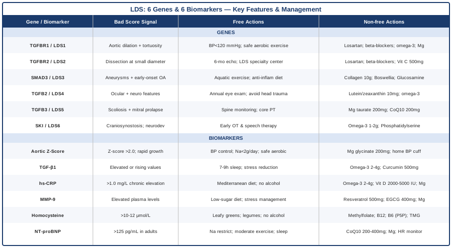
What follows covers two complementary strategies. The primary section focuses on six specific biomarkers — measurable, trackable, and largely modifiable — that offer a more complete picture of LDS disease activity. The second section examines the six main LDS-associated genes individually: what each one does, and what the evidence supports in terms of lifestyle, supplementation, and targeted support. Neither section replaces specialist care, but both are designed to make that care more precise.
6 Biomarkers Worth Tracking in Loeys-Dietz Syndrome
Biomarker tracking in LDS has historically been almost entirely visual — aortic diameters on imaging, followed by a surgical decision threshold. That remains essential, but it leaves a significant gap. A patient can have rising inflammatory burden, accelerating matrix breakdown, and worsening cardiac strain well before any of it appears on an echocardiogram. The six biomarkers below reflect the biological systems most directly relevant to LDS pathology. Some are standard clinical tests; others require more deliberate ordering. All of them are actionable.
1. Aortic Root Diameter and Z-Score
Why it matters: This remains the most critical surveillance parameter in LDS. Aortic dissection and rupture are the primary causes of premature death in the syndrome, and aortic root dilation is the chief warning sign. Critically, LDS behaves differently from Marfan syndrome in this respect: dissection in LDS can occur at smaller absolute diameters and at younger ages, making the frequency and precision of measurement even more important. A z-score — which adjusts the raw measurement for body surface area — provides a more accurate comparison across time and across patients than a raw diameter alone.
What it may reveal: A z-score above 2.0 in adults warrants close monitoring. The threshold for prophylactic surgical intervention in LDS is typically set more aggressively than in Marfan syndrome, often around 4.0–4.5 cm for aortic root depending on the specific genetic subtype, rate of change, and family history of dissection. Rate of change over consecutive studies is often the most informative single variable.
How to measure it
Transthoracic echocardiography (TTE) is the primary tool, performed every 6–12 months. For complete arterial surveillance — which LDS specifically requires given the risk of dilation beyond the aortic root — cardiac MRI or CT angiography of the entire arterial tree from head to pelvis is recommended at baseline and periodically thereafter. Cost: TTE typically runs $200–$800 out of pocket in the United States; cardiac MRI $500–$3,000. Most insurance carriers cover these for confirmed LDS patients, and in publicly funded healthcare systems they are standard of care.
If the score is bad, the plan without supplements
Blood pressure management and activity modification are the two most impactful non-pharmacological interventions. Isometric exercise — any activity involving sustained muscle tension against resistance (heavy lifting, straining, breath-holding under load) — is contraindicated because it sharply elevates aortic wall stress. Competitive sports and activities with collision risk are off the table. What remains accessible and beneficial: walking, flat-terrain cycling, swimming at a comfortable pace, and recreational non-competitive activities that keep heart rate in a conversational range. Sodium restriction (targeting below 2,000 mg/day) helps maintain resting blood pressure. Daily home blood pressure measurement provides data that a clinic visit every few months simply cannot replicate.
If the score is bad, the plan with supplements or equipment
Beta-blockers (atenolol, metoprolol succinate) reduce heart rate and aortic wall pulsatile stress; losartan reduces both blood pressure and TGF-β signaling downstream. Both are first-line pharmacological approaches in LDS and are medically prescribed. As adjuncts, magnesium glycinate at 200–400 mg/day supports vascular smooth muscle relaxation and modest blood pressure reduction. A validated upper-arm home blood pressure cuff ($30–$80) is arguably the most cost-effective monitoring tool available for daily aortic protection — surges detected at home can prompt medication adjustments far earlier than periodic clinic readings would allow. Continuous magnesium use is appropriate; recheck serum magnesium at 3-month intervals.
2. TGF-β1 Serum Level
Why it matters: Transforming growth factor beta-1 is the central signaling molecule whose dysregulation defines LDS at a molecular level. All six LDS-associated gene mutations converge on TGF-β pathway overactivation, and circulating TGF-β1 levels provide a functional read on how active that pathway is at any given moment. This is not yet a fully standardized clinical biomarker for LDS with established reference ranges, but it has been studied in Marfan syndrome and related connective tissue disorders, where higher circulating TGF-β correlates with more aggressive vascular outcomes — consistent with the mechanistic work originally described in Loeys et al. (2005).
What it may reveal: Elevated TGF-β1 alongside stable imaging may indicate active arterial wall remodeling below the resolution threshold of echocardiography. Serially elevated values — especially if they are rising — may support a decision to increase monitoring frequency or optimize pharmacological management. They can also flag inflammatory triggers (infections, dietary patterns, stress) that are driving pathway overactivation at a particular time.
How to measure it
TGF-β1 is measured from a blood draw using ELISA-based assays. It is not part of standard cardiology panels and requires specific ordering, typically available through specialty or academic reference labs. Cost: approximately $100–$300. Sample handling significantly affects results — blood must be processed promptly and under controlled temperature. A single result should always be interpreted in clinical context rather than in isolation.
If the score is bad, the plan without supplements
Sleep quality is one of the most powerful modifiable drivers of systemic cytokine levels including TGF-β1. Poor sleep architecture — particularly fragmented REM sleep and insufficient total duration — raises inflammatory signaling across multiple pathways. Prioritizing 7–9 hours per night in a cool, dark environment with consistent timing is high-yield and zero-cost. Sustained psychological stress activates the HPA axis and feeds inflammatory cascades; structured stress management (see the complementary approaches section) directly targets this mechanism. Moderate-intensity aerobic exercise, maintained consistently, attenuates chronic inflammatory signaling over time through its effects on endothelial function and immune regulation.
If the score is bad, the plan with supplements or equipment
Losartan, medically prescribed, remains the most direct pharmacological modifier of TGF-β downstream signaling in LDS — its use is often motivated by this mechanism as much as by blood pressure control. Among nutritional adjuncts with relevant evidence, omega-3 fatty acids (EPA+DHA at 2–4 g/day) from high-quality fish oil have demonstrated the ability to reduce TGF-β1 in several cardiovascular and inflammatory contexts. Curcumin in a highly bioavailable form (such as a phytosome preparation at 500–1,000 mg/day) modulates TGF-β expression in tissue studies; LDS-specific clinical trial data is absent, but its anti-inflammatory mechanism makes it a reasonable adjunct under medical guidance. Continuous use with quarterly biomarker reassessment is a practical approach.
3. High-Sensitivity C-Reactive Protein (hs-CRP)
Why it matters: Chronic low-grade systemic inflammation accelerates arterial remodeling and matrix breakdown — two processes already dysregulated in LDS. hs-CRP is the most accessible, best-validated marker of vascular inflammation available in routine clinical practice. Peter Attia consistently emphasizes it as a front-line cardiovascular risk indicator in Outlive, particularly when tracked longitudinally. For LDS patients, where the arterial wall is structurally vulnerable, any sustained inflammatory burden compounds the underlying risk.
What it may reveal: hs-CRP above 1.0–2.0 mg/L in a non-acutely ill individual suggests chronic inflammatory activity. In LDS patients, even modest chronic elevation may warrant identification of dietary, behavioral, or infectious drivers rather than acceptance as background noise.
How to measure it
Standard blood test, widely available at essentially any lab. Cost: typically $10–$50, often included in expanded cardiovascular panels. Should be measured fasting, and not within two weeks of any acute illness, which transiently spikes CRP independently of chronic inflammation. Serial measurements (every 3–6 months) are far more useful than a single data point.
If the score is bad, the plan without supplements
Dietary pattern is the most modifiable driver of chronic hs-CRP elevation. A Mediterranean-style diet — emphasizing vegetables, legumes, whole grains, fatty fish, olive oil, and minimizing ultra-processed food and refined carbohydrates — has robust evidence for reducing hs-CRP by 20–40% over 3–6 months in cardiovascular populations. Eliminating alcohol and significantly reducing refined sugar are additional high-yield behavioral changes that directly lower inflammatory burden. Regular moderate-intensity aerobic activity (150 minutes/week of LDS-safe exercise) has a dose-dependent anti-inflammatory effect. Sleep, again, is a critical lever: poor sleep raises circulating inflammatory markers including CRP consistently across studies.
If the score is bad, the plan with supplements or equipment
Omega-3 fatty acids (2–4 g/day EPA+DHA) have the strongest evidence base among available supplements for reducing hs-CRP. Vitamin D supplementation (2,000–5,000 IU/day) with levels targeted to 40–60 ng/mL serum 25-OH vitamin D has consistent inverse associations with hs-CRP across multiple large studies; test and dose accordingly rather than using a fixed protocol. Magnesium (200–400 mg/day) corrects a common deficiency that is independently associated with elevated inflammatory markers. These are generally taken continuously; recheck hs-CRP and vitamin D at 3-month intervals when actively managing elevated values.
4. Matrix Metalloproteinase-9 (MMP-9)
Why it matters: MMP-9 is an enzyme responsible for degrading structural components of the extracellular matrix — specifically collagen and elastin, which form the mechanical backbone of arterial walls. In aortic aneurysm disease, elevated MMP-9 reflects active wall remodeling and accelerating structural breakdown. For LDS patients, where connective tissue metabolism is already dysregulated, tracking MMP-9 provides a direct biochemical window into how aggressively that breakdown is occurring between imaging appointments.
What it may reveal: Higher plasma MMP-9 has been associated with faster rates of aneurysm progression in thoracic aortic disease and in Marfan syndrome. Critically, MMP-9 elevation appears to precede measurable diameter changes in several published series, suggesting it may serve as an early warning signal that imaging alone would miss.
How to measure it
Measured from plasma or serum via ELISA. Not part of standard clinical panels — requires deliberate ordering through specialty or reference labs. Cost: approximately $100–$300. Acute physical exertion and acute infection both transiently elevate MMP-9; testing should be performed in a resting state and at least two weeks after any acute illness for accurate interpretation.
If the score is bad, the plan without supplements
Dietary pattern has a meaningful effect on MMP activity. High-sugar, high-refined-carbohydrate diets drive MMP expression through inflammatory and advanced glycation end-product (AGE) pathways; reducing these categories is among the most accessible non-supplement interventions. Chronic psychological stress elevates glucocorticoids, which in turn increase MMP expression in vascular tissue — another argument for structured stress management. Moderate-intensity aerobic exercise, maintained over months, improves endothelial function in a way that tends to normalize MMP-9 in cardiovascular disease contexts; the same LDS intensity caveats apply.
If the score is bad, the plan with supplements or equipment
Sub-antimicrobial doxycycline (20–100 mg/day) is the most studied pharmacological MMP-9 inhibitor and has been investigated in thoracic aortic aneurysm patients — this remains experimental and requires a physician's prescription and monitoring. Among evidence-informed nutritional options, magnesium inhibits certain MMP activation pathways. Trans-resveratrol at 500 mg/day has demonstrated MMP-9 suppression in vascular cell and animal studies. EGCG from green tea extract (standardized to ~50% EGCG, approximately 400–800 mg/day) shows similar in vitro evidence for MMP inhibition. None of these have LDS-specific clinical trial data; they represent reasonable adjuncts when discussed with a managing physician. A cycling approach for resveratrol and EGCG — 8 weeks on, 2–4 weeks off — reduces the theoretical risk of tolerance, though long-term safety data in this population is limited.
5. Homocysteine
Why it matters: Elevated homocysteine is an independent vascular toxin. It damages endothelial cells directly, promotes arterial stiffness, activates MMP activity, stimulates smooth muscle proliferation, and increases oxidative stress within arterial walls — all processes that compound the existing vulnerability in LDS. Thomas Dayspring has consistently highlighted homocysteine as one of the most underrated and underused cardiovascular biomarkers in clinical practice, particularly because it is inexpensive and highly actionable. For LDS patients with already-compromised arterial walls, even modest homocysteine elevation deserves prompt attention.
What it may reveal: Homocysteine above 10–12 µmol/L is generally considered elevated; values above 15 µmol/L represent clinically significant risk. The MTHFR gene variant (C677T or A1298C) is a common genetic contributor to elevated homocysteine that is worth testing in LDS patients, as it impairs folate metabolism and can be specifically addressed.
How to measure it
Standard fasting blood draw, widely available. Cost: $20–$80. Often available as part of expanded cardiovascular panels. Renal function affects homocysteine metabolism, so a creatinine or eGFR level is a useful companion test. Results should always be interpreted alongside B-vitamin intake history.
If the score is bad, the plan without supplements
Dietary patterns affect homocysteine primarily through B-vitamin and folate supply. A diet rich in dark leafy greens, legumes, eggs, whole grains, and organ meats provides the methylation cofactors — folate, B6, B12 — needed to convert homocysteine to less harmful forms. Eliminating or significantly reducing alcohol is high-yield, because alcohol impairs B-vitamin absorption at the gut level and depletes folate stores. Adequate protein intake also supports the transsulfuration pathway, which provides an alternative route for homocysteine clearance.
If the score is bad, the plan with supplements or equipment
This is among the clearest supplement protocols in cardiovascular nutrition. Methylfolate (5-MTHF) at 400–800 mcg/day, methylcobalamin (B12) at 500–1,000 mcg/day, and pyridoxal-5-phosphate (P5P form of B6) at 25–50 mg/day together supply the activated cofactors that drive the methylation cycle responsible for clearing homocysteine. For individuals with confirmed MTHFR C677T variants, methylated forms are specifically necessary — standard synthetic folic acid may be poorly converted and can accumulate. Adding trimethylglycine (betaine) at 500–1,000 mg/day provides an alternative methylation pathway independent of B vitamins. These are taken continuously; recheck homocysteine at 8–12 weeks to confirm response, then annually once stable.
6. NT-proBNP
Why it matters: NT-proBNP (N-terminal pro-B-type natriuretic peptide) is a protein released by cardiac muscle cells in response to mechanical wall stress — it rises when the heart is under increased pressure or volume load. In LDS patients, the combination of aortic root dilation, potential valve dysfunction, arterial stiffness, and hypertension can impose cumulative hemodynamic strain on the heart that does not always show up early on imaging assessments of structure or function. NT-proBNP provides a functional biochemical signal of that strain in real time.
What it may reveal: NT-proBNP persistently above 125 pg/mL in adults under 75 suggests the heart is working harder than it should. In LDS patients, where cardiac demand is already elevated by arterial and valvular pathology, even moderate elevation warrants investigation rather than watchful waiting.
How to measure it
Standard blood draw, widely available. Cost: $30–$100. A single value requires clinical context for interpretation — renal function, body weight, and age all affect NT-proBNP. As with all biomarkers, serial measurements over time are far more informative than any single reading.
If the score is bad, the plan without supplements
Reducing cardiac afterload through blood pressure management, sodium restriction, and activity pacing is the primary non-pharmacological approach. Endurance-type activity within LDS-safe parameters — moderate intensity, no competitive breathlessness, no exertional ceiling — maintains cardiac conditioning without adding wall stress. Sleep quality, again, is relevant: nocturnal hypertension and sympathetic nervous system activation during poor sleep both increase myocardial workload.
If the score is bad, the plan with supplements or equipment
Beta-blockers and ARBs (medically prescribed) directly reduce NT-proBNP by lowering cardiac wall stress. As adjuncts, CoQ10 at 200–400 mg/day supports mitochondrial efficiency in cardiac cells and has demonstrated NT-proBNP reduction in heart failure studies; the evidence is not LDS-specific but the mechanism is compelling. Magnesium (200–400 mg/day) contributes to cardiac membrane stabilization and rhythm regulation. Wearable continuous heart rate monitors used at rest and during mild daily activity can provide context for trending NT-proBNP values — spikes in resting heart rate or rhythm irregularity may precede biomarker rises and warrant earlier evaluation. CoQ10 can be taken continuously; reassess NT-proBNP every 3–6 months when actively managing elevation.
The 6 LDS-Associated Genes: What Each One Does and How to Respond
Understanding which gene is mutated in a given LDS patient changes the clinical picture in meaningful ways. The six LDS subtypes differ in cardiovascular severity, in which non-cardiac organ systems are most prominently affected, and in the degree of risk attached to particular findings. Genetic diagnosis is not just taxonomic labeling — it shapes the monitoring protocol, the surgical threshold, and the lifestyle priorities. What follows is a practical overview of each gene, paired with evidence-informed compensatory strategies.
TGFBR1 — LDS Type 1
What it does: TGFBR1 encodes the type 1 TGF-β receptor subunit. Mutations here cause paradoxical overactivation of downstream TGF-β signaling despite receptor dysfunction — a counterintuitive result that reflects the complexity of feedback regulation in the pathway. LDS type 1 is classically associated with prominent craniofacial features (hypertelorism, bifid uvula, cleft palate) alongside cardiovascular and skeletal involvement.
Clinical profile: Aortic root dilation is present in the majority of patients. Arterial tortuosity — twisting and elongation of arteries throughout the body — is a hallmark and can affect the cervical, intracranial, and visceral arteries, not just the aorta. Full arterial imaging rather than aortic-only surveillance is therefore essential.
If the gene is bad, the plan without supplements
The core priorities are consistent blood pressure management (targeting systolic below 120 mmHg at rest), exclusion of isometric and high-intensity exertion, and sodium restriction. Low-to-moderate aerobic activity — walking, cycling on flat terrain, swimming — maintains cardiovascular fitness within safe parameters. Full arterial surveillance from head to pelvis via MRA or CTA at baseline and at regular intervals is a medical necessity. Awareness of symptoms suggesting arterial dissection in any location (sudden severe headache, back or chest pain, limb pallor or pain) and a clear emergency action plan are part of realistic preparedness.
If the score is bad, the plan with supplements or equipment
Losartan (medically prescribed) directly opposes the TGF-β overactivation caused by TGFBR1 mutation — its use in this context is mechanistically motivated, not just antihypertensive. Beta-blockers provide complementary aortic wall protection. Omega-3 fatty acids (2–4 g/day EPA+DHA) and magnesium glycinate (200–400 mg/day) are reasonable anti-inflammatory and vasculo-supportive adjuncts. Wearable blood pressure monitoring during typical daily activity can identify situational pressure spikes — particularly around physical effort, stress, or temperature changes — that clinic readings miss.
TGFBR2 — LDS Type 2
What it does: TGFBR2 encodes the type 2 TGF-β receptor subunit, which pairs with TGFBR1 to form the functional receptor complex. Mutations here produce a phenotype historically confused with Marfan syndrome — tall stature, arachnodactyly, aortic dilation — but with a clinically important distinction: the risk of aortic dissection in TGFBR2 mutation carriers is higher at smaller aortic diameters than in Marfan syndrome, and dissection frequently occurs in arteries beyond the aorta.
Clinical profile: TGFBR2 LDS is generally considered the most cardiovascularly aggressive subtype. Skin manifestations — translucency, easy bruising, wide atrophic scars — may accompany severe arterial disease. Surgical thresholds are typically set lower for TGFBR2 mutation carriers than for other LDS subtypes or Marfan syndrome.
If the gene is bad, the plan without supplements
All TGFBR1 precautions apply, often with heightened urgency. Imaging intervals of 6 months rather than 12 months may be appropriate in cases of active dilation or family history of early dissection. Clear communication with a Marfan/LDS specialty center (rather than general cardiology) is particularly important for TGFBR2 patients given the nuances of surgical timing. Realistic discussion about prophylactic surgical options — aortic root replacement — should happen before crisis rather than during one.
If the score is bad, the plan with supplements or equipment
The same losartan-plus-beta-blocker pharmacological framework applies. Some centers add an additional ARB or consider irbesartan depending on tolerability. Vitamin C at 500–1,000 mg/day supports collagen cross-linking and provides modest antioxidant protection of arterial wall structures — the mechanism is relevant to the connective tissue fragility characteristic of this subtype. L-arginine supplementation is generally avoided in aortic aneurysm contexts due to theoretical concerns about vasodilatory effects; this distinction matters when patients encounter general cardiovascular supplement recommendations.
SMAD3 — LDS Type 3 / Aneurysms-Osteoarthritis Syndrome
What it does: SMAD3 is an intracellular signaling protein that carries TGF-β's message from the cell surface receptor into the nucleus, where it regulates gene expression. Mutations here create a dual paradox: defective signal transmission paired with compensatory overproduction of TGF-β upstream. LDS type 3, also called aneurysms-osteoarthritis syndrome (AOS), is named for its characteristic combination of arterial aneurysms and premature, often polyarticular osteoarthritis.
Clinical profile: SMAD3 LDS may present with less dramatic aortic disease than types 1 and 2, but compensates with wider arterial involvement and earlier joint deterioration. Osteoarthritis often presents in the third or fourth decade, well before typical population onset.
If the gene is bad, the plan without supplements
Joint preservation becomes an equally important priority alongside cardiovascular monitoring. Aquatic exercise, cycling, and non-load-bearing movement preserve cardiovascular conditioning without joint impact. Physical therapy focused on periarticular muscle strengthening slows functional joint deterioration. Anti-inflammatory dietary strategies are doubly relevant here — the Mediterranean dietary pattern addresses both the arterial inflammatory load and the articular inflammatory drive. Low-impact movement maintained consistently is strongly preferable to episodic high-intensity activity.
If the score is bad, the plan with supplements or equipment
Collagen peptides (10–15 g/day) taken with vitamin C (200 mg) 30–60 minutes before activity have emerging evidence for cartilage support in osteoarthritis (Shaw et al., 2017, American Journal of Clinical Nutrition). Boswellia serrata extract standardized to 65% boswellic acids (300–500 mg twice daily) has clinical evidence for reducing articular inflammation with a favorable safety profile. Glucosamine sulfate at 1,500 mg/day has mixed but generally supportive evidence for symptom management in osteoarthritis. All three can be taken continuously; reassess joint symptoms and functional capacity every 3–6 months.
TGFB2 — LDS Type 4
What it does: TGFB2 encodes TGF-β2, one of the three main TGF-β ligand isoforms. Unlike mutations in the receptor genes, TGFB2 mutations cause haploinsufficiency — the cell produces approximately half the normal amount of the ligand. Counterintuitively, the downstream signaling network overcompensates, producing pathway overactivation despite lower ligand levels.
Clinical profile: LDS type 4 often features a milder skeletal phenotype but can involve ocular abnormalities including lens dislocation and progressive myopia. Neurological features such as Arnold-Chiari malformation have been described. Aortic involvement is present and follows the same surveillance requirements as other LDS types.
If the gene is bad, the plan without supplements
Regular ophthalmologic evaluation — annual minimum — is as important as cardiac monitoring in TGFB2 LDS. Contact sports, activities with head trauma risk, and any exercise that significantly raises intraocular pressure (heavy resistance movements, sustained inverted positions) warrant specific avoidance. Neurological symptom awareness — particularly symptoms consistent with Chiari (occipital headaches exacerbated by coughing or straining, balance difficulty) — should be part of routine self-monitoring.
If the score is bad, the plan with supplements or equipment
Lutein and zeaxanthin (10 mg / 2 mg per day) support retinal health as an accessible protective measure. Omega-3 fatty acids provide benefits for both cardiovascular and ocular health through shared anti-inflammatory pathways. For cardiovascular management, losartan and beta-blockers follow the same framework as other LDS types. Blue light–filtering lenses for screen use are a low-cost practical addition for patients with progressive myopia.
TGFB3 — LDS Type 5
What it does: TGFB3 encodes the TGF-β3 isoform, which plays a critical role in cardiac development, palate formation, and ongoing connective tissue homeostasis in aortic tissue. Haploinsufficiency produces a combination of structural developmental anomalies and dysregulated matrix remodeling throughout life.
Clinical profile: LDS type 5 tends toward prominent skeletal findings — severe scoliosis and pectus deformity are common. Mitral valve prolapse occurs at above-average rates. Cleft palate and other midline defects may be present, reflecting the gene's role in early facial development.
If the gene is bad, the plan without supplements
Scoliosis monitoring (spinal radiographs during growth phases, assessment for bracing or surgical consultation when curves progress beyond 25–30 degrees) is a component of care in TGFB3 LDS that is often underemphasized relative to cardiac surveillance. Supervised core stabilization physical therapy — emphasizing the muscles that reduce mechanical load on a scoliotic spine — should be initiated early and maintained. Mitral valve prolapse should be included in the echocardiographic protocol at each visit.
If the score is bad, the plan with supplements or equipment
Electrolyte balance is particularly relevant in mitral valve prolapse, where magnesium deficiency has been associated with symptomatic palpitations and increased arrhythmia burden. Magnesium taurate or glycinate (200–400 mg/day) addresses this directly with a favorable tolerability profile. CoQ10 at 200 mg/day has specific evidence for symptom improvement in mitral valve prolapse beyond its general cardiac support role. Ergonomic seating, lumbar support devices, and positional awareness strategies reduce daily mechanical load on a scoliotic spine — practical, inexpensive, and often overlooked.
SKI — LDS Type 6 / Shprintzen-Goldberg Overlap
What it does: SKI encodes a transcriptional repressor that normally holds TGF-β signaling in check by suppressing SMAD-mediated gene activation. Loss-of-function mutations result in unopposed TGF-β target gene expression, with downstream effects on cranial development, cognition, and connective tissue. This subtype overlaps significantly with Shprintzen-Goldberg syndrome and represents the most neurologically complex of the LDS-associated gene mutations.
Clinical profile: Craniosynostosis, intellectual disability, and characteristic facial dysmorphism are more prominent in SKI LDS than in other subtypes. Aortic involvement is present but may be less severe than in TGFBR1/2 subtypes. Neurodevelopmental support is as central to the management plan as cardiovascular surveillance.
If the gene is bad, the plan without supplements
Early and sustained neurodevelopmental support — speech therapy, occupational therapy, structured educational support — optimizes outcomes in ways that later intervention cannot fully replicate. Sensory support strategies tailored to the individual's profile, consistent routine, and cognitive load management are practical tools. Cardiovascular monitoring follows the standard LDS framework adapted to the patient's functional capacity.
If the score is bad, the plan with supplements or equipment
Omega-3 fatty acids (EPA+DHA at 1–2 g/day, in pediatric-appropriate doses for children) have accumulating evidence for supporting neurological function and reducing neuroinflammation across neurodevelopmental contexts. Phosphatidylserine (100–200 mg/day) has cognitive support evidence in certain populations, though LDS-specific data is absent. Both can be taken continuously; reassess cognitive and behavioral markers every 6 months with the developmental care team.
What Peter Attia's "Outlive" Teaches That Directly Applies to LDS Management
Peter Attia's Outlive: The Science and Art of Longevity (2023) is not a book about Loeys-Dietz syndrome. But for anyone managing a condition with elevated cardiovascular mortality risk beginning in young adulthood, its framework may be more practically useful than anything written specifically for LDS — because it teaches exactly the kind of rigorous, biomarker-anchored, anticipatory thinking that LDS management requires. Here are the ten most directly applicable insights.
1. Extend Healthspan, Not Just Lifespan
Attia argues that the goal of longevity medicine is not primarily to avoid death but to extend the period of physical, cognitive, and emotional vitality — what he calls the "Marginal Decade." For LDS patients managing lifelong risk, this reframe is practical: the question becomes not just "how do I avoid aortic dissection" but "how do I maintain cardiovascular, joint, and cognitive function at the highest possible level for as long as possible."
2. Track Rate of Change, Not Just Absolute Values
Throughout Outlive, Attia emphasizes that biomarker trends over time are typically more informative than any single measurement. This maps directly to aortic root tracking in LDS: a root that is 4.0 cm but growing 0.4 cm per year is more concerning than a root that is 4.2 cm but stable for three years.
3. hs-CRP and Homocysteine Are Actionable — Most Physicians Don't Routinely Order Them
Both appear in Outlive as systematically underused cardiovascular risk markers that are cheap, available, and highly responsive to lifestyle and supplementation. The fact that most cardiology appointments do not include these tests is a gap LDS patients can close by proactively requesting them.
4. Zone 2 Exercise Is the Correct Exercise Prescription for Vascular Health
Attia's central exercise recommendation — low-to-moderate intensity "Zone 2" cardio at a pace where speech is still comfortable — is precisely the exercise modality safe and beneficial for LDS patients. It improves mitochondrial density, endothelial function, and insulin sensitivity without raising aortic wall stress.
5. Protein Intake Is Dramatically Underestimated in Most Adults
For LDS patients managing joint deterioration (particularly relevant in SMAD3 types) and connective tissue fragility, adequate dietary protein (targeting 1.6–2.2 g/kg body weight/day) provides the amino acid substrate for collagen and structural protein synthesis. This is a simple, actionable dietary adjustment that most patients have not considered.
6. Sleep Is a Biological Intervention, Not a Lifestyle Preference
Outlive dedicates significant attention to sleep as a primary driver of cardiovascular and inflammatory outcomes. Attia frames sleep quality as a health behavior as important as exercise and diet — a position strongly supported by the evidence on blood pressure, cytokine production, and cardiac repair during sleep. For LDS patients, this framing elevates sleep from a nice-to-have to a clinical priority.
7. Emotional Health Directly Affects Physical Biomarkers
Attia dedicates a chapter to psychological wellbeing as a genuine health variable with measurable physiological effects — not a soft element of care. Chronic psychological distress raises blood pressure, inflammatory markers, and cortisol in ways that directly threaten LDS management targets. His openness about this intersection is particularly relevant for patients navigating the long-term psychological weight of a serious genetic diagnosis.
8. Look Beyond Standard Lipids: ApoB and Lp(a) Matter
Attia advocates expanding lipid assessment beyond LDL cholesterol to include ApoB (the primary atherogenic lipoprotein particle count) and Lp(a) (a genetically elevated lipoprotein variant associated with early arterial disease). LDS patients have structurally vulnerable arterial walls; adding lipid-driven plaque risk to structural aneurysm risk is a compounding burden worth specifically addressing.
9. Metabolic Health Modulates Systemic Inflammation
Attia discusses insulin resistance and metabolic dysfunction as central drivers of chronic inflammation — a mechanism that accelerates arterial disease even in the absence of other genetic risk. For LDS patients, maintaining metabolic health through dietary quality and regular moderate activity adds an anti-inflammatory layer that complements all other management strategies.
10. Preventive Action Taken Before Decline Is Orders of Magnitude More Effective
The book's core message — that the window for meaningful prevention is years before symptoms appear — is directly applicable to LDS. Patients who carry a known genetic diagnosis have a rare advantage: they know their risk landscape before damage accumulates. Acting on that knowledge with intelligent, consistent monitoring and lifestyle choices is the highest-value thing an LDS patient can do.
Complementary Approaches with Clinical Relevance for LDS
The following three modalities have the strongest available human evidence for managing the cardiovascular, inflammatory, and autonomic dimensions of LDS. High-impact physical modalities — vigorous yoga, spinal manipulation — are excluded due to contraindications related to arterial fragility and connective tissue hypermobility. The options selected here are safe, accessible, and mechanistically relevant.
Mindfulness-Based Stress Reduction (MBSR)
What it is and why it may be relevant: MBSR is a structured 8-week program combining mindfulness meditation, body scan practice, and gentle movement, developed at the University of Massachusetts Medical School. For LDS patients, the relevance goes beyond general wellbeing. Chronic psychological stress — which is a realistic and understandable response to living with serious genetic cardiovascular risk — is a physiological trigger that raises blood pressure, activates the HPA axis, and elevates inflammatory cytokines including TGF-β1. Managing this stress response through an evidence-based, structured program directly serves the biological goals of LDS management, not just quality of life.
Supporting evidence: Multiple randomized controlled trials have demonstrated that MBSR reduces circulating inflammatory markers including hs-CRP and IL-6. Separate meta-analyses of mindfulness-based interventions confirm reductions in systolic blood pressure of approximately 4–5 mmHg with sustained practice — clinically meaningful in the context of aortic wall protection. A 2013 review published in Psychoneuroendocrinology synthesized evidence showing consistent reductions in stress-related inflammatory biomarkers across multiple chronic health populations.
How to apply it realistically for LDS: The standard MBSR format is 2.5 hours per week for 8 weeks, available through hospitals, community centers, and validated online platforms. Post-course maintenance practice of 20–30 minutes daily sustains the physiological benefits. The movement component within MBSR should be adapted with a physician's guidance to avoid strong spinal extensions, inversions, or positions that increase aortic pressure — modifications are routinely made in clinical settings for medically complex participants.
Biofeedback and HRV Training
What it is and why it may be relevant: Biofeedback uses real-time monitoring of physiological signals — most commonly heart rate variability (HRV), blood pressure, and skin conductance — to teach patients voluntary regulation of those parameters through learned autonomic responses. For LDS patients, where blood pressure control is among the primary goals of all pharmacological intervention, biofeedback offers a direct, non-pharmacological complement to that control through physiological self-regulation. HRV biofeedback specifically trains the autonomic nervous system toward parasympathetic dominance — lowering both resting heart rate and vascular resistance.
Supporting evidence: Meta-analyses of biofeedback interventions for hypertension have found mean reductions in systolic blood pressure of 7–8 mmHg and diastolic pressure of 5–6 mmHg. HRV biofeedback studies have demonstrated persistent effects on autonomic regulation that outlast the training period itself. The mechanism — increased respiratory sinus arrhythmia and improved baroreflex sensitivity — is directly relevant to the vascular strain patterns in LDS.
How to apply it realistically for LDS: Clinical HRV biofeedback training (4–8 sessions of 30–50 minutes with a trained practitioner) produces the best-validated outcomes. At-home continuation using validated chest-strap HRV monitors with dedicated breathing apps allows daily practice at low cost. The core technique involves slow, paced breathing at approximately 5–6 breaths per minute synchronized to real-time HRV feedback. No cardiovascular contraindications apply; this is accessible to nearly all LDS patients regardless of disease severity.
Breathing-Based Therapies
What it is and why it may be relevant: Slow, controlled breathing — specifically at resonant frequency (approximately 5–6 breaths per minute) using techniques such as extended-exhale breathing and box breathing — directly modulates autonomic balance, lowering sympathetic tone and increasing parasympathetic activity. The physiological result includes lower resting blood pressure, reduced vascular resistance, improved cardiac vagal tone, and decreased inflammatory signaling. For LDS patients, this represents a daily accessible intervention requiring no equipment and no prescription that directly targets one of the central management objectives.
Supporting evidence: Multiple RCTs demonstrate that consistent slow breathing practice reduces systolic blood pressure by 4–10 mmHg in hypertensive populations, with effect sizes comparable to some single antihypertensive medications. Research published in Frontiers in Human Neuroscience (Russo et al., 2017) demonstrated significant HRV and blood pressure improvements from structured 5–6 breaths/minute protocols. The FDA-cleared RESPeRATE device, which guides pacing through audio biofeedback, has independent controlled evidence for blood pressure reduction.
How to apply it realistically for LDS: Begin with 10 minutes daily of slow-paced breathing: inhale for 4 seconds, exhale for 6 seconds, in a seated or reclined position. Progress to 20 minutes daily for sustained benefit. The RESPeRATE device provides real-time guidance for pacing and costs approximately $80–$150. Free smartphone apps (Paced Breathing, iBreathe, Breathwrk) offer similar functionality at no cost. This is among the safest, most accessible, and most mechanistically relevant daily practices available for LDS-related cardiovascular management.
Conclusion
Loeys-Dietz syndrome is a condition that responds well to informed engagement. The six biomarkers covered here — aortic dimensions, TGF-β1, hs-CRP, MMP-9, homocysteine, and NT-proBNP — give a richer, more continuous picture of disease activity than imaging alone. The six LDS-associated genes frame why different patients experience different risk profiles and may need different priorities in their monitoring and lifestyle approach.
None of this replaces the expertise of a specialized center, the imaging protocols your cardiologist has established, or the pharmacological management your physicians have recommended. What it can do is sharpen the quality of those conversations — and give you the vocabulary to ask more precise questions about which gaps in your current monitoring might be worth closing.
The most practical next step is simple: bring this information to your next appointment with your cardiologist or geneticist. Ask which of these biomarkers are currently being tracked, discuss whether any of the others are worth adding to your panel, and review whether your activity and dietary patterns are aligned with what the evidence supports. Better information, applied consistently, compounds over time — and in a condition where you already know your risk landscape, that is a genuine advantage.
Musculoskeletal: Joint Conditions Spine Conditions
Cardiovascular: Heart Conditions Blood Pressure Conditions Blood Vessel Conditions Vascular Conditions
Mental Health: Neurodevelopmental Conditions
Autoimmune: Inflammatory Conditions Connective Tissue Conditions
Eye: Vision Conditions