This article contains AI generated content.
· UpdatedLumbosacral Radiculopathy — 6 Genes And 7 Biomarkers To Track
Introduction
If you have lumbosacral radiculopathy, you already know the particular frustration of a pain that does not stay in one place. It radiates. It changes character. It interrupts your sleep, your concentration, your ability to sit through a meal or walk around the block. And most of the standard advice — rest, anti-inflammatories, maybe physical therapy — often helps a little, but rarely answers the question that matters most: why is this happening in my specific body, and what can actually change it?
The truth is that lumbosacral radiculopathy is not a single, uniform condition. For some people, it is driven primarily by mechanical compression of the nerve root. For others, the inflammatory environment around the disc and nerve is the dominant factor. For others still, a genetic predisposition to disc degeneration, abnormal pain signaling, or impaired nerve repair is quietly shaping the entire picture. Generic rehabilitation protocols are not designed to distinguish between these scenarios, which is why outcomes vary so widely between patients with nearly identical imaging findings.
This article takes a different approach. It focuses on two layers of biology that most clinicians do not discuss with patients: the measurable blood biomarkers that reflect what is actually happening at the inflammatory and metabolic level, and the genetic variants that influence how your discs age, how your nerves signal pain, and how efficiently your body resolves damage. Neither layer is a crystal ball. But together, they offer a sharper view of your specific situation than an MRI alone can provide.
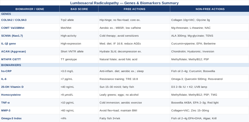
Better information does lead to better decisions. If your inflammatory markers are elevated, that is an actionable signal. If you carry a variant in a gene that reduces your natural anti-inflammatory capacity, there are practical steps — some requiring nothing more than dietary change — that may improve your trajectory. This article covers seven biomarkers worth tracking, six genetic variants worth knowing about, a summary of one of the most evidence-grounded books ever written on spine rehabilitation, and five complementary modalities with meaningful clinical support. None of this replaces a diagnosis or a treatment plan from a qualified clinician. But it gives you more to bring to that conversation.
7 Biomarkers Worth Monitoring If You Have Lumbosacral Radiculopathy
Biomarkers are measurable biological signals — proteins, metabolites, ratios — that reflect the underlying activity driving a condition. For lumbosacral radiculopathy, the most relevant biomarkers cluster around inflammation, nerve health, disc matrix integrity, and nutritional status. What follows are the seven most clinically actionable options, selected for their combination of strong evidence, practical accessibility, and modifiability.
1. High-Sensitivity C-Reactive Protein (hs-CRP)
Why it matters
C-reactive protein is produced by the liver in response to inflammatory signaling, primarily from interleukin-6. The high-sensitivity version of the test can detect low-grade chronic inflammation that standard CRP testing misses. In the context of lumbosacral radiculopathy, elevated hs-CRP reflects a systemic inflammatory environment that sustains nerve root irritation, slows healing, and amplifies pain signaling. Multiple studies have documented elevated CRP in patients with symptomatic lumbar disc herniation, and levels tend to correlate with symptom severity rather than disc size alone. Research indexed on PubMed consistently links systemic inflammation to both radiculopathy onset and persistence.
How to measure it
A standard blood draw ordered by any clinician. Cost: approximately $10–35 in the US. Target: below 1.0 mg/L for cardiovascular and general health purposes; above 3.0 mg/L indicates meaningful chronic inflammation. Many people with persistent radiculopathy test in the 2–5 mg/L range without being aware of it.
If the score is elevated — without supplements
The most powerful interventions are lifestyle-based. An anti-inflammatory diet removing refined carbohydrates, industrial seed oils, and ultra-processed foods can reduce hs-CRP by 30–50% within 8–12 weeks. Regular aerobic exercise (30–45 minutes, 4–5 days per week, low-impact options like walking or cycling are appropriate during active radiculopathy) lowers CRP through multiple mechanisms. Prioritizing 7–9 hours of sleep and managing chronic stress (which elevates cortisol and downstream inflammation) are both independently effective. Excess visceral fat is a major driver of elevated CRP, so even modest body composition change has a measurable impact.
If the score is elevated — with supplements or equipment
- Fish oil (EPA + DHA): 2–4g combined EPA/DHA daily, taken with a fat-containing meal. Reduces CRP by inhibiting pro-inflammatory eicosanoid production. No meaningful cycling required; monitor if on anticoagulants. Side effects at high doses: blood thinning, GI discomfort. - Curcumin with piperine: 500–1000mg standardized curcumin extract with 5–10mg piperine (black pepper extract) twice daily with meals. Meta-analyses support modest but consistent CRP reduction. Cycling: 8 weeks on, 2 weeks off is a common approach. Side effects: rare GI upset; may affect drug metabolism via CYP450 pathways. - Boswellia serrata (AKBA extract): 300–500mg of standardized extract (minimum 30% AKBA content) twice to three times daily. Directly inhibits 5-lipoxygenase, a key inflammatory enzyme. Can be used continuously. Side effects: occasional GI discomfort.
2. Interleukin-6 (IL-6)
Why it matters
IL-6 is one of the primary cytokines released by nucleus pulposus cells following disc herniation. It drives local nerve root sensitization and is a key upstream signal that tells the liver to produce CRP. Elevated IL-6 in the epidural space — and to a lesser extent in systemic circulation — correlates with the transition from acute to chronic radicular pain. While hs-CRP reflects the downstream effect, IL-6 is closer to the source. Research published on PubMed has demonstrated elevated IL-6 levels in the cerebrospinal fluid and serum of patients with symptomatic disc herniation.
How to measure it
A blood draw, less commonly ordered than CRP. Cost: approximately $40–80. Reference range varies by lab; most set normal below 7 pg/mL. IL-6 is more variable throughout the day than CRP, so a single measurement is less definitive. Trend over time or paired with hs-CRP is most informative.
If the score is elevated — without supplements
Resistance training (not during acute flare) is one of the most potent long-term reducers of baseline IL-6, primarily by increasing anti-inflammatory myokines. Time-restricted eating (compressing calories into an 8–10 hour window) has shown reductions in inflammatory cytokine profiles in human trials. Reducing psychological stress through structured practices is meaningful because psychological stress directly triggers IL-6 release.
If the score is elevated — with supplements or equipment
The same omega-3 and curcumin protocols described for hs-CRP apply here. Additionally: - Quercetin: 500–1000mg daily with meals. Quercetin inhibits NF-κB, a transcription factor that drives IL-6 gene expression. Cycling: 6–8 weeks on, 2 weeks off. Side effects: generally well-tolerated; rare headache or GI discomfort. - Resveratrol: 150–500mg/day with a fat-containing meal. Activates SIRT1, which downregulates pro-inflammatory pathways. Combine with quercetin or pterostilbene for additive effect. Side effects: mild GI effects at higher doses.
3. 25-OH Vitamin D
Why it matters
Vitamin D is not simply a bone-health nutrient. Its receptor is expressed in virtually every tissue, including peripheral nerves, spinal cord, and dorsal root ganglia. Deficiency is independently associated with heightened pain sensitivity, impaired nerve regeneration, and increased disc degeneration. In chronic low back pain and radiculopathy specifically, studies consistently find a higher prevalence of vitamin D insufficiency compared to pain-free controls. One mechanism: vitamin D suppresses prostaglandin synthesis and modulates the expression of nerve growth factor, both of which influence radicular pain intensity. Data on PubMed consistently support this relationship.
How to measure it
Standard serum 25-OH vitamin D blood test. Cost: $30–60. Target: 40–60 ng/mL (100–150 nmol/L) for optimal neuromuscular function, per most functional medicine practitioners including those aligned with Peter Attia's approach. Many clinicians consider levels above 20 ng/mL "sufficient" — a threshold increasingly regarded as too conservative for pain and neurological health.
If the score is low — without supplements
Midday sun exposure (arms and legs exposed) for 15–30 minutes 4–5 times per week can raise levels meaningfully, particularly for lighter skin types and in summer months. Dietary sources contribute modestly: fatty fish, egg yolks, and UV-exposed mushrooms. This approach is often insufficient to correct a significant deficiency, but meaningfully improves levels in mild cases.
If the score is low — with supplements or equipment
- Vitamin D3 (cholecalciferol): 2000–5000 IU/day for adults with confirmed insufficiency; retest in 90 days. Always pair with vitamin K2 (MK-7 form, 100–200mcg/day) to direct calcium to bone rather than soft tissue. Take with the largest fat-containing meal of the day for best absorption. No cycling required. Retest to avoid over-supplementation. Side effects at very high doses (>10,000 IU chronically): hypercalcemia; stay under 4000 IU without physician monitoring. - UVB lamp (narrowband 311nm): 3–5 minutes on skin 3–4x/week during winter months, following device-specific guidelines. A meaningful adjunct for those who cannot supplement orally.
4. Homocysteine
Why it matters
Elevated homocysteine is a direct marker of impaired methylation — the biochemical process that controls DNA expression, neurotransmitter balance, and cellular repair. In the context of radiculopathy, high homocysteine is relevant because it directly damages peripheral nerves, promotes oxidative stress in spinal structures, and correlates with accelerated disc degeneration. B-vitamin deficiencies (particularly B12, B6, and folate) are the primary driver of elevated homocysteine, and these same deficiencies impair nerve repair. Research on PubMed documents this relationship in peripheral nerve conditions, which shares mechanistic overlap with radiculopathy.
How to measure it
Serum homocysteine blood test. Cost: $20–50. Optimal range: below 9 μmol/L. Values above 15 μmol/L indicate hyperhomocysteinemia with meaningful cardiovascular and neurological risk. Many labs set the upper limit of normal at 15, but functional practitioners typically target below 10.
If the score is elevated — without supplements
A diet rich in leafy greens, legumes, eggs, and organ meats provides natural folate, B6, and B12. Avoiding excessive alcohol (a known B-vitamin depleter) and reducing coffee intake slightly may help. These dietary shifts are often sufficient for mild elevations (10–13 μmol/L).
If the score is elevated — with supplements or equipment
- Methylfolate (5-MTHF): 400–800mcg/day. This is the activated form of folate, critical for those with MTHFR gene variants who cannot efficiently convert folic acid. Avoid synthetic folic acid if MTHFR status is unknown or positive. - Methylcobalamin (B12): 1000mcg/day sublingual or oral. Methylcobalamin has superior neurological bioavailability compared to cyanocobalamin, particularly relevant for nerve repair in radiculopathy. - Pyridoxal-5-Phosphate (P5P, active B6): 25–50mg/day. Side effects: B6 toxicity (peripheral neuropathy paradoxically) at very high doses above 200mg/day; stay well below this. - Trimethylglycine (TMG / Betaine): 500–1500mg/day if homocysteine remains above 12 despite B-vitamin correction. Works as an alternative methyl donor. Side effects: fishy body odor at high doses, GI discomfort.
5. TNF-α (Tumor Necrosis Factor-alpha)
Why it matters
TNF-α is arguably the most important inflammatory cytokine in lumbosacral radiculopathy. It is directly released by herniated nucleus pulposus tissue in contact with the nerve root and has been shown to chemically sensitize and demyelinate nerve fibers even without mechanical compression — which explains why some patients with modest disc herniation experience severe pain while others with larger herniations remain symptom-free. The success of TNF-blocking biologics in rheumatoid arthritis has prompted serious research into their role in radiculopathy, and several clinical trials have explored epidural TNF antagonists. This makes it one of the most biologically specific biomarkers available for this condition.
How to measure it
Serum TNF-α testing via ELISA. Less routinely ordered; may require a specialty or functional medicine lab. Cost: $50–100. Normal levels are typically below 8–10 pg/mL; reference ranges vary by lab. Results should be interpreted alongside hs-CRP and IL-6 for a more complete inflammatory picture.
If the score is elevated — without supplements
Cold water immersion (10–15 minutes in 14–18°C water, 3–4x per week) has demonstrated reductions in circulating TNF-α in human studies. Regular moderate-intensity aerobic exercise and time-restricted feeding both reduce TNF-α independently. Eliminating trans fats and reducing saturated fat from ultra-processed sources are dietary priorities.
If the score is elevated — with supplements or equipment
- Boswellia serrata (AKBA): As described above; AKBA specifically inhibits TNF-α production. 300–500mg 2–3x/day. - Omega-3 fatty acids (EPA specifically): EPA competes with arachidonic acid and directly reduces TNF-α production. 2–3g EPA/day (not total omega-3; look at the EPA fraction specifically). - Low-Level Laser Therapy (LLLT / Photobiomodulation): 810–980nm class IV laser or home-grade 650nm red light devices applied to the lumbar region. Studies document reduction in local TNF-α expression following photobiomodulation. A 10–15 minute session per day is typical for home devices. This is a technology investment ($200–800 for a quality home device) with a growing evidence base. Side effects: minimal; avoid direct eye exposure.
6. Matrix Metalloproteinase-3 (MMP-3)
Why it matters
MMP-3 is an enzyme responsible for breaking down extracellular matrix components, including aggrecan, collagen, and fibronectin — the structural proteins that give intervertebral discs their resilience and hydration capacity. Elevated MMP-3 is a marker of active disc matrix degradation and has been documented in both disc tissue and serum of patients with disc herniation and radiculopathy. It reflects a catabolic environment in which repair is outpaced by destruction. While this biomarker is less commonly ordered than CRP or vitamin D, it provides unique information about the structural dimension of the condition and whether disc repair strategies are likely to be relevant.
How to measure it
Serum MMP-3 via immunoassay, available through specialty labs and some rheumatology-oriented panels. Cost: $60–120. Normal serum range: approximately 17–60 ng/mL (varies by lab). This test is more commonly ordered in rheumatoid arthritis contexts but can be requested specifically. Evidence for its use in this specific condition is still growing — treat it as a useful signal rather than a definitive marker.
If the score is elevated — without supplements
Avoiding excessive spinal compression loading (especially combined with flexion, such as heavy deadlifting with rounded back or long periods in a flexed-seated posture) reduces mechanical triggers of MMP expression in disc cells. Maintaining an optimal body weight reduces compressive load and inflammatory drive. Resistance training at appropriate intensity improves overall tissue quality and reduces the catabolic dominance.
If the score is elevated — with supplements or equipment
- Collagen peptides (hydrolyzed): 10g/day marine or bovine collagen, ideally taken with 500–1000mg vitamin C to maximize collagen synthesis. Provides the amino acid substrate (glycine, proline, hydroxyproline) for matrix repair. No cycling needed. Side effects: minimal; rare digestive discomfort. - Zinc: 15–30mg/day elemental zinc (from zinc glycinate or picolinate). MMP-3 is a zinc-dependent enzyme; adequate zinc supports regulatory control of MMP activity. Take separate from high-fiber meals. Side effects: nausea if taken on empty stomach; copper depletion at high doses (>40mg/day) — supplement 1–2mg copper if using long-term. - Vitamin C: 500–1000mg ascorbic acid twice daily. Acts as a cofactor for collagen crosslinking and a direct antioxidant reducing oxidative triggers of MMP expression.
7. Omega-3 Index
Why it matters
The Omega-3 Index measures the percentage of EPA and DHA in red blood cell membranes — a reflection of your actual tissue-level omega-3 status over the past 2–3 months, not just recent diet. A low Omega-3 Index (below 4%) is associated with a pro-inflammatory cellular environment, heightened pain sensitivity, and impaired nerve function. In contrast, an index above 8% corresponds to the lowest inflammatory profile and best nerve membrane fluidity for signal transmission. Unlike measuring omega-3 intake or even serum levels, the red blood cell index reflects true incorporation into cell membranes — the functional measure that matters for nerve health. Thomas Dayspring and others in lipidology have advocated for this test as one of the most underused yet actionable cardiovascular and inflammatory markers available.
How to measure it
A finger-prick test available from specialist labs such as OmegaQuant or through some physician panels. The OmegaQuant Home Test kit costs approximately $50–70 and requires no prescription. Target: above 8%, ideally 8–12%. Most Western adults test in the 4–6% range.
If the score is low — without supplements
Eating fatty fish (salmon, mackerel, sardines, anchovies, herring) at least 3 times per week can raise the Omega-3 Index by 1–2 percentage points over 12 weeks. This approach is sufficient to move from very low (below 4%) to the acceptable range for some individuals, but is usually insufficient to reach the optimal range without supplementation.
If the score is low — with supplements or equipment
- Fish oil: 2–4g combined EPA + DHA daily with meals. Quality matters significantly — choose a molecularly distilled product with third-party IFOS certification. Results in the Omega-3 Index are visible after 8–12 weeks. No cycling required. Side effects: fishy breath (minimize by freezing capsules), increased bleeding time at high doses. - Algae-based omega-3 (DHA + EPA): The plant-based equivalent derived from the same microalgae that fish consume. Approximately 250–500mg DHA + EPA per capsule. Better for those who avoid fish products. Equivalent efficacy for raising the Omega-3 Index. - Krill oil: Provides omega-3s in phospholipid form, which some research suggests has better membrane incorporation efficiency at lower doses. 1–2g/day. Cost is higher than fish oil per gram of omega-3.
Understanding what your inflammatory and nutritional baseline looks like is one of the most useful diagnostic steps you can take. But it is only half of the picture — the other half involves the genetic architecture that shapes how your body processes all of this in the first place.
The Genetic Side: 6 Variants That May Be Shaping Your Pain
Genetics does not determine your fate with lumbosacral radiculopathy. But specific genetic variants influence how your discs age, how your nervous system amplifies pain signals, how efficiently you resolve inflammation, and how well your nerve repair mechanisms function. Understanding which variants you carry can explain otherwise puzzling aspects of your situation — and, more importantly, can point toward targeted interventions. The following six genes have the strongest human evidence base for relevance to this condition.
A note on testing: consumer genetics platforms like 23andMe provide raw data that can be analyzed through third-party tools (Genetic Genie, SelfDecode, Rhonda Patrick's FoundMyFitness report). Clinical-grade whole exome or whole genome sequencing offers greater completeness but at higher cost ($300–1000+). For the variants below, most are detectable through standard SNP genotyping.
Gene 1: COL9A2 and COL9A3 — Collagen and Disc Structural Integrity
What it affects
COL9A2 and COL9A3 encode alpha chains of type IX collagen, a structural protein found in the annulus fibrosus of intervertebral discs. The Trp2 allele of COL9A2 (and similar variants in COL9A3) has been associated in multiple Scandinavian and Finnish cohort studies with a significantly elevated risk of disc herniation — one of the primary mechanical causes of lumbosacral radiculopathy. Type IX collagen functions as a linker between collagen type II fibrils and the proteoglycan matrix; when its function is compromised, disc structural integrity is reduced under normal mechanical loading. Individuals with these variants may experience disc problems earlier in life or with less obvious mechanical provocation than the general population.
If the variant is present — without supplements
The priority is load management and movement quality. Avoid combined lumbar flexion and axial loading (the movement pattern most damaging to the annulus fibrosus). Learning proper hip-hinge mechanics and using them consistently during daily activities — bending, lifting, sitting — reduces annular stress meaningfully. Maintaining strong core musculature (not simply "strong abs" but true spinal stabilizer function — multifidus, transversus abdominis, quadratus lumborum) reduces the load the disc must absorb per movement cycle. McKenzie-based extension exercises, when appropriate, may reduce disc displacement. Work with a physiotherapist familiar with spinal mechanics to establish a safe movement baseline.
If the variant is present — with supplements or equipment
- Collagen peptides: 10g/day hydrolyzed marine or bovine collagen, taken with vitamin C. The amino acid substrate directly supports collagen synthesis in disc tissue. This is the most directly relevant supplement for a COL9 variant. - Vitamin C (ascorbic acid): 500–1000mg twice daily. Cofactor for hydroxylation of proline and lysine residues in collagen — essential for crosslink formation and structural integrity. - Glycine: 3–5g/day (additional glycine beyond collagen peptides). The most abundant amino acid in collagen; dietary intake from standard diets is typically insufficient for optimal collagen synthesis. Side effects: generally excellent tolerability. Modest sleep improvement is a common secondary benefit.
Gene 2: COMT Val158Met — Pain Sensitivity and Stress Response
What it affects
The COMT gene encodes catechol-O-methyltransferase, an enzyme that degrades catecholamine neurotransmitters — dopamine, adrenaline, and noradrenaline — in the prefrontal cortex and other regions. The Val158Met polymorphism (rs4680) results in three functional phenotypes: Val/Val (high COMT activity, fast catecholamine breakdown), Val/Met (intermediate), and Met/Met (low COMT activity, slow breakdown). Met/Met carriers have chronically elevated synaptic dopamine and norepinephrine in the prefrontal cortex, which paradoxically correlates with lower pain thresholds and greater vulnerability to chronic pain conditions — a counterintuitive but well-replicated finding. Studies show that Met/Met individuals who experience acute back pain are more likely to transition to chronic pain compared to Val/Val carriers, even when controlling for injury severity. Gary Brecka has highlighted COMT as one of the key genetic factors in pain sensitivity and stress response, and the research literature supports this emphasis.
If the variant is present (Met/Met) — without supplements
Regular aerobic exercise is the most effective lifestyle modifier for COMT Met/Met individuals: it normalizes dopamine turnover and reduces the prefrontal oversensitivity that amplifies pain. Mindfulness-based stress reduction (MBSR) has been specifically studied in chronic pain populations and reduces the cortical amplification of pain signals — particularly relevant for Met/Met individuals. Avoiding excessive caffeine (which competes with catecholamine metabolism pathways) and limiting high-stress, unpredictable environments where possible reduce the acute catecholamine spikes that COMT Met/Met carriers clear more slowly.
If the variant is present — with supplements or equipment
- Magnesium L-threonate: 1500–2000mg/day (providing approximately 144mg elemental magnesium). Crosses the blood-brain barrier efficiently; supports prefrontal cortex function and modulates NMDA receptor activity, which is directly implicated in central sensitization — a major feature of chronic radicular pain in COMT Met/Met individuals. Take in the evening. Side effects: mild drowsiness. - L-theanine: 100–200mg, 1–2x daily. Promotes alpha brainwave activity, reduces anxiety-driven catecholamine release without sedation. Synergistic with magnesium. No cycling required. - NAC (N-Acetyl Cysteine): 600–1200mg/day in two divided doses. Modulates glutamate signaling, which is dysregulated in chronic pain states partly mediated by catecholamine imbalance. Cycling: 5 days on, 2 days off to prevent tolerance. Side effects: GI upset at high doses if taken on empty stomach.
Gene 3: SCN9A (Nav1.7) — Peripheral Pain Signaling
What it affects
SCN9A encodes the Nav1.7 sodium channel, which plays a central role in the initial transmission of pain signals from peripheral nociceptors to the spinal cord. This gene is sometimes called the "pain volume dial" of the peripheral nervous system — gain-of-function mutations cause extreme pain disorders, while loss-of-function mutations result in congenital insensitivity to pain. In the context of lumbosacral radiculopathy, common SCN9A variants that slightly increase channel activity are associated with greater pain intensity and a higher likelihood of developing chronic pain following disc herniation. This channel is also expressed on dorsal root ganglion neurons — the exact cell bodies compressed or irritated in lumbosacral radiculopathy. Ali Torkamani's work on genetic pain risk has highlighted Nav1.7 as one of the most pharmacogenomically relevant targets in pain research.
If the variant is present — without supplements
Cold therapy reduces Nav1.7 channel conductance directly by lowering the temperature of superficial tissues near affected nerve roots. Cold packs (15–20 minutes, wrapped in cloth to prevent skin damage) applied to the lumbar region during acute flares may reduce pain signaling more effectively in high-activity Nav1.7 variants. Avoiding central sensitization triggers — irregular sleep, excessive alcohol, dehydration, and psychological trauma — is important because Nav1.7 overactivity is amplified in a sensitized central nervous system.
If the variant is present — with supplements or equipment
- Alpha-lipoic acid (ALA): 300–600mg/day (R-lipoic acid form is more bioavailable). Has direct neuroprotective effects on dorsal root ganglion neurons and documented efficacy in reducing peripheral neuropathic pain, which overlaps mechanistically with radiculopathy. Best taken on an empty stomach. Side effects: rare B1 depletion at very high chronic doses (>600mg); GI discomfort in some individuals. At doses above 600mg, supplement with B1 (thiamine, 50–100mg). - Magnesium glycinate: 300–400mg elemental magnesium/day. Magnesium acts as a physiological NMDA receptor antagonist; NMDA receptor activation is downstream of Nav1.7 signaling. Take before bed. Side effects: loose stools if dose is increased too quickly. - TENS (Transcutaneous Electrical Nerve Stimulation): A non-invasive, well-established method to modulate peripheral pain signaling. Standard units are available for $30–100. For radiculopathy, place electrodes along the dermatome of pain, not over the lumbar spine directly. Use 80–100 Hz for pain relief sessions of 20–30 minutes. Evidence supports short-term pain reduction; best used as an adjunct.
Gene 4: IL-1β — Amplifying the Local Inflammatory Response
What it affects
Interleukin-1 beta is a potent pro-inflammatory cytokine produced in response to disc injury and nucleus pulposus exposure to the epidural space. The IL-1β gene contains well-characterized polymorphisms (including rs1143634 and rs16944) that increase the baseline transcriptional rate of this inflammatory cytokine, resulting in a chronically more reactive inflammatory response to the same mechanical stimulus. Individuals with high-expression IL-1β variants experience more sustained and severe nerve root inflammation following disc herniation, and are more likely to develop long-lasting symptoms. This gene is part of the reason two people with identical disc findings on MRI have entirely different pain experiences. Research catalogued on PubMed links IL-1 gene variants to both disc degeneration progression and radiculopathy severity.
If the variant is present — without supplements
Dietary interventions targeting the NF-κB pathway — the transcription factor that activates IL-1β expression — are directly relevant. A Mediterranean-pattern diet, which is rich in polyphenols, monounsaturated fats, and omega-3 fatty acids, has demonstrated specific reductions in IL-1β production in human trials. Reducing ultra-processed food intake and added sugar is a high-priority first step, as advanced glycation end-products (AGEs) from processed food are potent NF-κB activators. Intermittent fasting protocols (16:8 or similar) reduce IL-1β via ketone body production; beta-hydroxybutyrate directly inhibits the NLRP3 inflammasome, which is IL-1β's main activating complex.
If the variant is present — with supplements or equipment
- Curcumin: 500–1000mg with piperine, twice daily. Specifically inhibits NLRP3 inflammasome activation and NF-κB — the two key activating mechanisms for IL-1β. This makes it more mechanistically targeted here than for other inflammatory genes. - Omega-3 (EPA-dominant): As above. EPA-derived resolvins directly inhibit IL-1β production and promote resolution of local inflammation. - Berberine: 500mg 2–3x daily with meals. Activates AMPK while inhibiting NF-κB, reducing IL-1β expression. Do not combine with metformin without medical supervision. Cycling: 8 weeks on, 4 weeks off. Side effects: GI discomfort, particularly in the first 1–2 weeks.
Gene 5: ACAN (Aggrecan) — Disc Hydration and Cushioning Capacity
What it affects
Aggrecan is the primary proteoglycan in intervertebral discs, responsible for retaining water within the nucleus pulposus and providing the hydraulic cushioning capacity that allows discs to absorb spinal load. The ACAN gene contains a variable number tandem repeat (VNTR) polymorphism in which shorter alleles are associated with reduced disc height, accelerated disc dehydration, and earlier onset of degenerative disc disease — all of which increase the mechanical vulnerability of the lumbosacral spine to nerve root compression. While this variant does not cause radiculopathy directly, it establishes the structural substrate in which nerve root compression becomes more likely with age and loading. Individuals with shorter ACAN VNTR alleles tend to develop disc degeneration a decade earlier than average.
If the variant is present — without supplements
Hydration is directly relevant: the nucleus pulposus requires adequate systemic hydration to maintain its osmotic water-retention capacity. Drinking 2–3 liters of water daily — not dependent on thirst — directly supports disc hydration. Avoiding prolonged static postures (particularly seated lumbar flexion) reduces compressive unloading of the nucleus pulposus. Decompression exercises — hanging from a pull-up bar, supine knee-to-chest stretching, traction — may provide brief periods of negative pressure that facilitate fluid reabsorption into the disc. Swimming and aquatic exercise are low-load aerobic options that maintain disc nutrition (which is avascular and depends on diffusion from mechanical loading-unloading cycles).
If the variant is present — with supplements or equipment
- Chondroitin sulfate: 800–1200mg/day. Chondroitin is a major sulfated glycosaminoglycan in aggrecan; supplemental chondroitin may support the proteoglycan content of disc tissue. Effects take 8–12 weeks to emerge. Combine with glucosamine sulfate (1500mg/day) for potentially additive effect. Side effects: generally minimal; rare GI discomfort. - Hyaluronic acid (oral high-molecular-weight form): 80–200mg/day. Emerging evidence supports some benefit to joint and disc hydration markers. Side effects: minimal. - Inversion table or lumbar traction device: A moderate evidence base supports brief mechanical lumbar decompression for disc-related radiculopathy. 5–10 minutes at 30–60 degrees inversion (not fully inverted) 2x/day. Contraindicated in hypertension, glaucoma, and recent spinal surgery. Inversion tables cost $150–400.
Gene 6: MTHFR — Methylation and Nerve Repair Capacity
What it affects
The MTHFR C677T and A1298C variants reduce the activity of methylenetetrahydrofolate reductase, the enzyme that converts dietary folate into 5-methyltetrahydrofolate — the active form used for DNA methylation, neurotransmitter synthesis, and homocysteine recycling. MTHFR variants directly impair B12 and folate activation, raise homocysteine, and reduce the methyl donor supply needed for nerve repair and neurogenesis. In the context of lumbosacral radiculopathy, this matters because damaged nerve roots rely heavily on the methylation cycle for axonal repair and Schwann cell function. Homocysteine elevation — the downstream consequence of impaired MTHFR — is independently neurotoxic. The C677T homozygous variant (TT genotype) reduces enzyme activity by approximately 70%. This is estimated to affect 10–15% of Northern European populations. Research on PubMed supports the connection between this variant and impaired neurological repair.
If the variant is present — without supplements
Emphasize dietary sources of natural folate (not folic acid, which requires MTHFR activity to activate): dark leafy greens, lentils, chickpeas, asparagus, avocado, and beets. Eggs and liver provide natural methylcobalamin (B12). Avoiding folic acid-fortified processed foods is advisable for TT homozygotes, as unmetabolized folic acid may actually competitively inhibit the already-limited residual MTHFR function.
If the variant is present — with supplements or equipment
- Methylfolate (5-MTHF): 400–800mcg/day (some TT homozygotes benefit from 1000–2000mcg under physician guidance). This bypasses the dysfunctional MTHFR step entirely and provides the active form directly. Start low — some individuals with MTHFR and low B12 can experience anxiety or overmethylation symptoms if methylfolate is introduced too quickly before B12 is adequate. - Methylcobalamin B12: 1000mcg/day sublingually. Directly activates the methionine synthase step in the methylation cycle. Foundational for nerve repair. - P5P (Pyridoxal-5-Phosphate): 25–50mg/day. The active B6 cofactor, bypassing B6 conversion steps that may also be impaired in MTHFR variants. Side effects: above 200mg/day, peripheral neuropathy risk paradoxically increases; stay well below this threshold.
The genetic picture provides the context; the biomarkers show the current state. Together, they point toward a more personalized approach than generic recommendations allow. The next section draws from one of the most rigorously evidence-based books on spinal rehabilitation — one that challenges much of what conventional medicine tells patients about back pain and disc problems.
Back Mechanic by Stuart McGill: 10 Things That May Change How You Think About Lumbosacral Radiculopathy
Back Mechanic by Dr. Stuart McGill — a spine biomechanics researcher who spent three decades at the University of Waterloo — is arguably the most practically rigorous book ever written on back pain and disc-related conditions. McGill challenges the standard advice that patients with disc herniation and radiculopathy receive, and replaces it with specific, testable principles grounded in biomechanical research and clinical observation. The following ten points represent its most important and often counterintuitive insights.
1. Pain triggers are individual — and findable
Most patients are told to avoid pain but are given no method for identifying which specific movements and postures cause it. McGill shows that every person has a specific set of "pain triggers" — usually a combination of posture and load — that can be systematically identified through careful movement assessment. Knowing your triggers with precision is more valuable than any generic restriction.
2. Flexion is the main culprit for most disc herniations
Decades of cadaveric and in-vivo research show that repeated lumbar flexion under load — bending forward while lifting, or sustained slouching — is the primary mechanical mechanism behind disc herniation. Most patients continue these patterns unknowingly. McGill's core recommendation is to eliminate provocative flexion before attempting any rehabilitation.
3. Stretching your back may be making it worse
The near-universal advice to "stretch your tight back" is, for most disc-related radiculopathy patients, counterproductive. Lumbar flexion stretches (knees to chest, toe touches, spine twists) increase nuclear pressure and annular stress in already-compromised discs. McGill recommends replacing these with hip mobility work and neural flossing to address hamstring tightness without loading the lumbar spine.
4. The Big Three exercises form the foundation
McGill's "Big Three" — the curl-up (a modified crunch that does not flex the lumbar spine), the bird-dog, and the side bridge — are the foundational rehabilitation exercises for anyone with disc-related pain. They build spinal endurance and stability without generating the compressive and shear forces that aggravate radiculopathy. These are non-negotiable starting points before progressing to any other movement.
5. Core stability, not core strength, is the goal
McGill makes an important distinction: spinal stability depends on muscular endurance and coordination, not maximum strength. The ability to maintain a stable spine throughout a full day of activity is more protective than the ability to perform heavy lifts. Patients who focus on the wrong metric (strength) while neglecting endurance often worsen their condition.
6. Walking is the best therapeutic activity
McGill identifies spine-neutral walking as one of the most consistently effective therapeutic activities for disc-related radiculopathy. It loads the spine rhythmically, promotes disc hydration through alternating compression and decompression, and normalizes movement patterns without exceeding the tissue's tolerance. For many patients, the prescription is simply: walk more, daily.
7. Imaging findings and pain are weakly correlated
McGill echoes what radiological research has established clearly: MRI findings of disc herniation, bulge, and even moderate stenosis correlate poorly with pain intensity or prognosis. Many pain-free adults have identical imaging findings. Treating the image rather than the patient and their specific movement pattern is one of the most common clinical errors.
8. The pain threshold concept is actionable
McGill introduces the concept of a "tissue tolerance threshold": accumulating stress on already-sensitized spinal structures (from posture, repetitive motion, or sustained loading) eventually exceeds a threshold and triggers pain, even from movements that seem innocuous. The strategy is not to avoid all activity but to stay consistently below the threshold while building tolerance gradually.
9. Neural flossing addresses the nerve component specifically
For lumbosacral radiculopathy, McGill emphasizes sciatic nerve flossing as a specific technique to restore nerve mobility without loading the disc: lying supine, gently alternating ankle dorsiflexion and plantarflexion while the hip is flexed, creating a gentle gliding motion along the sciatic nerve's anatomical path. This directly addresses the neural tension component of radiculopathy without mechanical risk.
10. Recovery is a process of desensitization, not elimination
Perhaps McGill's most important clinical insight: the goal is not to eliminate all pain before resuming activity, but to progressively desensitize the nervous system by staying consistently within tolerance, gradually expanding the envelope of tolerated movement, and building a new relationship between motion and pain. This framing shifts patients from passivity to agency.
Complementary Approaches With Meaningful Clinical Evidence
Several non-pharmacological modalities have accumulated meaningful human evidence for lumbosacral radiculopathy specifically, or for the combination of disc-related mechanical pain, nerve irritation, and chronic pain sensitization that defines the condition. The following five are chosen for evidence quality and practical applicability.
Yoga
Yoga for lumbosacral radiculopathy works through two mechanisms simultaneously: improving functional flexibility in the hip and posterior chain — reducing compensatory lumbar loading — and downregulating the sympathetic nervous system, which is chronically activated in chronic pain states and amplifies pain signaling. For disc-related radiculopathy, the key distinction from generic yoga classes is avoiding poses involving deep lumbar flexion (forward folds, happy baby, seated spinal twists with flexion), which can increase disc pressure and worsen nerve root irritation. A specifically modified practice is both safer and more effective than a standard class.
A randomized controlled trial published in Annals of Internal Medicine (Cherkin et al., 2016) compared yoga, physical therapy, and education for chronic low back pain with leg pain components, finding that yoga participants had comparable pain and functional outcomes to physical therapy at 12 and 26 weeks, with significantly better patient satisfaction. Meta-analyses on PubMed support yoga as a clinically relevant option for this population.
In practice: seek a teacher experienced in therapeutic or adaptive yoga. Begin with supine postures (reclined mountain, constructive rest, gentle bridge pose without lumbar flattening, pigeon pose modification using a chair rather than floor). Avoid any pose that reproduces leg pain during or after practice. Frequency: 2–3 sessions per week, 30–45 minutes each. Allow 6–8 weeks before assessing effectiveness.
Spinal Manipulation / Chiropractic Manipulation
Spinal manipulation — performed by chiropractors, osteopaths, or trained physiotherapists — involves applying a controlled force to the lumbar vertebral joints to restore segmental mobility and reduce reflex muscle guarding. For lumbosacral radiculopathy, its primary role is in addressing the segmental dysfunction and paraspinal muscle hypertonicity that often accompany disc herniation, reducing compressive loading on the nerve root. The evidence is specifically stronger for disc-related low back pain with mild-to-moderate radicular symptoms than for severe or progressive neurological deficits. Contraindications include cauda equina syndrome, significant neurological deterioration, fracture, or significant disc protrusion with progressive motor loss.
A systematic review and meta-analysis by Rubinstein et al. (2011) in the British Medical Journal found that spinal manipulation was more effective than sham treatment and equivalent to first-line medical treatments for low back pain with leg pain at short-term follow-up. The NICE guidelines in the UK include manual therapy (including manipulation) as a recommended component of care for chronic low back pain with radicular features, when combined with exercise. PubMed evidence accessible at PubMed.
In practice: choose a practitioner who performs a thorough clinical assessment (neurological exam, rule-out of red flags) before treating, and who combines manipulation with exercise prescription rather than offering manipulation alone. Initial frequency is typically 2–3 sessions per week for 3–4 weeks, reassessing progress at each visit. Expect mild post-treatment soreness. Discontinue if symptoms worsen neurologically.
Low-Level Laser Therapy / Photobiomodulation
Photobiomodulation (PBM) uses red and near-infrared light (typically 630–980nm) to stimulate mitochondrial function, reduce local inflammation, and accelerate tissue repair in peripheral nerves and paraspinal muscles. Its mechanism in radiculopathy is directly relevant: PBM reduces local TNF-α and IL-6 expression, promotes Schwann cell activity supporting nerve repair, and reduces prostaglandin E2 — all documented in human tissue studies. Both class IV therapeutic lasers (clinic-based, 10–60W, faster treatment delivery) and consumer-grade red light panels (60–300mW/cm², requiring longer exposure times) have evidence supporting efficacy.
A randomized controlled trial by Glazov et al. and a systematic review published in Photomedicine and Laser Surgery documented significant pain reduction and improved functional outcomes in patients with lumbar radiculopathy treated with low-level laser therapy compared to sham controls. Meta-analyses on PubMed support this evidence base. The evidence is stronger for clinic-grade devices; consumer devices have weaker but growing support.
In practice: clinic-based PBM treatments cost $30–80/session; a typical protocol is 8–12 sessions over 4–6 weeks. For home devices (Joovv, MitoRed, or similar), apply the panel to the lumbar region and affected dermatome for 10–20 minutes per day at the recommended distance. Wavelengths of 660nm and 830nm applied to the lumbar paravertebral region and along the sciatic nerve distribution are the most studied. Side effects are minimal; avoid direct eye exposure.
Mindfulness-Based Stress Reduction (MBSR)
MBSR, developed by Jon Kabat-Zinn at the University of Massachusetts Medical School, is an 8-week structured program combining mindfulness meditation, body scan practices, and gentle yoga. Its relevance to lumbosacral radiculopathy extends well beyond "stress reduction": chronic pain fundamentally involves central sensitization — the nervous system's amplification of incoming signals — and MBSR directly targets this amplification process through changes in prefrontal cortex activity, reduced amygdala reactivity, and altered pain processing pathways. Structural brain changes in pain-processing regions have been documented in experienced meditators and MBSR graduates.
A landmark JAMA Internal Medicine study by Cherkin et al. (2016) — a rigorous randomized controlled trial — found that MBSR produced significantly greater reductions in functional disability and chronic low back pain (including radicular components) compared to usual care, with effects maintained at 52 weeks. This is one of the strongest trial designs applied to a mind-body intervention for this condition. The study is indexed on PubMed.
In practice: the standard MBSR program involves weekly 2.5-hour group sessions plus a full-day retreat, with 45 minutes of daily home practice. Formal programs are available through hospitals, community centers, and online (Palouse Mindfulness offers a free full MBSR course). The most common barrier is time commitment; even abbreviated versions (10–15 minutes of daily body scan practice) produce measurable benefit at 8 weeks. Expect improvement in sleep, anxiety, and pain catastrophizing before direct pain reduction becomes apparent — typically weeks 4–6.
Massage Therapy
Massage therapy for lumbosacral radiculopathy works primarily through reducing paraspinal muscle hypertonicity, improving local circulation to ischemic muscles, and decreasing the mechanosensitivity of trigger points that can refer pain into the gluteal and posterior thigh regions in patterns overlapping with radicular pain. Massage does not directly address the compressed nerve root, but by reducing the muscular guarding and secondary myofascial pain patterns that develop around the primary injury, it meaningfully reduces overall symptom burden and allows greater participation in therapeutic exercise.
A Cochrane systematic review on massage for low back pain (Furlan et al., 2015) found that massage was superior to sham massage and as effective as exercise therapy for short-term pain relief, with effects maintained at 52 weeks when combined with exercise. For radiculopathy specifically, deep tissue massage along the piriformis, quadratus lumborum, and gluteus medius — muscles that commonly develop secondary tension in response to altered gait and posture from radicular pain — has the most targeted rationale. Evidence from PubMed supports this as a component of multimodal care.
In practice: choose a registered massage therapist or licensed massage therapist with experience in musculoskeletal and sciatic presentations. Standard session: 60 minutes, focusing on lumbar erectors, quadratus lumborum, piriformis, and hamstrings. Frequency: 1–2 sessions per week during active symptoms; maintenance monthly once stability is established. Communicate pain intensity and radiation patterns at each session so the therapist can adjust depth and technique. Avoid deep percussive work directly over the lumbar spine during acute nerve irritation.
Conclusion
Lumbosacral radiculopathy is not simply a mechanical problem waiting for the disc to reabsorb. It exists within a biological context — an inflammatory environment, a genetic background, a nervous system shaped by both heredity and experience — and understanding that context is one of the most practical things you can do to improve your situation.
The seven biomarkers covered here give you actionable signals about what is driving your condition at this moment. The six genetic variants offer a framework for understanding why your pain behaves the way it does — why recovery feels slow, why inflammation persists, why certain interventions help you more than others. Stuart McGill's principles offer a behavioral roadmap that is more specific and more challenging than generic advice but also more reliably effective. And the complementary modalities above are not alternatives to medical care — they are additions that, chosen thoughtfully and applied consistently, make the rest of the treatment plan more likely to work.
The next smart step is identifying where you currently stand. Order an hs-CRP, vitamin D, and homocysteine panel as a starting point — three of the most modifiable and most commonly abnormal markers in this population. Review your genetics if you have 23andMe or similar data. And if you have not yet read Back Mechanic and identified your personal pain triggers, that is the most direct investment you can make in understanding your spine's specific vulnerabilities. Discuss findings with a clinician who is willing to go beyond the imaging report.