This article contains AI generated content.
· UpdatedLymphedema — 5 Genes and 7 Biomarkers to Track
Introduction
Living with lymphedema means navigating a condition that is still widely misunderstood — even in medical settings. The standard management protocol — compression garments, manual drainage, exercise, skin care — remains the backbone of treatment for good reason. But it was designed for the average patient. If your lymphedema is progressing despite following the standard playbook, or if you want to catch deterioration before it becomes irreversible fibrosis, the average approach will only take you so far.
The gap between generic management and genuinely personalized care is largely an information problem. Most patients do not know which biological signals to track. They do not know whether their lymphatic failure is primarily a structural issue, an inflammatory one, a fibrotic one, or a combination of all three. Without that clarity, it becomes difficult to match interventions to the actual mechanism driving the problem.
This article takes a more specific approach. The first half explores 7 biomarkers that can meaningfully track the state of your lymphatic system — measuring everything from extracellular fluid accumulation to the inflammatory and fibrotic processes that drive long-term tissue damage. The second half covers 5 genes with well-established roles in lymphatic function, along with what each genetic variant actually does and how it can be partially compensated for. Beyond that, you will find a breakdown of what research on movement, sleep, and recovery reveals about lymphatic function, and four complementary approaches with real clinical protocols.
No single biomarker or gene tells the whole story. But together, they shift the conversation from managing symptoms to understanding the system driving those symptoms. That is a better place to start.
Summary
Biomarkers covered in this article:
- L-Dex score (bioimpedance spectroscopy): can detect lymphedema 6–10 months before visible swelling — the most powerful early-warning tool available, and one of the few with an actual RCT behind its use in surveillance programs. - VEGF-C: reveals whether your lymphatic vessels have capacity to regenerate — critically relevant after cancer surgery, and measurable from a blood draw. - hsCRP: tracks systemic inflammation, which both drives and results from lymphedema — an affordable and actionable starting point for anyone managing the condition. - IL-6: a key cytokine that identifies patients at risk of advancing from reversible edema to permanent fibrotic change before that transition becomes visible. - TGF-β1: the fibrosis alarm — when elevated, the window to prevent permanent tissue remodeling is closing, and specific interventions become urgent. - Tissue Dielectric Constant (TDC): a non-blood marker that maps tissue fluid content spatially, detecting accumulation in areas where tape measure changes are not yet visible. - Lymphocyte-to-neutrophil ratio and CD4/CD8 panel: reveals the immune dysfunction underlying chronic lymphatic failure — often overlooked but directly tied to fibrotic progression.
Genes covered in this article:
- FLT4 (VEGFR3): the lymphangiogenesis receptor — mutations explain why some patients cannot regenerate lymphatic vessels after disruption, and why their response to standard CDT is limited. - FOXC2: governs lymphatic valve formation and responds to AMPK signaling — one of the few genetic factors with modifiable epigenetic expression through lifestyle. - GATA2: ties lymphatic development to immune function — variants here often explain unusual infections or disproportionate inflammatory responses in affected limbs. - PIEZO1: the mechanosensory channel — directly activated by physical movement, making it uniquely responsive to specific exercise modalities including rebounding. - CCBE1: required for VEGF-C activation — and dependent on zinc-containing metalloproteinases, meaning nutritional status directly impacts this gene's downstream function.
The article also covers Andrew Huberman's research-backed breakdown of what sleep, breathing, and movement do to lymphatic flow — including the finding that the lymphatic system is up to 10 times more active during sleep — and four complementary modalities (manual lymphatic drainage, low-level laser therapy, breathing therapy, and yoga) with specific protocols and study references.
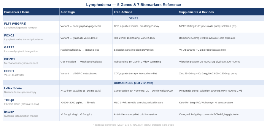
7 Biomarkers to Track in Lymphedema
Biomarkers are measurable biological signals that reflect what is happening inside your body. For lymphedema, they serve three purposes: early detection before visible swelling appears, monitoring of disease progression, and evaluation of how well your current treatment approach is working. The 7 markers below were selected for clinical relevance, practical measurability, and strength of evidence. They range from simple blood tests available at any laboratory to specialized devices requiring a clinical referral. Used together, they give a more complete picture than limb circumference alone.
1. L-Dex Score — Bioimpedance Spectroscopy (BIS)
Why it matters: The L-Dex score is the most direct clinical biomarker for subclinical lymphedema. Generated by bioimpedance spectroscopy, it measures the ratio of extracellular to intracellular fluid in the at-risk limb compared to the contralateral side. A value more than 10 points above your personal baseline is considered clinically significant. What makes BIS uniquely powerful is that it detects extracellular fluid accumulation months before visible swelling appears, providing a genuine early-intervention window. Prospective data published in Cancer demonstrated that BIS-guided surveillance in breast cancer patients significantly reduced rates of progression to chronic lymphedema compared to standard tape-measure monitoring — one of the few biomarker strategies in lymphedema with an actual randomized prospective trial supporting it.
How to measure it: The ImpediMed L-Dex U400 is the FDA-cleared device for this measurement. It is available at lymphedema clinics, some cancer centers, and physical therapy practices trained in oncology rehabilitation. Baseline should be established before any surgical procedure if possible, and tracked every 3–6 months thereafter. Cost: typically $50–$150 per session as part of a clinic visit.
If the score is bad — plan without supplements: Early BIS elevation (L-Dex +7 to +10 from baseline) is the critical intervention point. Immediate steps: fit for a gradient compression sleeve (30–40 mmHg) worn during waking hours, begin a Complete Decongestive Therapy (CDT) program with a certified lymphedema therapist, and start low-intensity aerobic exercise — walking or swimming 30 minutes, 5 days per week — which drives muscular pump activity without overloading lymphatic capacity. Elevate the affected limb above heart level for 20–30 minutes twice daily. Avoid prolonged heat exposure, tight clothing, and heavy lifting involving the affected limb.
If the score is bad — plan with supplements or equipment: - Sequential pneumatic compression device: multi-chamber devices (Flexitouch or similar) used 1–2 hours daily at home have strong evidence for reducing L-Dex scores and are FDA-cleared. Cost: $1,000–$5,000, often partially covered by insurance with a documented lymphedema diagnosis. - Selenium (selenomethionine): 200 mcg/day has shown benefit in reducing lymphedema volume in post-radiation patients. Selenium is a cofactor for glutathione peroxidase with anti-inflammatory and anti-fibrotic properties. Cycle: 3–6 months, then reassess. Do not exceed 400 mcg/day due to toxicity risk. - Micronized Purified Flavonoid Fraction (MPFF / Daflon): diosmin/hesperidin combination at 500–1000 mg twice daily improves lymphatic vessel tone and reduces protein-rich edema. Well-tolerated with minimal side effects. Cycle: 3–6 months with one-month breaks.
2. VEGF-C — Vascular Endothelial Growth Factor C
Why it matters: VEGF-C is the primary driver of lymphangiogenesis — the growth and maintenance of lymphatic vessels. It signals through the VEGFR-3 receptor (encoded by FLT4) on lymphatic endothelial cells to build and repair lymphatic networks. When VEGF-C is chronically low, the lymphatic system loses its capacity to regenerate after injury or surgical disruption. This is particularly relevant in secondary lymphedema following cancer surgery, where damaged lymphatic anatomy requires new collateral vessel formation to compensate. Studies have shown that patients with lower plasma VEGF-C after axillary lymph node dissection have higher rates of lymphedema development at two-year follow-up (PubMed).
How to measure it: Measured via ELISA assay from a standard blood draw. Reference ranges vary by lab, but most studies use 500–1000 pg/mL as a clinically meaningful range. Available through research labs and an increasing number of specialized oncology centers. Cost: $80–$200. Measure at baseline and every 6 months if at risk.
If the score is bad — plan without supplements: Moderate aerobic exercise is the most consistent non-pharmacological driver of endogenous VEGF-C production. Zone 2 cardio (60–70% of maximum heart rate), 150–180 minutes per week, stimulates lymphatic endothelial signaling and upregulates lymphangiogenic pathways. Resistance training at moderate load (not heavy lifting in the affected limb) also activates VEGF-C production. Avoid sedentary periods longer than 90 minutes — intermittent movement throughout the day sustains baseline lymphangiogenic signaling.
If the score is bad — plan with supplements or equipment: - Quercetin: 500–1000 mg/day has shown VEGF-C upregulation in endothelial cell models alongside documented anti-inflammatory effects. Human trials remain limited but the safety profile is strong. Take with bromelain for improved absorption. - Red clover isoflavones: modest evidence suggests phytoestrogens support lymphangiogenic signaling in post-menopausal women with lymphedema. Discuss with a physician if hormone-sensitive conditions are present. - High-intensity interval training (HIIT) twice weekly: provides a strong VEGF-C stimulus through high-magnitude shear stress on vascular endothelium. Two sessions per week (20 minutes each, work-rest intervals of 30 seconds on, 90 seconds off) complement Zone 2 base training.
3. High-Sensitivity CRP (hsCRP)
Why it matters: Chronic low-grade inflammation is both a driver and a consequence of lymphedema. The protein-rich interstitial fluid accumulating in lymphedematous tissue creates a pro-inflammatory microenvironment that, in turn, accelerates fibrosis and impairs lymphangion contractility. High-sensitivity CRP is a reliable, affordable marker of systemic inflammation — one of the key biomarkers Peter Attia recommends for long-term health tracking — and it is equally relevant here because inflammation drives the fibrotic progression that converts reversible edema into permanent tissue change. Studies of patients with breast cancer-related lymphedema consistently show elevated hsCRP compared to controls without lymphedema (PubMed), and reducing hsCRP through lifestyle interventions correlates with reduced lymphedema severity in observational data.
How to measure it: Standard blood test available at any laboratory. Target: below 1.0 mg/L (optimal); 1–3 mg/L (elevated risk); above 3.0 mg/L (high inflammation). Cost: $15–$40. Measure every 3–6 months when managing active lymphedema.
If the score is bad — plan without supplements: Anti-inflammatory lifestyle protocol: eliminate processed seed oils (linoleic acid-rich oils), reduce refined carbohydrates, prioritize 7–9 hours of sleep (the glymphatic and lymphatic systems clear inflammatory debris during rest), and implement stress management consistently. Cold water immersion at 50–60°F (10–15°C) for 2–5 minutes, 3–5 times per week, has robust evidence for reducing systemic inflammatory markers. Walking 30 minutes after meals blunts postprandial inflammation and activates the muscular lymphatic pump.
If the score is bad — plan with supplements or equipment: - Omega-3 fatty acids (EPA + DHA): 2–4 grams/day from high-quality fish oil — one of the most evidence-backed anti-inflammatory interventions available and a consistent recommendation from Peter Attia. Allow 6–12 weeks to see measurable hsCRP reduction. No cycling needed for long-term use; monitor LDL-C at higher doses. - Curcumin (phospholipid-complexed, BCM-95 or Meriva): 400–500 mg twice daily. Inhibits NF-κB and reduces pro-inflammatory cytokines including IL-1β and TNF-α. Some studies show 40–50% hsCRP reduction over 8 weeks in inflammatory conditions. Take with food. Cycle: 3–6 months, then one-month break. - Magnesium glycinate: 300–400 mg at night. Magnesium deficiency is strongly linked to elevated CRP. The glycinate form has the best tolerability. Long-term use is safe when kidney function is normal.
4. Interleukin-6 (IL-6)
Why it matters: IL-6 is a key pro-inflammatory cytokine with a dual role in lymphedema. It promotes the inflammatory cascade that drives lymphatic endothelial dysfunction, and it activates the STAT3 signaling pathway responsible for fibroblast-to-myofibroblast transformation — the cellular mechanism that turns reversible edema into permanent fibrotic tissue. Elevated IL-6 in lymphedematous tissue has been documented in multiple human studies (PubMed) and correlates with both severity of swelling and the rate of fibrotic progression. Tracking IL-6 alongside hsCRP identifies patients whose inflammatory driver has not been adequately addressed by current treatment.
How to measure it: Serum IL-6 via standard blood test. Normal range typically below 7 pg/mL (lab reference ranges vary). Available from commercial labs including LabCorp and Quest. Cost: $30–$80. Pair with hsCRP for a fuller inflammatory profile.
If the score is bad — plan without supplements: IL-6 responds strongly to consistent aerobic exercise. Skeletal muscle produces IL-6 during exercise, but this produces downstream anti-inflammatory effects by upregulating IL-10 and IL-1Ra. Five days per week of 30-minute moderate walks consistently lowers resting IL-6 over 8–12 weeks. Reduce visceral adiposity through caloric moderation and time-restricted eating (10–12 hour eating window) — visceral fat is a major IL-6 source independent of edema. Sleep deprivation acutely spikes IL-6; 7–9 hours is non-negotiable.
If the score is bad — plan with supplements or equipment: - Boswellia serrata (AKBA extract): 100–200 mg of AKBA — specifically inhibits the 5-lipoxygenase (5-LOX) pathway and reduces IL-6 in inflammatory conditions. Evidence in inflammatory arthritis and bowel disease is solid; lymphedema-specific trials are limited but the mechanism is directly relevant. Cycle: 6–12 weeks with two-week breaks. - Resveratrol (trans-resveratrol): 200–500 mg/day — inhibits NF-κB and reduces IL-6 in multiple clinical studies. Take with a fat-containing meal or in liposomal form for absorption. Cycle: 12 weeks. - Infrared sauna: 120–140°F, 15–20 minutes, 3–4 times per week. Regular sauna use has been associated with significant reductions in IL-6 and systemic inflammatory markers, and the heat-stress response upregulates heat shock proteins with lymphoprotective effects.
5. TGF-β1 — Transforming Growth Factor Beta-1
Why it matters: TGF-β1 is arguably the most important single biomarker for predicting whether a patient's lymphedema will progress to irreversible fibrosis. It is the master regulator of fibrosis in most tissues, and its role in lymphedema is well-established: elevated TGF-β1 drives conversion of lymphatic endothelial cells into a dysfunctional phenotype, promotes fibroblast activation, and degrades the extracellular matrix in ways that block lymphatic regeneration (PubMed). Research by Rockson's group at Stanford has demonstrated that TGF-β1 blockade in animal models of lymphedema dramatically reduces fibrotic progression. While direct anti-TGF-β1 therapy is not yet clinically available, measuring TGF-β1 identifies the patients for whom anti-fibrotic strategies should be implemented urgently — before the Stage II to Stage III transition becomes visible.
How to measure it: Plasma TGF-β1 via ELISA blood test. Normal range approximately 200–2000 pg/mL (highly lab-dependent). Elevated values above 2000–3000 pg/mL in most lab contexts signal active fibrotic signaling. Available through specialized and academic laboratories. Cost: $100–$200. Trending over 6 months is more valuable than a single time point.
If the score is bad — plan without supplements: TGF-β1 signaling in lymphatic tissue is reduced by regular aerobic exercise, as mechanotransduction from lymph flow mechanically downregulates TGF-β1 gene expression in lymphatic endothelium. Manual lymphatic drainage (MLD) performed by a certified therapist twice weekly has been shown to reduce fibrotic markers in connective tissue. Strict skin care is critical: even minor scratches, sunburn, or insect bites in the affected limb activate TGF-β1 through the wound-healing cascade. Any dermal inflammatory trigger should be minimized aggressively.
If the score is bad — plan with supplements or equipment: - Ketotifen (prescription): a mast cell stabilizer that has been specifically studied for lymphedema by Rockson's group. Mast cells are a key source of TGF-β1 in lymphedematous tissue. Dose: 1 mg twice daily. Side effect: sedation, particularly in the first weeks. Requires a physician's prescription; this is one of the most promising pharmacological leads for anti-fibrotic lymphedema management. - Systemic enzyme therapy (Wobenzym N): contains bromelain, papain, rutin, and trypsin. European studies support benefit in post-surgical edema with fibrotic components. 2–3 tablets 3 times daily on an empty stomach. Avoid if on anticoagulants. - Serrapeptase + Nattokinase: 100 mg serrapeptase on empty stomach in the morning; 100 mg nattokinase 30 minutes before meals. Both have fibrinolytic and connective-tissue anti-fibrotic activity. Evidence in lymphedema specifically is preliminary. Three-month trial, with monitoring for any unusual bruising.
6. Tissue Dielectric Constant (TDC)
Why it matters: The Tissue Dielectric Constant measures the water content of superficial tissue non-invasively using a dielectric probe placed against the skin. In lymphedema, accumulation of protein-rich interstitial fluid increases the TDC value relative to the contralateral limb. A TDC ratio greater than 1.20 between affected and unaffected sides is considered clinically significant. TDC adds something that L-Dex cannot provide: spatial resolution — you can map values at multiple points along the limb, identifying exactly where fluid accumulation is most concentrated (PubMed). This is particularly useful for monitoring localized treatment response and identifying areas that require more focused manual drainage.
How to measure it: Measured with the MoistureMeterD or similar dielectric probe devices. Available in lymphedema therapy clinics and research centers. Some therapists use it alongside BIS as a combined early-detection protocol. Cost: $100–$200 per evaluation. Not widely reimbursed but increasingly available at academic lymphedema centers.
If the score is bad — plan without supplements: Elevated TDC responds most rapidly to intensive CDT. During the intensive phase (first 2–4 weeks of treatment), focus on: supervised MLD 5 days per week, multi-layer compression bandaging applied between sessions, daily skin care to maintain barrier integrity (a damaged skin barrier facilitates additional fluid egress), and segment-specific exercises taught by your lymphedema therapist to activate the lymphangion pump in the highest-TDC areas.
If the score is bad — plan with supplements or equipment: - Sequential pneumatic compression pump: 60–90 minutes daily. Multi-chamber pumps mimicking natural lymphatic flow direction are superior to single-chamber devices. Shown in multiple RCTs to reduce TDC by improving lymphatic transit velocity. - Kinesiology taping (Kinesio Tape): applied in specific lymphedema patterns over the affected limb between therapy sessions. Studies show TDC reduction of approximately 8–12% over 4 weeks with regular application. Cost: $20–$40 for materials. Application technique must be learned from a trained lymphedema therapist to be effective.
7. Lymphocyte-to-Neutrophil Ratio and Immune Panel
Why it matters: Lymphedema is not purely a mechanical drainage problem — it is a profoundly immunological one. The lymphatic system is central to immune surveillance, and disruption of lymphatic flow creates aberrant T-cell trafficking and innate immune dysregulation. Research has demonstrated that CD4+ Th2 cells accumulate specifically in lymphedematous tissue and drive the inflammatory-fibrotic cascade through IL-4 and IL-13 secretion — creating a distinct, self-sustaining immune phenotype in affected limbs (PubMed). The lymphocyte-to-neutrophil ratio (LNR) from a standard complete blood count provides an accessible proxy for immune balance. A low LNR signals immune stress or inflammatory dominance.
How to measure it: LNR is derived from a standard CBC: lymphocytes divided by neutrophils. Normal LNR approximately 1.5–3.0. For deeper profiling: CD4/CD8 ratio and NK cell count from a lymphocyte subset panel. Cost: CBC $15–$30; lymphocyte subsets $80–$150. Measure every 6 months alongside other inflammatory markers.
If the score is bad — plan without supplements: Restore immune balance through consistent aerobic exercise (shifts the Th1/Th2 balance toward homeostasis), 7–9 hours of sleep (NK cell activity decreases sharply with even one night of poor sleep), and reduction of chronic psychological stress (cortisol chronically suppresses lymphocyte counts). Protect gut health aggressively — the gut-associated lymphoid tissue (GALT) accounts for approximately 70% of total immune system activity, and disrupted gut microbiome diversity directly impairs lymphocyte differentiation.
If the score is bad — plan with supplements or equipment: - Vitamin D3 + K2: target serum 25(OH)D between 50–80 ng/mL. Vitamin D is a potent immunomodulator, reducing Th2 dominance and supporting regulatory T-cell (Treg) function directly relevant to lymphedema's immune phenotype. Dose: 3,000–5,000 IU D3 daily with 100 mcg MK-7 K2. Test before and 3 months after starting to avoid hypercalcemia. - Zinc glycinate or zinc picolinate: 15–30 mg/day. Zinc deficiency impairs T-cell development and NK cell activity. Do not exceed 40 mg/day long-term (risk of copper depletion); add 2 mg copper if using for more than 3 months. - Medicinal mushroom beta-glucans: Lion's mane (1–3 g/day) or reishi (1–2 g/day) from dual-extracted preparations have documented immunomodulatory effects, specifically supporting NK cell activity and T-cell balance toward a less Th2-dominant phenotype. Cycle: 12 weeks on, 2–4 weeks off.
5 Key Genes That Shape Lymphatic Function
Understanding the genetic architecture of lymphedema explains why some people develop the condition after relatively minor surgical disruption while others tolerate significant lymph node removal with no lasting consequences. The genes below include those directly responsible for primary hereditary lymphedema syndromes and those whose variants modulate risk and treatment response across broader populations. Even if you have secondary lymphedema, genetic context can clarify why your biological response has been more severe or treatment-resistant than expected.
Gene 1: FLT4 (VEGFR3) — The Lymphangiogenesis Receptor
FLT4 encodes VEGFR-3, the primary receptor for VEGF-C and VEGF-D on lymphatic endothelial cells. Mutations in FLT4 cause Milroy disease (hereditary lymphedema type I), characterized by bilateral lower limb lymphedema from birth or early childhood. The causal link was established in Nature Genetics (PubMed). Beyond Milroy disease, reduced-function FLT4 variants have been identified in patients with secondary lymphedema who respond poorly to standard CDT. When VEGFR-3 signaling is impaired, lymphatic endothelial cells cannot respond adequately to VEGF-C, preventing new vessel formation and compromising lymphatic valve integrity.
If the gene is bad — plan without supplements: Since the regenerative lymphangiogenesis pathway is impaired, physical therapies that mechanically drive lymph flow become essential compensations: CDT with compression bandaging, daily aquatic exercise (water pressure provides natural full-body lymphatic support), and diaphragmatic breathing exercises 10–15 minutes twice daily to engage the thoracic pump. Avoid prolonged limb dependency (no standing or sitting for more than 90 minutes). Elevate the limb 8–12 inches above heart level at night.
If the gene is bad — plan with supplements or equipment: - MPFF (Daflon 500): diosmin/hesperidin 500 mg twice daily supports lymphatic vessel tone through mechanisms independent of VEGFR-3 transcription — relevant when the receptor pathway itself is compromised. Cycle: 3–6 months. - Multi-chamber pneumatic compression device: when lymphangiogenesis is structurally limited, mechanical assistance is not optional. Daily use of 1–2 hours replaces what impaired vessel formation cannot provide. - Ketotifen (with physician guidance): mast cell stabilization reduces the inflammatory load on lymphatic endothelium, partially compensating for impaired VEGFR-3 signaling in the surrounding microenvironment. 1 mg twice daily as prescribed.
Gene 2: FOXC2 — The Lymphatic Valve Transcription Factor
FOXC2 encodes a transcription factor essential for lymphatic valve development and proper differentiation of lymphatic endothelial cells. Mutations in FOXC2 cause lymphedema-distichiasis syndrome — lymphedema combined with an extra row of eyelashes — as identified by Fang et al. (PubMed). Beyond this syndrome, FOXC2 variants are associated with varicose veins, venous insufficiency, and late-onset lymphedema. Importantly, FOXC2 expression is regulated by the AMPK signaling pathway — which responds to exercise and caloric restriction — making this one of the few lymphedema-relevant genes with modifiable epigenetic expression through lifestyle.
If the gene is bad — plan without supplements: Activate FOXC2 expression through AMPK pathway stimulation: intermittent fasting (16:8 minimum) activates AMPK and has downstream effects on FOXC2 expression in endothelial tissues. High-intensity interval training (HIIT), two sessions per week, provides strong AMPK activation. Avoid chronic hyperglycemia — elevated blood glucose suppresses FOXC2 expression through insulin signaling interference. Consistent daily aerobic exercise is the other major AMPK driver.
If the gene is bad — plan with supplements or equipment: - Berberine: 500 mg 2–3 times daily before meals. Berberine is one of the most potent natural AMPK activators, comparable in several studies to metformin. It activates FOXC2 indirectly through AMPK signaling. Start at 250 mg to minimize GI side effects and increase gradually. Cycle: 12 weeks on, 4 weeks off. Do not combine with metformin without physician guidance. - Resveratrol: 200–500 mg/day activates SIRT1, which works synergistically with AMPK to upregulate FOXC2 in endothelial tissue. Take with a fat-containing meal or in liposomal formulation. - Cold exposure: cold water immersion or 1–3 minute cold showers at 50–60°F activate AMPK and epigenetically support genes governing vascular endothelial function. Three to five times per week.
Gene 3: GATA2 — Immune-Lymphatic Integration
GATA2 encodes a transcription factor critical for both blood cell formation (hematopoiesis) and lymphatic vascular development. GATA2 haploinsufficiency causes Emberger syndrome: primary lower limb lymphedema combined with severe immunodeficiency, susceptibility to myelodysplastic syndrome, and recurrent infections (PubMed). Beyond Emberger syndrome, partial GATA2 loss-of-function variants are increasingly identified in patients with lymphedema complicated by unusual infections or atypical inflammatory responses. The immune component makes GATA2-related lymphedema a distinct clinical challenge: you may be simultaneously managing lymphatic drainage failure and an underlying immunological vulnerability.
If the gene is bad — plan without supplements: Strict infection prevention is the priority. Meticulous skin care to prevent even minor abrasions in at-risk areas — cellulitis in the setting of GATA2 dysfunction can be disproportionately severe and rapid in progression. Prompt antibiotic treatment at the very first signs of infection is essential. Avoid skin procedures (acupuncture, tattoos, manicures, blood draws) in affected limbs. If a GATA2 variant is confirmed, co-management with both a lymphedema therapist and a hematologist or immunologist is strongly recommended.
If the gene is bad — plan with supplements or equipment: - Vitamin D3 (3,000–5,000 IU daily) and Vitamin C (500–1,000 mg twice daily, buffered form): foundational immune support in documented immunodeficiency. Monitor serum 25(OH)D levels; target 50–80 ng/mL. - High-diversity probiotics (50–100 billion CFU, multi-strain) combined with prebiotic fiber (inulin or FOS, 5–10 g/day): given that GATA2 affects gut-associated immune tissue, maintaining a robust and diverse gut microbiome is a long-term priority, not a short-term fix. Requires 3–6 months minimum for measurable microbiome shifts. - Emergency antibiotic standby (physician-prescribed): for confirmed GATA2 mutations with documented immunodeficiency, maintaining a ready antibiotic course (e.g., amoxicillin-clavulanate) for early cellulitis management is critical medical infrastructure. This is a discussion to have with your prescribing physician, not a self-managed strategy.
Gene 4: PIEZO1 — The Mechanosensory Ion Channel
PIEZO1 is a mechanosensitive ion channel expressed in endothelial cells that converts physical forces — shear stress and pressure — into cellular signals. Gain-of-function mutations in PIEZO1 cause hereditary lymphatic dysplasia type 3, where lymphatic vessels fail to regress normally during development (PubMed). Beyond this syndrome, PIEZO1 variants affect how endothelial cells respond to exercise-driven shear stress, with direct implications for how well lymphatic vessels adapt to physical loading. What makes PIEZO1 uniquely interesting is that it is directly activated by mechanical stimulation — meaning that how you move, exercise, and apply pressure to the affected limb has molecular consequences at the PIEZO1 level.
If the gene is bad — plan without supplements: Exploit PIEZO1's mechanosensitivity directly. Rhythmic mechanical compression through exercise — especially walking, swimming, and rebounding on a mini-trampoline — activates PIEZO1 signaling in lymphatic endothelial cells. Rebounding (10–20 minutes twice daily) is particularly effective because the gravitational changes at each bounce create optimal mechanical stimulation for lymphatic propulsion. Whole-body vibration platforms (25–50 Hz) provide an alternative mechanical stimulus for those unable to rebound, with a 2021 trial showing significant lymphedema volume reduction with vibration therapy.
If the gene is bad — plan with supplements or equipment: - Whole-body vibration platform (Power Plate or equivalent, 25–50 Hz, low amplitude): 1–3 minutes per session, 2–3 times per week. Provides mechanical stimulation that activates PIEZO1 in lymphatic endothelium. Cost: $300–$5,000 depending on device quality; clinical units are available in some physiotherapy centers. - Magnesium glycinate: 300–400 mg at night. PIEZO1 channel function is influenced by cellular magnesium concentration. Ensuring optimal magnesium status (red blood cell magnesium 5.5–6.5 mg/dL) supports normal mechanosensory signaling. Long-term use is safe with normal kidney function. - Mini-trampoline (rebounder): $50–$250; a low-cost mechanical therapy specifically targeting PIEZO1 activation. Ten to twenty minutes twice daily. Begin gently, wearing compression, and progress as limb tolerance improves.
Gene 5: CCBE1 — The VEGF-C Activator
CCBE1 (Collagen- and Calcium-Binding EGF Domains 1) encodes a protein required for the proteolytic activation of VEGF-C. Without functional CCBE1, VEGF-C cannot be fully processed and cannot signal through VEGFR-3. Biallelic CCBE1 mutations cause Hennekam syndrome (generalized lymphatic dysplasia) — one of the most severe forms of primary lymphedema (PubMed). Heterozygous carriers with one abnormal copy may have subtle lymphatic functional impairment that manifests as an exaggerated response to secondary lymphedema triggers such as surgery or radiation. CCBE1 also works in concert with ADAMTS3, a zinc-dependent metalloproteinase required for VEGF-C processing — directly linking zinc nutritional status to this gene's downstream function.
If the gene is bad — plan without supplements: Since CCBE1 dysfunction impairs VEGF-C activation — the same lymphangiogenic pathway as FLT4 — the physical management priorities are closely aligned: maximize mechanical lymph transport through CDT, aquatic therapy, and diaphragmatic breathing; reduce lymphatic load through a low-sodium diet and avoiding prolonged limb dependency. Work with a lymphedema specialist to establish a baseline lymphoscintigraphy to map the full extent of lymphatic dysfunction and identify functional collateral pathways.
If the gene is bad — plan with supplements or equipment: - Zinc bisglycinate (25–30 mg) plus 2 mg copper: ADAMTS3, the metalloproteinase that collaborates with CCBE1 to activate VEGF-C, is zinc-dependent. Maintaining optimal zinc status may partially support VEGF-C activation downstream. Monitor serum zinc and copper levels at baseline and 3 months. - N-Acetylcysteine (NAC): 600–1,200 mg/day. NAC reduces oxidative stress in the extracellular matrix, which can impair metalloproteinase function broadly. Supporting ECM redox homeostasis indirectly supports CCBE1/ADAMTS3 activity. Well-tolerated long-term. Cycle: 8–12 weeks on, 2–4 weeks off. - Daily pneumatic compression device combined with weekly professional MLD: when the lymphangiogenic mechanism is impaired at the VEGF-C activation step, the mechanical transport pathway is the primary compensation. This combination is the therapeutic cornerstone.
What the Science of Sleep and Movement Reveals About Lymphatic Flow
Andrew Huberman has discussed the lymphatic and glymphatic systems across multiple Huberman Lab episodes, particularly in the context of sleep, inflammation, and exercise physiology. While not exclusively focused on lymphedema, the convergence of his research discussions provides a powerful mechanistic framework for understanding how daily choices affect lymphatic function at a cellular level. The following are 10 of the most impactful insights directly relevant to lymphedema management.
1. Sleep Is the Single Most Powerful Driver of Lymphatic Clearance
The brain's lymphatic system — the glymphatic system — is up to 10 times more active during sleep than during waking hours. During slow-wave sleep (deep NREM), interstitial space expands and cerebrospinal fluid flushes inflammatory debris. The same principle governs peripheral lymphatics: most lymph flow regulation and inflammatory clearance occurs during rest. Chronic sleep deprivation of even 1–2 hours disrupts lymphatic drainage cycles throughout the body. For lymphedema patients, protecting sleep quality is not optional — it is part of the treatment.
2. Sleeping Position Affects Lymphatic Clearance Efficiency
Research cited in Huberman Lab content found that lateral (side) sleeping significantly improves glymphatic clearance efficiency compared to supine or prone positions. For lymphedema patients with lower limb involvement, side sleeping with the affected limb slightly elevated by a small pillow between the knees may optimize both local limb drainage and systemic lymphatic activity simultaneously.
3. Nasal Breathing Activates the Thoracic Lymphatic Pump
Diaphragmatic breathing driven by nasal inhalation creates rhythmic pressure differentials in the thoracic cavity that drive lymph through the thoracic duct and into venous circulation. Habitual mouth breathing reduces this mechanical pumping effect. For lymphedema patients, 10 minutes of deliberate slow nasal breathing (5-second inhale, 5-second exhale) twice daily activates the thoracic lymphatic pump without any limb loading.
4. Morning Sunlight Resets the Circadian Clock That Governs Lymphatic Rhythm
Lymphatic transport follows a circadian rhythm, peaking during active periods and declining during rest — regulated by core circadian clock genes. Ten to twenty minutes of outdoor light exposure within the first hour of waking sets the circadian clock via the suprachiasmatic nucleus. For lymphedema patients, aligning therapy sessions, exercise, and compression wear with the body's natural lymphatic peak (mid-morning) may measurably improve treatment outcomes.
5. Zone 2 Cardio Is the Optimal Stimulus for Sustained Lymph Flow
Zone 2 aerobic training (conversational pace, 60–70% maximum heart rate) drives continuous lymphatic pumping through muscular contractions without generating excessive inflammatory metabolites or swelling-provoking venous pressure. This is the sweet spot for lymphedema patients who need to support lymph flow without triggering limb overload. 45 minutes of Zone 2 cardio, 4–5 times per week, represents the minimum effective dose for meaningful lymphatic benefit.
6. Cold Exposure Reduces Systemic Inflammatory Load on Lymphatic Tissue
Cold water immersion at 50–60°F reduces IL-6 and pro-inflammatory cytokines acutely and chronically over time. Eleven minutes total per week (in 1–3 minute sessions) produces significant anti-inflammatory effects. For lymphedema patients, avoid total immersion of severely swollen limbs in cold water — temperature extremes can trigger reactive vasodilation — but upper-body cold exposure or face immersion provides the systemic benefit while protecting the affected limb.
7. Heat Stress Upregulates Lymphangiogenic Growth Factor Expression
Heat stress from sauna use upregulates heat shock proteins and increases VEGF expression broadly in endothelial tissue. For lymphedema, sauna use should be discussed with a therapist: heat can temporarily worsen acute swelling, but in stable, well-controlled lymphedema, 15–20 minutes at 175–200°F three to four times per week may support VEGF-C signaling and provide systemic anti-inflammatory benefits.
8. Chronic Stress Hormones Directly Impair Lymphatic Flow
Chronically elevated cortisol disrupts lymphatic endothelial barrier function and promotes a pro-fibrotic cellular environment. The physiological sigh — a double nasal inhale followed by a long, slow exhale — rapidly activates the parasympathetic nervous system and is one of the fastest-acting acute stress-reduction techniques available. For lymphedema patients, chronic psychological stress is an under-recognized driver of inflammatory and fibrotic progression, and managing it is part of managing the condition.
9. Resistance Training in Compression Does Not Worsen Lymphedema
Fear of exercise-induced swelling leads many lymphedema patients to avoid resistance training entirely — a decision supported by outdated guidance. Multiple RCTs now confirm that supervised resistance training performed while wearing compression garments does not worsen lymphedema and may improve lymphatic contractility and quality of life. Progressive loading over 8–12 weeks at moderate intensity is the safe and evidence-based approach.
10. Gut Microbiome Diversity Determines the Systemic Inflammation Setpoint
The gut-associated lymphoid tissue (GALT) is the largest lymphatic organ in the body. Disruption of gut microbiome diversity impairs lymphatic immune function and raises the systemic inflammatory baseline that accelerates lymphedema progression. Fermented foods (2–4 servings daily) and dietary diversity (30+ different plant species per week) represent the most consistently supported microbiome interventions in current research — with direct downstream effects on lymphatic immune homeostasis.
Complementary Approaches With Clinical Support
The following four modalities were selected specifically for meaningful clinical evidence in lymphedema, not general wellness claims. Each has at least one human study, RCT, or evidence-based guideline supporting its use in this condition.
Manual Lymphatic Drainage (MLD)
Manual Lymphatic Drainage is a specialized massage technique developed by Emil Vodder specifically to stimulate lymphatic vessel contractions and redirect lymph around blocked or damaged areas. Unlike conventional massage, MLD uses extremely light pressure — approximately 30 mmHg — applied in slow, rhythmic, circular motions following the anatomical lymphatic pathways. It is the most evidence-backed complementary intervention in lymphedema and forms the core of Complete Decongestive Therapy (CDT) alongside compression bandaging.
Systematic reviews and multiple RCTs confirm that MLD combined with compression bandaging produces significantly greater volume reduction than compression alone (PubMed). The International Society of Lymphology consensus document recognizes MLD as a standard adjunct in breast cancer-related lymphedema management. Both the Casley-Smith and Foldi schools have published specific protocols for upper and lower limb applications with documented outcomes.
In practice: seek a therapist certified by the Lymphology Association of North America (LANA) or an equivalent national body. Intensive phase: 5 sessions per week for 2–4 weeks. Maintenance phase: 1–2 professional sessions per month, supported by 15–20 minutes of self-MLD daily. Your therapist should teach self-MLD technique. Performing MLD approximately 30 minutes before exercise enhances lymphatic capacity for the increased lymph load that activity generates.
Low-Level Laser Therapy (LLLT) / Photobiomodulation
LLLT uses specific light wavelengths (630–1000 nm) at low power density to stimulate mitochondrial activity, reduce inflammation, and promote lymphangiogenesis in damaged lymphatic tissue. In lymphedema, it targets fibrotic obstruction of lymphatic channels and supports regeneration of damaged lymphatic endothelium by increasing ATP production and nitric oxide via cytochrome c oxidase activation.
A landmark RCT by Carati et al. published in Cancer demonstrated that LLLT produced significant reductions in arm volume in breast cancer-related lymphedema compared to sham treatment (PubMed), with 31% of treated subjects achieving sustained reduction. A subsequent meta-analysis in Breast Cancer Research and Treatment further supported LLLT for upper limb lymphedema, with consistent findings across trials.
Practical application: LLLT should be administered by a trained practitioner using 830 nm wavelength, approximately 100 mW, applied to the affected limb and regional lymph nodes in 10–15 minute sessions, 2–3 times per week for 3–4 weeks. At-home photobiomodulation panels can supplement clinic treatment at lower intensity. Cost: $50–$150 per clinic session; home devices $200–$800. LLLT is most effective during the early inflammatory stage and less effective for advanced fibrotic lymphedema. It is contraindicated directly over active cancer sites.
Breathing-Based Therapies
Structured breathing exercises exploit the mechanics of the thoracic lymphatic pump: the pressure differentials created by diaphragmatic movement drive lymph through the thoracic duct and back into venous circulation. The thoracic duct returns approximately 3 liters of lymph per day to the bloodstream, and this flow is substantially dependent on the pressure gradients generated by each breath cycle.
A 2011 study demonstrated improvements in lymphatic transport in patients who practiced structured abdominal breathing for 20 minutes daily (PubMed). The physiological sigh — a double nasal inhale followed by a long extended exhale — maximally inflates the alveoli and creates the strongest thoracic pump stimulus available without equipment.
Practical protocol: 10–15 minutes of slow diaphragmatic breathing (5-count inhale through the nose, brief pause, 7-count exhale through pursed lips) twice daily — once in the morning and once before evening compression removal. During MLD sessions, breathing is coordinated with drainage strokes to amplify the drainage effect. For lower limb lymphedema, add 5 physiological sighs every 30–60 minutes during sedentary work to sustain intermittent thoracic pump activation throughout the day.
Yoga
Yoga for lymphedema combines multiple beneficial mechanisms: muscle contractions that drive lymphatic flow, deep breathing that activates the thoracic pump, gravity-assisted positions that facilitate lymph drainage, and stress reduction that lowers cortisol and systemic inflammation. Several trials have examined yoga specifically in breast cancer-related lymphedema with objective outcome measures.
A pilot RCT published in Cancer found that breast cancer survivors with lymphedema who completed an 8-week yoga program showed improvements in arm volume and lymphedema symptom burden compared to controls (PubMed). The evidence base is still limited in scale but methodologically sound and consistent with the known physiological mechanisms.
Practical application: seek an instructor with oncology yoga training (CYT-500 or IAYT-credentialed). Always wear compression during practice. Avoid heated yoga studios — temperature extremes can transiently worsen swelling. Prioritize poses that elevate the affected limb (legs up the wall, supine leg raises), integrate slow coordinated breathing throughout, and avoid inversions or positions that concentrate pressure on at-risk areas. A 30-minute gentle flow with these modifications, 3–5 times per week, is a realistic and evidence-consistent maintenance protocol.
Conclusion
Lymphedema responds better to management when you understand what is actually driving it in your case. Whether your primary challenge is impaired lymphangiogenesis (low VEGF-C, FLT4 variant), fibrotic progression (elevated TGF-β1, persistent IL-6), immune dysregulation (GATA2, low lymphocyte ratio), or inadequate early detection (L-Dex not yet tracked), the biomarkers and genes covered here give you a far more specific target than limb circumference measurements alone.
No single intervention works for everyone, and this article is not a substitute for working with a certified lymphedema therapist and a physician who understands your full clinical picture. What it offers is a framework: identify the right signals, understand what each one is telling you, and match your interventions — lifestyle, physical, nutritional, and where appropriate pharmacological — to what your biology is actually doing right now.
The practical next step is identifying which of these biomarkers you can access and establishing a baseline. Start with what is most affordable and available: hsCRP, IL-6, a standard CBC for LNR, and a conversation with your care team about L-Dex monitoring if you are post-surgical or at established risk. Build your picture from there. Better data leads to better decisions, and better decisions applied consistently over time produce better outcomes.
Cardiovascular: Vascular Conditions
Autoimmune: Inflammatory Conditions Connective Tissue Conditions