This article contains AI generated content.
· UpdatedLymphocytic Vasculitis: 7 Biomarkers and 5 Genes To Track
Introduction
Lymphocytic vasculitis is not the kind of diagnosis that comes with a clear roadmap. It arrives — often through a skin biopsy, or after months of unexplained symptoms — and suddenly you are holding a histological finding that describes what is happening in your vessel walls, but says almost nothing about why. That gap between a label and an explanation is where most patients spend far too much time.
What makes this condition particularly difficult is how many different things it can represent. In some people, lymphocytic vasculitis is a mild, recurring skin-limited process. In others, it signals an active autoimmune disease — lupus, Sjögren's syndrome, mixed connective tissue disease — or a systemic trigger like cryoglobulinemia driven by chronic hepatitis C. The same histological pattern can have dramatically different causes, which is why generic protocols often fall short. A treatment or lifestyle strategy that works well for one root cause may be irrelevant — or even counterproductive — for another.
That is where objective data becomes genuinely useful. Tracking specific biomarkers allows you to see your inflammatory burden quantitatively, monitor whether your interventions are working, and detect flares before they peak. Understanding your genetic landscape adds another layer: certain variants are now known to significantly alter immune regulation in ways that predispose to exactly the kind of lymphocytic-driven vascular inflammation seen in this condition. This is not fringe science — it is the direction that evidence-based personalized medicine is moving.
This article walks through both dimensions in practical terms. The biomarker section covers the seven most informative tests — why each one matters for lymphocytic vasculitis specifically, how to get them measured affordably, and what to do when results are out of range. The genetics section explores five key variants linked to autoimmune susceptibility and inflammatory dysregulation, with actionable compensation strategies for each. Further sections cover evidence-based complementary approaches and summarize one of the most important books in autoimmune research. Better information does not guarantee remission, but it gives you something far more valuable than generic advice: a basis for making smarter decisions.
Summary
This article is structured around four layers of information that standard medical consultations rarely combine. First, 7 biomarkers you can actually track — including hsCRP, ESR, cryoglobulins, the ANA panel, complement levels (C3/C4), lymphocyte subsets, and IL-6 — each explained with cost ranges, optimal targets, and specific plans (with and without supplements) for when results are abnormal. Second, 5 genes — HLA-DRB1, PTPN22, IRF5, STAT4, and TNFAIP3 — that research has linked to autoimmune vasculitis risk, including practical compensation strategies for each variant. Third, a summary of Sarah Ballantyne's The Paleo Approach, distilled into 10 high-impact insights on reversing autoimmune inflammation through diet, gut repair, and lifestyle change — with mechanisms most physicians do not discuss. Finally, four complementary modalities with actual human evidence for inflammatory and autoimmune conditions: the autoimmune protocol, mindfulness-based stress reduction, microbiome-directed therapies, and breathing-based techniques. If you have been cycling through standard treatments without a clear sense of what is actually driving your inflammation, the combination of biomarker tracking and genetic awareness described here may be the most useful reframe you have encountered.
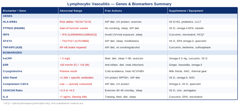
7 Biomarkers to Track in Lymphocytic Vasculitis
Understanding your inflammatory status through objective measurement is one of the most practical things you can do when managing a condition as variable as lymphocytic vasculitis. These seven biomarkers offer a layered picture — from broad inflammation signals to immune cell imbalances and specific triggers. Used together over time, they reveal whether your condition is stable, improving, or flaring, and what may be driving it at the biological level.
1. High-Sensitivity C-Reactive Protein (hsCRP)
C-reactive protein is produced by the liver in response to inflammatory cytokine signaling — particularly interleukin-6 (IL-6). In lymphocytic vasculitis, elevated hsCRP reflects systemic inflammatory burden, even when the condition appears to be skin-limited. It does not tell you why the inflammation is present, but it tells you how much — and trending it over time is one of the most reliable ways to know whether an intervention is working.
The high-sensitivity version of this test matters. Standard CRP misses low-grade chronic inflammation in the 1–3 mg/L range, which is exactly the territory relevant to autoimmune conditions. Physicians like Peter Attia have consistently emphasized hsCRP as a core tracking marker for systemic inflammatory risk. In vasculitis, a patient who feels "okay" but has persistently elevated hsCRP is often heading toward a flare.
How to Measure It
Standard blood draw, available through any physician or direct-to-consumer labs. Cost: $10–40. Request hsCRP specifically — not standard CRP. Target: below 1 mg/L for low inflammatory risk. Values above 3 mg/L indicate meaningful inflammation and warrant further investigation. Above 10 mg/L usually suggests acute infection or significant autoimmune activity.
If the Score Is High: The Plan Without Supplements
The strongest lifestyle interventions for elevated hsCRP are dietary and sleep-based. Adopting a Mediterranean-style or autoimmune protocol diet — eliminating ultra-processed foods, refined seed oils, added sugar, and refined grains — reduces inflammatory load measurably within 4–8 weeks. Moderate aerobic exercise (150 minutes per week at 60–70% of maximum heart rate) reduces chronic CRP over 8–12 weeks in most populations. Sleep is non-negotiable: targeting 7–9 hours with consistent sleep/wake timing has a measurable anti-inflammatory effect independent of diet. Cold exposure (brief cold shower, not prolonged) may transiently reduce CRP via sympathetic activation, though evidence for this application is still early.
If the Score Is High: The Plan With Supplements or Equipment
Omega-3 fatty acids (EPA + DHA): 2–4g daily of combined EPA/DHA from fish oil or algae oil. This dose range consistently reduces CRP in clinical trials. Long-term use is well-tolerated. Side effects: minor GI upset at high doses, mild blood-thinning effect (relevant if on anticoagulants). Curcumin with piperine: 500–1000mg standardized curcumin extract plus 20mg piperine, twice daily. Evidence for CRP reduction is solid in systematic reviews. Cycle: 8 weeks on, 2 weeks off. Side effects: GI discomfort at high doses; interactions with anticoagulants and some chemotherapy agents. Vitamin D (if below 30 ng/mL): 2000–5000 IU daily with vitamin K2 (100–200mcg MK-7). Deficiency is independently associated with elevated CRP and worse autoimmune outcomes. Recheck levels after 90 days; adjust dose to target 40–60 ng/mL.
2. Erythrocyte Sedimentation Rate (ESR)
ESR measures how quickly red blood cells settle in a test tube — a process slowed by inflammatory proteins that cause cells to clump and settle faster. It is less specific than hsCRP but captures a different dimension of the inflammatory process, and in vasculitis it often tracks disease activity over a slightly longer time horizon. Both ESR and hsCRP elevated simultaneously strengthens the case for active inflammatory disease more than either marker alone.
ESR has important confounders: anemia, pregnancy, and normal aging can all artificially elevate it. This is why it must be interpreted alongside the clinical picture and other markers — not in isolation. Despite its limitations, it is inexpensive, universally available, and has decades of clinical validation in inflammatory and autoimmune disease monitoring.
How to Measure It
Standard blood draw. Cost: $10–30. Reference ranges vary by age and sex. For adults under 50: values above 20 mm/hr (women) or 15 mm/hr (men) are generally flagged. Significantly elevated values above 50–60 mm/hr in any age group without obvious explanation (acute infection, surgery) deserve investigation. Trend over 3–6 month intervals.
If the Score Is High: The Plan Without Supplements
Anti-inflammatory diet, elimination of processed foods and alcohol, adequate hydration, and aerobic exercise form the lifestyle foundation. If hepatitis C is an underlying driver — a common trigger for cryoglobulin-mediated lymphocytic vasculitis — successful antiviral treatment typically normalizes ESR within months. Treating any concurrent infection, including dental or sinus infections, which are often overlooked sources of chronic inflammation, can produce meaningful ESR improvements.
If the Score Is High: The Plan With Supplements or Equipment
Ginger extract: 1–2g standardized extract daily with meals. ESR-reducing effects have been shown in rheumatoid arthritis and other inflammatory conditions. Generally safe long-term; GI irritation is the main side effect. Boswellia serrata: 300–500mg three times daily of standardized extract (containing AKBA). Evidence from inflammatory conditions is solid; anti-inflammatory without COX-2 side effects. Cycle: 12 weeks on, 4 weeks off. Avoid if on anticoagulants. Omega-3 fatty acids: same protocol as hsCRP — 2–4g EPA+DHA daily. Both markers respond to this intervention.
3. Cryoglobulins
Cryoglobulins are immunoglobulins that precipitate at low temperatures and dissolve when rewarmed to body temperature. Their presence in the blood — a condition called cryoglobulinemia — is one of the most specific and underdiagnosed drivers of lymphocytic vasculitis. When cryoglobulin immune complexes deposit in the walls of small to medium blood vessels, they provoke the lymphocytic infiltration that defines this histological pattern.
Type II and III mixed cryoglobulinemia — most commonly driven by chronic hepatitis C infection — is the best-characterized form. But cryoglobulins can also arise from hepatitis B, HIV, Epstein-Barr virus, Sjögren's syndrome, systemic lupus, lymphoproliferative disorders, and other causes. If your lymphocytic vasculitis has no clear explanation, cryoglobulins should be among the first specific tests ordered. In one cohort study of cutaneous vasculitis, cryoglobulinemia accounted for a substantial proportion of initially idiopathic cases once systematic testing was performed.
How to Measure It
The cryoglobulin test requires special handling — blood must be kept warm during collection and transport, at body temperature, to prevent premature precipitation and false negatives. Not all labs perform this correctly. Request it from a laboratory experienced with cryoglobulin handling, or ask your physician to specify the transport protocol. Cost: $100–300. A positive result should be followed by immunofixation electrophoresis to type the cryoglobulin (Type I, II, or III).
If the Score Is Positive: The Plan Without Supplements
Identifying and treating the underlying cause is the priority. For hepatitis C-driven cryoglobulinemia, modern direct-acting antiviral therapy (DAA) achieves sustained viral response in over 95% of patients and typically resolves cryoglobulin production within 6–12 months of viral clearance. Cold avoidance — warm clothing layers, heated environments, avoiding cold water — reduces the frequency of vasospastic episodes. Alcohol elimination is important if liver disease is co-present.
If the Score Is Positive: The Plan With Supplements or Equipment
No supplement directly clears cryoglobulins — the goal is reducing immune complex formation and supporting underlying organ systems. Milk thistle (silymarin): 200–400mg twice daily if liver involvement is present (hepatitis-associated). Long-term use is safe; mild GI effects possible. NAC (N-acetylcysteine): 600mg twice daily for antioxidant and liver-protective effects. Cycle: 8–12 weeks on, 2–4 weeks off; generally well-tolerated. Thermal garments and heated gloves: genuinely effective practical equipment for managing cold-induced symptoms in cryoglobulinemic vasculitis, particularly when Raynaud's phenomenon co-occurs. Avoid cold-plunge or cryotherapy protocols entirely — they are contraindicated here.
4. ANA Panel and Specific Autoantibodies
The antinuclear antibody (ANA) screen detects antibodies directed against cell nucleus components — a hallmark of many autoimmune diseases that drive lymphocytic vasculitis. A positive ANA at titers above 1:160, especially in the context of skin vasculitis, significantly raises the probability of an underlying connective tissue disease. But the ANA titer alone is insufficient; specific antibody patterns reveal the likely diagnosis.
Anti-Ro/SSA and anti-La/SSB antibodies point toward Sjögren's syndrome, which has a well-documented association with lymphocytic small vessel vasculitis. Anti-dsDNA and anti-Smith antibodies are highly specific for systemic lupus erythematosus (SLE), another major underlying cause. Anti-RNP antibodies suggest mixed connective tissue disease. Tracking these over time — monitoring titer changes with clinical status — provides longitudinal information about disease activity that a single ANA result cannot.
How to Measure It
ANA screen: $30–80. Full extended ANA panel including anti-dsDNA, anti-Ro/SSA, anti-La/SSB, anti-Sm, anti-RNP, anti-Scl-70, and anti-Jo-1: $100–300 depending on the panel configuration and laboratory. A rheumatologist referral is recommended when the ANA is positive at significant titers, as interpretation requires clinical correlation.
If the Score Is Abnormal: The Plan Without Supplements
A meaningful ANA with specific antibodies warrants rheumatology evaluation. Lifestyle priorities: rigorous UV protection (broad-spectrum SPF 50+, protective clothing, sun avoidance during peak hours — UV light is an established lupus flare trigger), an anti-inflammatory or autoimmune protocol diet, consistent sleep, and structured stress management. Avoiding alfalfa sprouts — which contain L-canavanine, a compound that can worsen lupus activity — is a commonly overlooked but specific dietary recommendation.
If the Score Is Abnormal: The Plan With Supplements or Equipment
Vitamin D: deficiency is strongly associated with higher ANA titers and more aggressive lupus activity. Target 40–60 ng/mL serum level. Dose: 2000–5000 IU daily with K2 (MK-7 form, 100–200mcg). Omega-3 fatty acids: 2–4g EPA+DHA daily; evidence suggests modest reduction in SLE disease activity over 12–24 weeks. UV-blocking blue-light glasses: indoors, reduce photosensitivity reaction risk — practical equipment with no side effects. NAC: 600mg twice daily; some positive signals in small lupus trials for reducing oxidative stress and flare frequency. Use with physician awareness, as effects in active autoimmune conditions require monitoring.
5. Complement Levels (C3, C4, and CH50)
The complement system is a central arm of the innate immune response. In most inflammatory conditions, markers go up. With complement, the opposite is often true in autoimmune vasculitis: C3 and C4 go down because they are being actively consumed during immune complex-mediated inflammation. Low complement in a patient with vasculitis and a positive ANA is one of the most diagnostically important patterns in rheumatology.
C4 deserves special attention. Some individuals carry genetic C4A null alleles, which means they produce half the normal amount of C4 even without active disease. This baseline C4 deficiency itself predisposes to lupus-like autoimmunity and can make the C4 level appear falsely reassuring (it was never high to begin with). If C4 is persistently low despite other markers normalizing, genetic complement testing may be warranted.
How to Measure It
C3 and C4 together: $50–150. CH50 (total hemolytic complement activity, a functional assay): $80–200. Trending these values over 3–6 month intervals alongside clinical status is more meaningful than a single snapshot, since fluctuations correlate with disease activity cycles.
If the Score Is Low: The Plan Without Supplements
Low complement reflects active immune complex consumption — which means the underlying disease is the target, not the complement level itself. An anti-inflammatory or autoimmune protocol diet reduces systemic immune activation and immune complex formation over time. Eliminating identified dietary triggers and managing infection promptly (which can amplify complement consumption) are the primary lifestyle levers. Protect against UV exposure, which can trigger complement-activating reactions in lupus.
If the Score Is Low: The Plan With Supplements or Equipment
No supplement directly raises complement. The goal is reducing the inflammation consuming it. Omega-3 fatty acids: 2–4g EPA+DHA daily; evidence suggests EPA specifically modulates immune complex-related complement activation. Vitamin D: 2000–5000 IU daily; deficiency correlates with more aggressive complement-consuming autoimmune disease. Quercetin: 500–1000mg daily, shown in some studies to modulate complement pathway activation. Cycle: 8 weeks on, 2 weeks off. Generally well-tolerated; mild GI effects possible. For complement-mediated disease, hydroxychloroquine (prescription only) remains the most evidence-backed intervention — discuss with your rheumatologist if not already prescribed.
6. Lymphocyte Subset Panel (CD4/CD8 Ratio)
Since lymphocytic vasculitis is defined by lymphocytic infiltration of vessel walls, understanding the composition and balance of your lymphocyte populations is directly relevant — not just incidentally. A lymphocyte subset panel (flow cytometry) measures absolute counts and percentages of total T cells (CD3+), T helper cells (CD4+), cytotoxic T cells (CD8+), NK cells (CD56+), and B cells (CD19+). The CD4/CD8 ratio is the most clinically informative single value.
A low CD4/CD8 ratio (below 1.0) can suggest chronic viral immune activation, immunodeficiency, or an overactive cytotoxic immune response — patterns seen in hepatitis-associated or HIV-associated vasculitis. A high ratio may reflect Th1-driven autoimmunity. The blood panel provides a systemic picture of immune balance; biopsy immunohistochemistry of the affected tissue gives a local picture. Both together give the most complete view.
How to Measure It
Lymphocyte subset panel by flow cytometry: $150–400. This is more specialized than routine blood work and typically requires a physician order. Immunology or rheumatology departments often include it in complex inflammatory workups. For normal adults, the CD4/CD8 ratio typically falls between 1.5 and 2.5; ratios below 1.0 or above 4.0 are clinically significant.
If the Score Is Abnormal: The Plan Without Supplements
Regular moderate aerobic exercise is among the best-documented interventions for improving lymphocyte function and natural killer cell activity. 30–45 minutes at 60–70% of maximum heart rate, 4–5 days per week, normalizes lymphocyte trafficking and NK cell function over 8–12 weeks. Sleep quality is foundational — NK cell cytotoxicity drops measurably after a single night of poor sleep, and chronic sleep deprivation produces CD4+ T cell imbalances. Chronic psychological stress independently shifts CD4+ T helper cells toward a pro-inflammatory Th1 phenotype, which is directly relevant to the lymphocytic infiltration pattern seen in vasculitis.
If the Score Is Abnormal: The Plan With Supplements or Equipment
Vitamin D: directly modulates T regulatory cell development and CD4+/CD8+ balance. Target 40–60 ng/mL; dose 2000–5000 IU daily with K2. Zinc: 15–25mg daily with food (to avoid nausea). Required for thymic hormone synthesis and T-cell maturation; deficiency significantly impairs lymphocyte production. Cycle: 8 weeks on, 2 weeks off. Long-term high-dose zinc depletes copper — supplement 1–2mg copper if using for extended periods. Astragalus membranaceus extract: 400–800mg daily. Evidence suggests enhancement of NK cell activity and support for T-cell normalization in immune dysregulation contexts. Use for 8–12 week cycles. Avoid during active immunosuppressive therapy.
7. Interleukin-6 (IL-6)
IL-6 is the cytokine that triggers the liver to produce CRP, drives fever, promotes plasma cell differentiation (leading to autoantibody production), and sustains chronic inflammation. In autoimmune vasculitis, IL-6 is not a downstream consequence — it is a central orchestrator. Measuring it directly provides information about the inflammatory signal itself, before the downstream effects manifest in other markers.
IL-6 measurement is particularly valuable because it tracks closer to the source. CRP can remain elevated for days after the IL-6 signal has already changed, and it can be normal while IL-6 is quietly rising. Tracking both together — and noting their relationship — reveals the kinetics of your inflammatory process. IL-6 receptor blockade with tocilizumab is approved for giant cell arteritis and is being studied across other vasculitic conditions; understanding your IL-6 baseline is increasingly clinically relevant.
How to Measure It
Serum IL-6: $80–200 through specialty labs (Quest, LabCorp, academic medical center labs). Requires physician order in most regions. Normal: generally below 7 pg/mL, though lab-specific ranges vary. Critical practical note: IL-6 has significant diurnal variation and rises sharply with exercise. Always measure fasting, in the morning, with no intense exercise in the 24 hours prior. Inconsistent measurement conditions make trending unreliable.
If the Score Is High: The Plan Without Supplements
Chronic aerobic exercise training — as distinct from individual exercise sessions — is one of the most powerful known IL-6 suppressors. A single session transiently raises IL-6 (muscle-derived), but training over 8–12 weeks significantly reduces baseline circulating IL-6. This effect is independent of weight loss and is one of the strongest arguments for consistent, moderate exercise in autoimmune inflammatory conditions. A Mediterranean or low-glycemic diet reduces metabolic drivers of IL-6 (particularly visceral adipose tissue, which is a major IL-6 source). Sleep optimization reduces overnight inflammatory cytokine surges, including IL-6.
If the Score Is High: The Plan With Supplements or Equipment
Curcumin with piperine: 500–1000mg curcumin + 20mg piperine, twice daily. Several randomized controlled trials demonstrate direct IL-6 reduction at this dose. Cycle: 8–10 weeks on, 2–3 weeks off. Side effects: GI discomfort at high doses; interactions with anticoagulants, tacrolimus, and some chemotherapy drugs. Omega-3 fatty acids: EPA specifically downregulates IL-6 transcription in monocytes. 2–4g EPA+DHA daily is the effective dose. Resveratrol: 250–500mg daily with a fat-containing meal (improves bioavailability). Evidence for IL-6 suppression in inflammatory conditions exists, though studies are smaller. Cycle: 8 weeks on, 2 weeks off. Can interact with blood thinners; use with awareness. Infrared sauna: 2–3 sessions per week, 15–25 minutes at 55–65°C. Repeated mild thermal stress appears to improve endothelial function and modulate chronic cytokine levels in some protocols. Start conservatively and discuss with your physician — contraindicated during active flares.
The Genetic Factors Behind Your Risk
Genetics does not determine your fate with lymphocytic vasculitis, but it shapes the terrain. Certain variants in immune-regulatory genes can lower the threshold for autoimmune activation, impair tolerance mechanisms, amplify inflammatory signaling, or reduce the brakes on inflammatory pathways. Understanding which variants you carry — through consumer genetic testing or clinical genomics — allows you to target compensatory strategies more precisely. The research here is strongest for autoimmune vasculitis broadly; direct lymphocytic vasculitis genetic studies are more limited, but the underlying immune biology is shared.
HLA-DRB1
What it does: The human leukocyte antigen system encodes proteins that present antigens to T cells — the molecular handshake that determines whether the immune system recognizes something as self or foreign. Certain HLA-DRB1 alleles, particularly DRB1*03:01 and DRB1*15:01, are consistently associated with increased risk for lupus, Sjögren's syndrome, and related autoimmune conditions that underlie many lymphocytic vasculitis cases. These alleles may present self-antigens in a way that is more likely to activate autoreactive T cells, contributing to the lymphocytic inflammation seen in vessel walls. Evidence is strong across multiple GWAS studies and meta-analyses involving thousands of patients.
If the Gene Is Unfavorable: The Plan Without Supplements
Since HLA variants shape how the immune system responds to environmental antigens — including dietary proteins, microbial components, and UV-generated oxidized lipids — reducing antigen exposure and immune stimulation is the core strategy. An AIP-style elimination diet removes dietary proteins (gluten, lectins, dairy proteins) known to sometimes trigger cross-reactive T cell responses in genetically susceptible individuals. UV protection is particularly important for DRB1 risk allele carriers with lupus tendency. Consistent aerobic exercise at moderate intensity supports regulatory T cell function, which provides a natural counterbalance to HLA-driven autoreactive T cell activation.
If the Gene Is Unfavorable: The Plan With Supplements or Equipment
Vitamin D: arguably the most important supplement for HLA risk allele carriers, as it promotes T regulatory cell differentiation and suppresses autoreactive T cell responses. Target 40–60 ng/mL serum level; dose 2000–5000 IU daily with K2. Probiotics (Lactobacillus rhamnosus GG and Bifidobacterium species): gut microbiome composition modulates the mucosal immune education that shapes how T cells respond to environmental antigens. Use diversified multi-strain probiotics with prebiotic support. Cycle: 12 weeks continuous, then reassess. Low-level laser therapy (LLLT) for skin protection: for HLA-associated photosensitive autoimmune conditions, LLLT protocols using 630–660nm wavelengths have shown anti-inflammatory skin effects in some small trials. Equipment cost: $100–400 for a consumer-grade device. Use 2–3x weekly, 5–10 minutes per site.
PTPN22 (R620W Variant)
What it does: PTPN22 encodes a phosphatase enzyme called LYP that dampens T cell activation signals. The R620W variant (rs2476601) creates a gain-of-function mutation that makes LYP hyperactive — which paradoxically lowers the T cell activation threshold and increases autoimmune risk. This seems counterintuitive, but the excess LYP activity disrupts normal T cell tolerance mechanisms, particularly the deletion of autoreactive T cells during thymic development. This variant is one of the most replicated non-HLA autoimmune risk variants across lupus, rheumatoid arthritis, type 1 diabetes, and Graves' disease. Evidence is strong and replicated in multiple large GWAS studies. Bottini et al., Nature Genetics 2004 identified this variant's role in autoimmunity, and subsequent research has confirmed its relevance in multiple populations.
If the Gene Is Unfavorable: The Plan Without Supplements
The primary approach is reducing the environmental triggers that can activate autoreactive T cells in genetically susceptible individuals. A strict anti-inflammatory diet, adequate sleep (T cell tolerance mechanisms are restored during sleep), avoidance of smoking (an independent T cell activation trigger and strong risk modifier for PTPN22-related autoimmune disease), and management of chronic low-grade infections all matter here. Moderate-intensity exercise supports regulatory T cell populations, which provide a natural check on the autoreactive T cells that PTPN22 variants fail to adequately suppress.
If the Gene Is Unfavorable: The Plan With Supplements or Equipment
Vitamin D: promotes T regulatory cell differentiation, directly compensating for the impaired tolerance seen with PTPN22 risk variants. 2000–5000 IU daily with K2. Omega-3 fatty acids (EPA dominant): EPA modulates T cell signaling pathways, with some evidence for specifically dampening autoreactive T cell responses. 2–4g EPA+DHA daily, EPA-dominant formulation. Luteolin: 100–200mg daily. This flavonoid has shown T cell regulatory effects in some autoimmune models, potentially compensating for PTPN22-related signaling dysregulation. Evidence is preliminary (largely animal studies); use with low expectations and cycle 8 weeks on, 4 weeks off. Side effects: generally mild; potential for mild GI effects.
IRF5 (Interferon Regulatory Factor 5)
What it does: IRF5 is a transcription factor that drives the production of pro-inflammatory cytokines, particularly type I interferons, TNF-alpha, and IL-12. Risk variants in IRF5 — particularly rs2004640 and rs10954213 — increase IRF5 activity, leading to an amplified interferon signature and heightened innate immune activation. This is directly relevant to lymphocytic vasculitis: an overactive interferon response promotes the kind of innate immune activation that initiates and sustains lymphocytic vascular inflammation. IRF5 variants are among the most consistently replicated risk loci in SLE and Sjögren's syndrome GWAS studies.
If the Gene Is Unfavorable: The Plan Without Supplements
Reducing the triggers that activate IRF5-driven interferon responses is central. Viral infections — even mild or subclinical ones — are potent interferon inducers, and in IRF5 risk variant carriers, this response may be excessive and self-sustaining. Minimizing infection exposures (handwashing, avoiding sick contacts, keeping vaccinations current), UV avoidance (UV activates DNA damage pathways that trigger IRF5-dependent interferon production), and consistent sleep (which modulates circadian IFN production) are the key lifestyle levers.
If the Gene Is Unfavorable: The Plan With Supplements or Equipment
Curcumin: 500–1000mg standardized extract twice daily. Evidence suggests curcumin modulates IRF5-driven NF-κB and cytokine pathways. Cycle: 8 weeks on, 2 weeks off. Resveratrol: 250–500mg daily with fat-containing food. Sirtuin activation via resveratrol has been shown to attenuate IRF5-mediated interferon signaling in some models. Cycle: 8 weeks on, 2 weeks off; may interact with anticoagulants. Hydroxychloroquine (prescription): directly interferes with the intracellular signaling pathways that activate IRF5 and type I interferon production in plasmacytoid dendritic cells. It is the most evidence-backed pharmacological intervention for IRF5-driven autoimmunity and should be discussed with your rheumatologist if not already prescribed.
STAT4 (Signal Transducer and Activator of Transcription 4)
What it does: STAT4 mediates signaling downstream of IL-12 and IL-23, cytokines that drive Th1 and Th17 T cell differentiation — exactly the T helper subsets most associated with autoimmune tissue damage, including lymphocytic vasculitis. Risk variants in STAT4 (particularly rs7574865) have been associated with SLE, rheumatoid arthritis, Sjögren's syndrome, and systemic vasculitis in GWAS studies. A STAT4 risk variant essentially means your immune system is more sensitive to IL-12/IL-23 signals and more prone to producing the kind of cytotoxic Th1 response that may mediate lymphocytic vascular inflammation.
If the Gene Is Unfavorable: The Plan Without Supplements
Factors that drive IL-12 and IL-23 production — including gut dysbiosis, sleep deprivation, chronic psychological stress, and high-fat diets — become especially important to address when STAT4 risk variants are present. An AIP or anti-inflammatory diet reduces the intestinal inflammatory signals that stimulate IL-12 production from gut-resident macrophages. Consistent sleep reduces overnight IL-12 surges. Mindfulness or structured relaxation practices lower the cortisol-driven immune shifts that favor Th1 polarization.
If the Gene Is Unfavorable: The Plan With Supplements or Equipment
Vitamin D: one of the most potent physiological modulators of Th1/Th2 balance, shifting immune responses away from the IL-12/Th1 axis that STAT4 variants amplify. 2000–5000 IU daily with K2. EPA-dominant omega-3 fatty acids: EPA specifically reduces IL-12 production and Th1 polarization. 2–4g EPA+DHA daily, EPA-to-DHA ratio above 2:1 preferred. Quercetin: 500–1000mg twice daily. Evidence for modulating Th1-driving IL-12 signaling; also shows STAT pathway inhibitory activity in some studies. Cycle: 8 weeks on, 2 weeks off. Generally well-tolerated; take with food to reduce GI effects.
TNFAIP3 (A20 Protein — Negative Regulator of NF-κB)
What it does: TNFAIP3 encodes A20, a ubiquitin-modifying enzyme that terminates NF-κB activation — one of the master switches of inflammation. When NF-κB is activated, it drives the transcription of hundreds of pro-inflammatory genes, including cytokines, chemokines, and adhesion molecules that recruit lymphocytes to vessel walls. A20 is the brake. Loss-of-function or reduced-expression variants in TNFAIP3 — found in SLE, Sjögren's syndrome, and some systemic vasculitides — mean this brake is impaired, and NF-κB-driven inflammation runs longer and harder than it should. Variants around rs5029939 and rs2230926 have been replicated in autoimmune GWAS studies.
If the Gene Is Unfavorable: The Plan Without Supplements
Identifying and eliminating NF-κB activating triggers is the most impactful lifestyle strategy. Known NF-κB activators include: saturated fats and refined carbohydrates, gut-derived lipopolysaccharide (LPS — released from gram-negative bacteria during intestinal permeability), chronic psychological stress (via cortisol pathways), smoking, excessive alcohol, and visceral adiposity. An AIP or Mediterranean diet with attention to gut barrier integrity directly targets the LPS-NF-κB pathway, which is particularly relevant when TNFAIP3 braking is impaired.
If the Gene Is Unfavorable: The Plan With Supplements or Equipment
Curcumin: the most directly NF-κB-targeted natural compound with meaningful human evidence. 500–1000mg standardized extract with 20mg piperine, twice daily. Cycle: 8 weeks on, 2 weeks off. Berberine: 500mg twice daily with meals. Berberine independently inhibits NF-κB activation and has a good evidence base for reducing systemic inflammation. Cycle: 12 weeks on, 4 weeks off; can affect gut microbiome composition with extended use. Interacts with cyclosporine — avoid combination. Sulforaphane (from broccoli sprout extract): activates the Nrf2 pathway, which counters NF-κB activity. 20–30mg sulforaphane equivalents daily. Cycle: 8 weeks on, 2 weeks off. Food-based delivery (freshly prepared broccoli sprouts) is the most bioavailable form.
What Sarah Ballantyne's Research Reveals About Reversing Autoimmune Inflammation
The Paleo Approach by Sarah Ballantyne (PhD, immunologist) is one of the most scientifically dense books available on reversing autoimmune disease through lifestyle change. Unlike popular wellness books that offer vague dietary suggestions, Ballantyne's work is rooted in peer-reviewed immunology, gastroenterology, and nutrient biochemistry. For conditions like lymphocytic vasculitis — where autoimmune dysregulation of the immune system is often central — the framework she outlines is arguably more mechanistically relevant than most clinical management protocols that focus exclusively on pharmacology. The following are the ten most impactful insights from her work.
1. Leaky Gut Is Not a Fringe Concept — It Has a Biological Mechanism
Intestinal permeability — colloquially called "leaky gut" — refers to the breakdown of tight junction proteins in the intestinal epithelium, which allows microbial components (particularly LPS from gram-negative bacteria) and partially digested food proteins to enter systemic circulation. Once in the bloodstream, these antigens trigger immune activation that can become self-sustaining. In individuals with TNFAIP3, PTPN22, or HLA risk variants, this systemic antigen exposure is particularly dangerous because the immune tolerance mechanisms that would normally contain the response are already compromised. Restoring gut barrier integrity is therefore not optional — it is mechanistically foundational.
2. Gluten and Dairy Proteins Deserve Special Attention in Autoimmune Conditions
Gliadin (from gluten) binds to the zonulin receptor in intestinal epithelial cells, triggering zonulin release and tight junction opening — even in people without celiac disease. Ballantyne cites research showing this occurs universally, not just in genetically susceptible individuals, though the magnitude varies. Casein (dairy protein) can cross-react with tissue antigens in autoimmune conditions, a phenomenon called molecular mimicry. The elimination of both during an AIP trial period — minimum 30–90 days — is not about avoiding discomfort; it is about removing two of the most potent gut-permeability and cross-reactivity triggers.
3. Lectins and Saponins Impair Gut Integrity Through Distinct Mechanisms
Beyond gluten and casein, Ballantyne details how lectins (found in legumes, grains, nightshades) and saponins (found in legumes, quinoa, nightshades) damage the intestinal epithelium through different pathways — lectins by binding glycosylated epithelial surface proteins, saponins by inserting into cholesterol-containing cell membranes. These are not theoretical harms: the evidence she cites includes intestinal permeability assays showing measurable changes after dietary lectin and saponin exposure. This explains why AIP restricts categories beyond just gluten.
4. Nutrient Density Is the Most Underrated Anti-Inflammatory Strategy
Ballantyne emphasizes that AIP is not simply an elimination diet — it is a nutrient-repletion protocol. The most nutrient-dense foods — organ meats (particularly liver), wild-caught seafood, bone broth, fermented vegetables, and deep-pigmented produce — provide fat-soluble vitamins (A, D, E, K2), minerals (zinc, magnesium, selenium), and long-chain omega-3 fatty acids at concentrations that support immune regulation, gut repair, and antioxidant defense. In autoimmune vasculitis, where inflammatory consumption of micronutrients is ongoing, the conventional diet typically cannot deliver sufficient nutrient density to support recovery.
5. Selenium Is Particularly Critical for Autoimmune Conditions
Selenium is required for the synthesis of selenoproteins — including glutathione peroxidases, which protect against oxidative damage to vessel walls — and for the conversion of T4 to active T3 thyroid hormone. Ballantyne notes that selenoprotein P in the gut is required for tight junction maintenance, making selenium directly relevant to gut permeability in autoimmune settings. Selenium deficiency amplifies autoimmune thyroid disease, and some evidence suggests broader effects on autoimmune regulation. Food sources: 1–2 Brazil nuts daily provides adequate selenium; supplemental selenium (100–200mcg as selenomethionine) is an option but toxicity risk exists above 400mcg.
6. Sleep Is an Immune Regulatory Intervention, Not Just Rest
Ballantyne dedicates significant space to the immunology of sleep — because it is not optional background context. During slow-wave sleep, the brain glymphatic system clears metabolic waste; simultaneously, the immune system shifts into a regulatory mode characterized by increased T regulatory cell activity, increased growth hormone (which supports tissue repair), and reduced inflammatory cytokine production. Chronic sleep deficiency drives CD4+ T cell imbalances, reduces NK cell cytotoxicity, elevates IL-6, and reduces T regulatory cell counts — all directly relevant to lymphocytic vasculitis. Her protocol recommendation is 8–10 hours per night, with a dark, cool, quiet sleep environment and consistent sleep/wake timing anchored by light exposure.
7. Cortisol Dysregulation Is a Driver of Autoimmune Flares, Not a Consequence
Chronic psychological stress does not simply worsen autoimmune disease by making people feel worse — it directly dysregulates the HPA axis in ways that impair immune tolerance. Ballantyne explains how chronically elevated cortisol eventually causes cortisol resistance in immune cells, which paradoxically increases inflammatory cytokine production because the anti-inflammatory signaling of cortisol stops working. Stress management is therefore not a soft add-on to the AIP protocol — it is a core biological intervention with measurable immune effects.
8. Cold and Heat Stress Modulate Immunity in Measurable Ways
Ballantyne briefly covers hormetic stress responses, including cold exposure and mild heat stress, as tools for improving mitochondrial function and regulatory immune activity. For lymphocytic vasculitis patients specifically: cold exposure is contraindicated in cryoglobulinemia (see biomarker section 3) but may be appropriate for other subtypes. Mild heat stress (sauna, warm baths) supports HSP70 production, which has documented anti-inflammatory effects. The caution here is applying protocols without knowing which subtype of vasculitis is present — another reason biomarker testing matters before lifestyle protocol choices.
9. The Gut Microbiome Shapes Whether the Immune System Inflames or Tolerates
The composition of gut bacteria determines the ratio of short-chain fatty acid (SCFA)-producing bacteria to LPS-producing bacteria, which in turn shapes the balance between regulatory immune activity (promoted by SCFAs, particularly butyrate) and inflammatory activity (promoted by LPS). Ballantyne cites research linking low-diversity gut microbiomes to higher rates of autoimmune disease. Fermented foods (sauerkraut, kimchi, kefir if tolerated), prebiotic fiber (from vegetables and resistant starch), and avoidance of gut-disrupting substances (alcohol, NSAIDs, proton pump inhibitors when not medically necessary) are the practical applications.
10. The Reintroduction Protocol Reveals Individual Triggers
One of the most clinically useful aspects of Ballantyne's protocol is its reintroduction methodology. After a 30–90 day elimination period, foods are systematically reintroduced one at a time, with a 72-hour observation window for symptom return. This transforms the AIP from a permanent restrictive diet into a diagnostic tool for identifying individual food triggers — which vary meaningfully between people with the same autoimmune diagnosis. The goal is not lifelong elimination of all AIP-excluded foods; it is the identification of your specific triggers, allowing a personalized, evidence-based long-term eating pattern. This is where most online discussions of AIP miss the point entirely.
Complementary Approaches With Human Evidence
Beyond biomarkers and genetics, several evidence-based modalities deserve consideration for their ability to modulate inflammation, support immune regulation, and improve quality of life in autoimmune conditions. The following approaches are selected for having meaningful human evidence, not just plausibility.
The Autoimmune Protocol (AIP) From Sarah Ballantyne
The autoimmune protocol is a dietary and lifestyle framework specifically designed to reduce intestinal permeability, lower systemic inflammatory load, and support immune tolerance in autoimmune conditions. It removes grains, legumes, dairy, nightshades, eggs, nuts, seeds, refined oils, food additives, and alcohol during an elimination phase, while emphasizing nutrient-dense animal proteins, organ meats, bone broth, non-starchy vegetables, and fermented foods. The lifestyle pillars — sleep optimization, stress management, and moderate movement — are as central as the dietary component.
Human evidence for AIP in autoimmune conditions has accumulated meaningfully in recent years. A published pilot study in Inflammatory Bowel Diseases (Konijeti et al., 2017) showed significant clinical remission in active Crohn's and ulcerative colitis after a 6-week AIP trial, with endoscopic improvement confirmed in a subset. While direct RCTs for vasculitis are absent, the mechanistic relevance — gut permeability, immune complex formation, lymphocyte regulation — is strong enough to make AIP a reasonable first-line lifestyle intervention for autoimmune-driven lymphocytic vasculitis.
In practice: begin with the 30-day elimination phase strictly adhered to, then use Ballantyne's structured reintroduction protocol to identify individual triggers. Work with a dietitian familiar with AIP to ensure nutritional completeness during the elimination phase. This is particularly important for patients on immunosuppressive medications, where dietary interactions and nutritional status have therapeutic relevance.
Mindfulness-Based Stress Reduction (MBSR)
MBSR is an 8-week structured program combining mindfulness meditation, body scan, and gentle yoga, developed by Jon Kabat-Zinn at the University of Massachusetts Medical Center. It is the most studied mind-body intervention in clinical research, with over 1,000 published trials. In the context of lymphocytic vasculitis, its relevance lies in the well-documented connection between chronic stress, cortisol dysregulation, and autoimmune flare mechanisms — pathways covered in detail in the genetics section above.
Specific evidence for inflammatory biomarker improvement through MBSR is robust. A randomized controlled trial published in Brain, Behavior, and Immunity (Rosenkranz et al., 2013) showed that MBSR produced significantly greater reductions in inflammatory markers compared to an active control intervention in healthy adults under stress. Studies in rheumatoid arthritis populations have shown improvements in disease activity scores and pro-inflammatory cytokine profiles. For autoimmune vasculitis specifically, the evidence is extrapolated rather than direct — but the mechanisms are highly plausible and the risk is negligible.
Practical application: enroll in a structured 8-week MBSR program (available in person through many hospitals and meditation centers, or via validated online platforms). The full 8-week format matters — shorter interventions show weaker and less durable effects. After completing the initial program, maintain a daily practice of 20–30 minutes, which is the minimum associated with sustained inflammatory benefit in longitudinal studies.
Microbiome-Directed Therapies
The gut microbiome is increasingly recognized as a major regulator of systemic immune activity. In autoimmune conditions, gut dysbiosis — reduced microbial diversity, overgrowth of LPS-producing gram-negative bacteria, and depletion of SCFA-producing species like Faecalibacterium prausnitzii and Akkermansia muciniphila — correlates with disease activity and treatment response. For lymphocytic vasculitis driven by autoimmune mechanisms, restoring a regulatory gut environment is mechanistically relevant.
Human evidence for microbiome-directed interventions in autoimmune disease includes a 2019 meta-analysis in Nutrients showing that multi-strain probiotic supplementation reduces CRP and IL-6 across inflammatory conditions. In lupus patients specifically, probiotic interventions have shown disease activity score improvements in pilot trials. The intervention strategy combines three elements: dietary prebiotic fiber (targeting 25–35g daily from diverse vegetable sources), multi-strain probiotic supplementation (targeting Lactobacillus rhamnosus GG, Bifidobacterium longum, and Bifidobacterium infantis), and fermented food intake (50–150mL of unpasteurized sauerkraut or kimchi daily, if tolerated).
Realistic implementation: begin with dietary changes before adding supplements, since dietary fiber shifts produce the largest and most durable microbiome changes. Add a well-characterized multi-strain probiotic (ensure colony counts above 10 billion CFU at expiration, not manufacture date). If gastrointestinal symptoms flare during probiotic introduction, reduce the dose and build more slowly. Those on biologic immunosuppressive agents should discuss probiotic use with their physician first.
Breathing-Based Therapies
Controlled breathing practices — including diaphragmatic breathing, coherent breathing (5 breaths per minute), and extended exhale techniques — activate the parasympathetic nervous system through vagal nerve stimulation, which has documented anti-inflammatory effects. The vagus nerve directly modulates macrophage cytokine production (the "cholinergic anti-inflammatory pathway"), reducing TNF-alpha and IL-6 release in peripheral immune cells. This mechanism was identified by Kevin Tracey at the Feinstein Institutes and has been replicated in multiple human trials.
A randomized controlled trial in Journal of Alternative and Complementary Medicine (Brown and Gerbarg, 2012) showed that Sudarshan Kriya yoga breathing (a structured pranayama-based protocol) reduced cortisol and inflammatory markers over 8 weeks. In rheumatoid arthritis, slow deep breathing studies have shown reductions in disease activity scores. For vasculitis, the evidence is indirect but the vagal anti-inflammatory mechanism is directly relevant to the lymphocytic-driven inflammation pattern.
Practical protocol: 10–20 minutes of coherent breathing (inhale for 5 seconds, exhale for 5 seconds — exactly 5 breaths per minute) twice daily. This specific frequency is associated with maximum heart rate variability and vagal tone increase. Morning and evening sessions anchor the practice around circadian inflammatory peaks. Apps that guide breathing rhythm (including Resonance Coherent Breathing) make the practice accessible. No side effects; begin immediately.
Conclusion
Lymphocytic vasculitis sits at the intersection of immunology, genetics, and lifestyle medicine — and the most useful thing you can do as a patient is refuse to treat those dimensions as separate. The biomarkers covered in this article give you an objective, trackable picture of your inflammatory burden and its most likely drivers. The genetic variants provide a framework for understanding why your immune system behaves the way it does, and which compensatory strategies are most logically targeted at your biology. The AIP framework and complementary approaches offer evidence-grounded levers that pharmacology alone cannot provide.
None of this replaces rheumatological care, biopsy-based diagnosis, or — when indicated — pharmacological immunomodulation. What it does is give you the tools to participate intelligently in your own management: tracking what changes, identifying what helps, and arriving at clinical appointments with data rather than impressions. The next smart step is to identify which of these biomarkers you have not yet measured, discuss them with your physician, and begin building a longitudinal picture. That picture, built incrementally, is where the most useful decisions get made.
Cardiovascular: Blood Vessel Conditions Vascular Conditions
Skin: Inflammatory Skin Conditions
Autoimmune: Inflammatory Conditions Connective Tissue Conditions