This article contains AI generated content.
Medial Collateral Ligament Bursitis: 4 Genes And 7 Biomarkers To Track
Introduction
Medial knee pain that lingers after a workout, a twist, or months of low-grade overuse rarely fits the tidy story most people are given. You've probably heard some version of "rest it, ice it, strengthen the quad" — advice that isn't wrong, but also isn't built for you specifically. It doesn't explain why the same activity level leaves one person symptom-free and another dealing with a swollen, tender bursa along the inner knee for months.
Generic advice treats medial collateral ligament (MCL) bursitis as a single mechanical event: too much friction, not enough recovery. In reality, how well that bursa calms down and stays calm depends on a mix of local mechanics and internal biology — how your body regulates inflammation, how efficiently it rebuilds collagen, and whether background metabolic or genetic factors are quietly working against you. Two people doing the same rehab program can heal at very different speeds because their internal chemistry isn't the same.
This article looks past the generic advice and into the biology that actually shapes bursa and ligament health. It focuses first on a set of measurable biomarkers — bloodwork you can actually order and track over time — that reveal whether your internal environment is helping or hindering recovery. It then looks at a small number of genes tied to connective tissue resilience, a mobility-focused podcast episode worth knowing about, and a handful of complementary approaches with real, condition-relevant evidence behind them.
None of this promises a quick fix, and it shouldn't. But better information about what's actually happening in your tissue — rather than a one-size-fits-all recovery script — gives you a real basis for decisions, and that's a reasonable place to find some grounded hope.
Summary
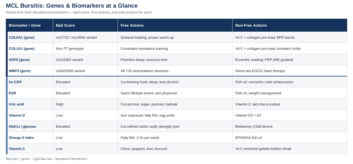
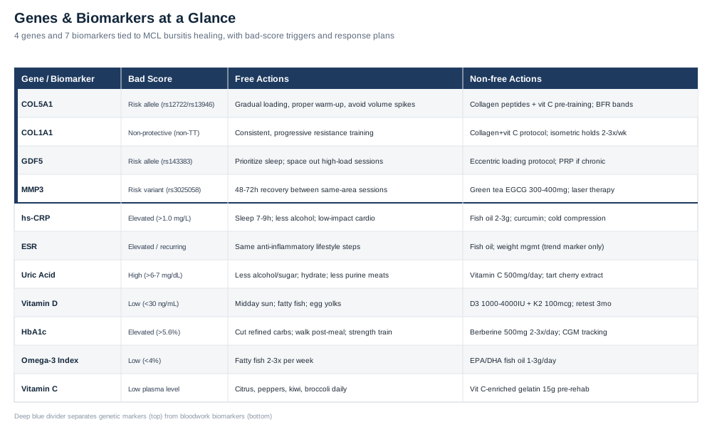
Before you dive in: this article walks through seven bloodwork markers that quietly influence how well a bursitis flare along the MCL resolves — from familiar ones like CRP and vitamin D to less obvious ones like your omega-3 index and plasma vitamin C, each with a concrete plan for improving it, with and without supplements. It then covers four genes tied to collagen strength and tissue remodeling that may explain why some knees are simply more injury-prone, plus what a person can realistically do about a "bad" variant. There's also a breakdown of a widely discussed mobility podcast episode challenging how most people think about joint stiffness and tissue capacity, and a short review of complementary approaches — from targeted massage to laser therapy — that have actual human evidence behind them for knee and soft-tissue conditions. The map below gives you the overview before you read the details.
The Bloodwork That Actually Explains Your Knee
Medial collateral ligament bursitis — inflammation of the bursa sac that cushions the MCL against the tendons and bone of the inner knee, closely related to what's clinically described as pes anserine bursitis — is usually triggered locally: tight hamstrings, repetitive friction, a direct blow, or knee osteoarthritis changing the mechanics of the joint, as detailed in the NCBI StatPearls review of pes anserine bursitis. But whether that local irritation resolves in three weeks or drags on for six months has a lot to do with what's happening systemically. The seven markers below are the ones worth tracking if you want an honest picture of your internal healing environment, not just the joint itself.
High-sensitivity CRP
C-reactive protein is the most widely used marker of systemic inflammation, and it matters here for two reasons. First, a persistently elevated hs-CRP suggests your body is running "hot" in general, which tends to slow local tissue healing and lower your pain threshold. Second, a markedly elevated CRP alongside warmth, redness, and fever raises concern for septic bursitis, a distinct and more urgent condition covered in the StatPearls entry on septic bursitis — a reason not to self-manage a hot, swollen bursa without medical evaluation. Peter Attia has long used hs-CRP as one of his core "inflammaging" markers precisely because it's cheap, stable, and predictive of how well tissue repair processes are functioning generally.
How to measure it: a standard blood draw, available through most primary care visits or direct-to-consumer labs (Quest, LabCorp, or similar) for roughly $15 to $40 without insurance.
If the score is bad, the plan without supplements: reduce total training and joint-loading volume until the local flare settles, prioritize 7 to 9 hours of sleep, address visceral fat if present (a major independent driver of baseline CRP), cut back on alcohol, and reintroduce low-impact aerobic activity like cycling or swimming once acute symptoms calm — this alone can meaningfully lower CRP over 8 to 12 weeks.
If the score is bad, the plan with supplements or equipment: EPA/DHA fish oil at 2 to 3 grams per day, and curcumin with piperine at 500 to 1,000 mg once or twice daily, cycled in 8- to 12-week blocks with a break in between. Side effects to watch: mild GI upset from curcumin, and increased bleeding risk when combined with blood thinners or before surgery. A cold-compression device (such as a Game Ready-style unit) can help manage local swelling during the same window. Retest CRP at 3 months, not sooner — it moves slowly.
Erythrocyte sedimentation rate (ESR)
ESR is a slower, blunter cousin of CRP, but it's useful precisely because it reflects a longer window of inflammatory activity. It's particularly relevant if bursitis keeps recurring, since a chronically elevated ESR can point toward an underlying inflammatory arthropathy (rheumatoid arthritis, gout, or another condition) rather than pure overuse.
How to measure it: a simple Westergren-method blood test, typically $10 to $20, often bundled with a basic metabolic or rheumatologic panel.
If the score is bad, the plan without supplements: the same anti-inflammatory lifestyle levers as CRP — sleep, weight management, alcohol reduction — plus a conversation with a physician if ESR stays elevated despite those changes, since it may point to something beyond the knee itself.
If the score is bad, the plan with supplements or equipment: fish oil and weight management as above; there's no equipment or supplement that moves ESR specifically, so treat it as a trend marker rather than a target, and recheck every 2 to 3 months during a flare-recovery period.
Serum uric acid
Uric acid deserves a place on this list because gouty or crystal-related bursitis is a genuine mimicker and sometimes co-driver of MCL-area bursa pain, as noted in the StatPearls chapter on gout. Even at sub-gout levels, higher uric acid correlates with more background inflammation.
How to measure it: included in most standard blood chemistry panels, roughly $10 to $15; home fingerstick uric acid meters exist for around $30 to $50 plus test strips if you want more frequent tracking.
If the score is bad, the plan without supplements: cut back on alcohol (beer in particular), reduce added sugar and high-fructose beverages, moderate purine-heavy processed meats and organ meats, and stay well hydrated.
If the score is bad, the plan with supplements or equipment: vitamin C at 500 mg per day has a modest uric-acid-lowering effect; tart cherry extract (480 to 960 mg per day) is a reasonable adjunct during flare periods, best cycled rather than used indefinitely. Avoid high-dose niacin, which raises uric acid. If true gout is confirmed by a physician, medication (allopurinol or similar) is a clinical decision, not a self-directed one.
25-hydroxyvitamin D
Tendons and ligaments express the vitamin D receptor, and vitamin D plays a real role in collagen remodeling and immune regulation within connective tissue, a relationship reviewed in a study on vitamin D and immunomodulation in tendon injury and a more recent scoping review on vitamin D and tendon healing. Deficiency doesn't cause bursitis on its own, but it may slow the repair process once irritation has started.
How to measure it: a standard blood test, roughly $30 to $50, widely available direct-to-consumer.
If the score is bad, the plan without supplements: 15 to 20 minutes of midday sun exposure several times a week (adjusted for skin tone and latitude), and more dietary fatty fish, egg yolks, and fortified foods.
If the score is bad, the plan with supplements or equipment: vitamin D3 at 1,000 to 4,000 IU per day depending on how deficient you are, ideally paired with 100 mcg of vitamin K2 to support proper calcium handling. Retest in 3 months. Avoid megadosing above 10,000 IU daily without physician monitoring — the main risk is hypercalcemia over time.
HbA1c and fasting glucose
Chronically elevated blood sugar drives the formation of advanced glycation end-products (AGEs) that stiffen collagen and impair its ability to slide and remodel normally, a mechanism demonstrated in tendon tissue in a study on AGEs and tendon collagen fiber sliding. This matters for bursitis recovery because the same collagen-remodeling machinery is what rebuilds a healthy bursa wall and surrounding ligament tissue.
How to measure it: HbA1c runs about $15 to $20, fasting glucose around $10; a continuous glucose monitor (roughly $50 to $100 per month) is a more advanced option if you want real-time feedback on how specific meals and activity affect your glucose control.
If the score is bad, the plan without supplements: reduce refined carbohydrates and added sugar, take a short walk after meals, add resistance training, and prioritize weight management and sleep quality.
If the score is bad, the plan with supplements or equipment: berberine at 500 mg two to three times daily, cycled in 8- to 12-week blocks with a break, can meaningfully improve glucose control; watch for GI side effects and check for interactions with other glucose-lowering medications before starting. A CGM is a useful (optional) equipment upgrade for personalizing the diet changes above. Retest HbA1c every 3 months.
Omega-3 index
The omega-3 index measures the EPA and DHA content of your red blood cell membranes, and it's a reasonable proxy for how well-equipped your body is to actively resolve inflammation rather than just suppress it — a distinction explained in research on the omega-3 index as a biomarker in physically active populations. A low index means fewer raw materials for the specialized pro-resolving mediators that actually shut down an inflammatory flare.
How to measure it: a specialized finger-prick test (OmegaQuant and similar labs), typically $50 to $75, usually out of pocket.
If the score is bad, the plan without supplements: eat fatty fish (salmon, sardines, mackerel) two to three times per week.
If the score is bad, the plan with supplements or equipment: combined EPA/DHA fish oil at 1 to 3 grams per day. Retest at 3 to 4 months, since red blood cell turnover is slow. Side effects are generally mild — fishy aftertaste, occasional GI upset — with a caution around high doses if you're already on blood thinners.
Plasma vitamin C
Vitamin C is a required cofactor for the enzymes that hydroxylate proline and lysine during collagen synthesis — a step that directly affects how well ligament and bursa wall tissue rebuilds itself. A well-known human study demonstrated this concretely: vitamin C-enriched gelatin taken before exercise nearly doubled markers of collagen synthesis compared with placebo.
How to measure it: plasma vitamin C testing is less commonly ordered and runs about $40 to $60 through specialty labs; for most people, ensuring adequate intake matters more than testing it directly.
If the score is bad, the plan without supplements: daily citrus fruit, bell peppers, kiwi, and broccoli generally cover baseline needs.
If the score is bad, the plan with supplements or equipment: following the study protocol, 15 grams of vitamin C-enriched gelatin or collagen peptides taken 30 to 60 minutes before physical therapy or rehab exercises, timed specifically around active loading sessions rather than used continuously. Mild GI upset is possible at higher doses; this is a targeted rehab-window strategy, not a daily supplement.
Tracking these seven markers together, rather than any single one in isolation, gives a much clearer picture of whether your body is set up to heal a stubborn bursitis flare — and it naturally raises the next question: are some of these tendencies baked in genetically to begin with?
What Your Genes May Be Telling You About Ligament Resilience
Direct-to-consumer genetic data has made it possible for people outside of sports science labs to look at variants tied to connective tissue injury risk. Researchers like Ali Torkamani, whose work on genomic risk interpretation emphasizes that most single variants carry small, probabilistic effects rather than deterministic ones, and public figures like Gary Brecka, who has popularized combining genetic and biomarker panels to personalize health decisions, are a reasonable starting point for understanding how to use this kind of data without over-interpreting it. The four genes below have the strongest human evidence specifically for tendon and ligament injury risk — though it's worth saying upfront that most of these associations come from moderate-sized cohort and meta-analysis studies, not large-scale definitive trials, and effect sizes are generally modest.
COL5A1
COL5A1 encodes the alpha-1 chain of type V collagen, a minor but critical collagen that regulates the diameter of the larger type I collagen fibrils making up ligament tissue. Certain variants (notably rs12722 and rs13946) are associated with altered soft tissue stiffness and a higher reported risk of musculoskeletal soft tissue injury, according to a meta-analysis of 21 observational studies on COL5A1 polymorphisms. In practical terms, a less favorable genotype may mean your ligament tissue is inherently a bit more extensible and slower to stiffen under load.
If the gene is unfavorable, the plan without supplements: build in more progressive, gradual loading for the muscles stabilizing the medial knee (hamstrings, adductors, quads), avoid sudden jumps in training volume, and never skip a proper warm-up before high-load activity.
If the gene is unfavorable, the plan with supplements or equipment: the vitamin C plus collagen peptide timing protocol described above, taken before rehab or training sessions, and blood flow restriction (BFR) training bands for lower-load tissue conditioning cycles — typically 2 to 3 sessions per week across 8-week blocks with a deload week built in.
COL1A1
COL1A1 encodes the primary structural collagen of ligament tissue, type I collagen. The Sp1 binding site polymorphism (rs1800012) shifts the balance between collagen type I and type III in tissue, which affects overall ligament stiffness, as shown in a meta-analysis on COL1A1 and sports-related tendon and ligament injuries. Interestingly, this is one of the few variants where a specific genotype (TT) appears protective rather than purely risk-increasing.
If the gene is unfavorable, the plan without supplements: consistent, ongoing resistance training is the single most reliable way to drive favorable tendon and ligament stiffness adaptations over time, alongside gradual progression in training load rather than large jumps.
If the gene is unfavorable, the plan with supplements or equipment: the same collagen and vitamin C pre-loading protocol, plus isometric holds (5 sets of 30 to 45 seconds) using resistance bands or a simple isometric rig, performed 2 to 3 times weekly — a loading style shown to favorably influence tendon and ligament stiffness without high mechanical strain.
GDF5
GDF5 (growth differentiation factor 5) is involved in joint and tendon development and repair signaling. The rs143383 variant has been linked to a higher risk of Achilles tendon pathology, though a study on GDF5 and anterior cruciate ligament rupture found no significant association with ACL injury specifically — a good example of why gene-injury links are often tissue-specific and shouldn't be over-generalized across every ligament in the body.
If the gene is unfavorable, the plan without supplements: prioritize sleep quality and duration, since much of the growth-factor signaling involved in tissue repair is concentrated during deep sleep stages, and avoid stacking high-load training sessions without adequate recovery windows between them.
If the gene is unfavorable, the plan with supplements or equipment: there's no supplement that reliably targets the GDF5 pathway directly, so the more useful lever is mechanical — a structured, low-load eccentric loading protocol for the affected tissue. If bursitis becomes chronic despite conservative care, a physician-guided discussion of regenerative options such as PRP is a reasonable next step, though this is a clinical decision rather than a self-directed one.
MMP3
MMP3 encodes matrix metalloproteinase 3, an enzyme responsible for breaking down and remodeling extracellular matrix. Certain variants (such as rs3025058) shift the balance toward more matrix breakdown relative to rebuilding, which may make tissue less resilient under repeated stress, per a study on the MMP3 gene in musculoskeletal soft tissue injury risk profiling.
If the gene is unfavorable, the plan without supplements: give tissue more time to remodel between intense loading sessions of the same area — generally 48 to 72 hours — rather than training the same movement pattern back-to-back.
If the gene is unfavorable, the plan with supplements or equipment: green tea catechins (EGCG), 300 to 400 mg per day, cycled 8 weeks on and 4 weeks off, may help modulate excess matrix breakdown; take with food to reduce mild liver-related concerns at higher doses. Local photobiomodulation device use (covered in more detail later in this article) is a reasonable complementary equipment option for supporting tissue remodeling in the affected area.
Genetics can explain some of the "why me" behind a stubborn bursitis case, but it's not the whole picture — daily movement habits and joint mechanics matter just as much, which is where a recent, widely discussed conversation on mobility becomes relevant.
What A Major Mobility Conversation Gets Right About Knee Health
The Huberman Lab episode "Dr. Kelly Starrett: How to Improve Your Mobility, Posture & Flexibility", released in December 2024, is one of the more practical, evidence-informed conversations available on how joint tissue actually adapts — and it challenges some assumptions that are still common in general medical advice about "just resting" an irritated joint. Below are ten of the most useful ideas from that conversation, framed specifically for someone managing MCL-area bursitis.
Tissue capacity is trainable, not fixed
The episode's central argument is that ligaments, tendons, and the surrounding bursa tissue respond to graded mechanical stress over time, much like muscle does. A joint that feels "fragile" today isn't necessarily fragile permanently — it's often under-conditioned for the demands being placed on it, and appropriately dosed loading is part of the fix, not the risk.
Small, frequent doses of movement outperform occasional intense sessions
Rather than one long stretching or mobility session per week, the discussion emphasizes short, frequent "movement snacks" spread through the day — a few minutes of targeted knee and hip mobility work several times daily tends to produce better tissue adaptation than a single 45-minute session.
Simple self-assessments reveal real restrictions
Basic movement screens — how a knee tracks during a bodyweight squat or a step-down — can reveal asymmetries or restrictions well before they show up as pain. For someone with recurring bursitis, this kind of self-check can catch a mechanical problem early, before the next flare.
Joint movement helps fluid exchange in and around connective tissue
Regular, pain-free range-of-motion work supports the exchange of synovial and interstitial fluid around joint structures, including bursae. Prolonged stillness — sitting for hours, or immobilizing a joint longer than necessary — can work against recovery rather than protect it.
Foam rolling is a warm-up primer, not a treatment
Self-myofascial work has a real but modest and short-lived effect — useful as preparation before activity, not as a stand-alone fix for a bursitis flare. The conversation is clear that this tool is being over-sold as a treatment when its actual role is much narrower.
Foot and ankle mechanics change knee loading
A meaningful portion of medial knee stress originates from how the foot and ankle move (or don't) during walking and standing. Addressing ankle mobility and footwear choices can meaningfully reduce the repetitive friction load on the medial knee structures, including the bursa.
Use temperature strategically, not habitually
Rather than defaulting to ice for everything, the discussion frames heat and cold as tools with different jobs — heat to prepare tissue for movement, cold to manage acute swelling after a flare — and cautions against using either one as a substitute for addressing the underlying mechanical cause.
Sleep position affects next-day joint stiffness
Extended time spent in one static position overnight can contribute to next-morning stiffness in the knee and surrounding tissue. Small position adjustments and a brief mobility routine upon waking are presented as a low-effort way to reduce this.
Train the ranges you actually use in daily life
Strength and mobility work should reflect real-world knee demands — squatting to sit, stepping down stairs, twisting to reach — rather than only training in the mid-range positions most gym routines default to. This is particularly relevant for a joint recovering from bursitis, since regaining confidence in full-range movement is part of full recovery.
Consistency beats intensity for long-term joint resilience
The compounding value of daily, moderate mobility input is emphasized repeatedly over occasional intense corrective sessions. For a chronic or recurring bursitis case, this reframes recovery as a long-term maintenance habit rather than a short-term fix to complete and forget.
Pain during movement isn't automatically a stop signal
One of the more contrarian points is that mild, non-worsening discomfort during controlled mobility work isn't automatically a reason to stop — a nuance that runs against the instinct to fully avoid any uncomfortable movement, though it's paired with a clear caveat to back off from anything that produces sharp, worsening, or swelling-associated pain.
Between the biology of genes and biomarkers and the day-to-day mechanics covered above, there's a third layer worth considering — adjunct therapies with actual clinical evidence behind them for knee and soft tissue conditions.
Complementary Approaches Worth Considering
These aren't replacements for addressing the mechanical cause of bursitis or for medical evaluation when needed, but each of the four below has meaningful human evidence relevant to knee and soft-tissue recovery.
Massage therapy
Manual soft tissue work can reduce muscle guarding and tension around an irritated knee, improving tissue pliability in the muscles surrounding — though not directly over — an inflamed bursa. This matters for bursitis that's driven partly by tight hamstrings or adductors pulling extra tension across the medial knee.
A randomized controlled trial on percussion massage therapy and recovery from muscle soreness and tissue stiffness found measurable improvements in recovery markers and perceived stiffness, supporting its use as a recovery adjunct rather than a primary treatment.
To apply this realistically: use 10 to 15 minute sessions targeting the hamstrings, adductors, and surrounding quadriceps 2 to 3 times per week during a flare, avoiding direct deep pressure over the bursa itself, and pair with icing afterward if swelling is present.
Tai chi
Tai chi is a slow, low-impact movement practice that improves proprioception and neuromuscular control — relevant because bursitis around the knee is often linked to poor joint stabilization patterns built up over months of overuse.
A pilot randomized controlled trial on Tai chi and knee proprioception in osteoarthritis patients found significant improvements in knee joint position sense after 12 weeks of twice-weekly, 60-minute sessions of modified Yang-style forms, though the same study noted the effect didn't clearly persist long-term without continued practice.
To apply this realistically: start with a beginner instructor-led class twice weekly, avoid deep lunging postures during an acute flare, and treat it as a maintenance and prevention-phase practice rather than an acute treatment.
Low-level laser therapy (photobiomodulation)
Photobiomodulation uses red or near-infrared light believed to influence local mitochondrial activity and inflammatory signaling, offering a plausible, non-invasive way to support a slow-healing bursa or surrounding tissue.
A randomized placebo-controlled trial combining low-level laser therapy with strength training in knee osteoarthritis found reduced reliance on NSAIDs and improved function at 52 weeks, though the study noted mixed results on primary pain scores alone — an honest caveat that this works best paired with active rehab, not as a stand-alone fix.
To apply this realistically: use a reputable clinic-grade or well-reviewed home device with red-to-near-infrared wavelengths (roughly 630–904 nm), directly over the affected area for a few minutes, 2 to 3 times weekly for several weeks, avoiding use over broken or acutely inflamed open skin.
EMG biofeedback
Surface EMG biofeedback retrains activation patterns in the muscles that stabilize the knee, particularly useful when bursitis develops after a ligament injury or surgery and the surrounding muscles have become inhibited or imbalanced.
A randomized controlled trial on EMG biofeedback for the vastus medialis after ACL reconstruction found meaningful improvements in knee extension and muscle activation over a 6-week rehab window compared with standard rehab alone.
To apply this realistically: this requires a physical therapist with sEMG equipment — it's best integrated into a structured rehab program rather than attempted with consumer devices alone, and is most valuable specifically when bursitis has followed a ligament injury or knee surgery.
Conclusion
Medial collateral ligament bursitis responds to more than rest and ice — it responds to the internal conditions that determine how well your body controls inflammation, rebuilds collagen, and remodels connective tissue. The seven biomarkers covered here give you a concrete, trackable way to see whether those internal conditions are working for you or against you, the four genes offer useful context for why some knees are simply more injury-prone to begin with, and the mobility and complementary approaches round out a realistic, evidence-aware recovery plan rather than a generic one.
The next useful step isn't a dramatic overhaul — it's picking one or two of the markers above to actually get tested, tracking your symptoms alongside them over a few months, and bringing that information to a physician or physical therapist who can help you interpret it in the context of your specific knee. That combination of data and professional judgment is a far better foundation for a decision than guesswork.
Musculoskeletal: Joint Conditions
Autoimmune: Inflammatory Conditions Connective Tissue Conditions