This article contains AI generated content.
Medial Gastrocnemius Strain: 6 Genes And 6 Biomarkers To Track
Introduction
A calf strain rarely announces itself with drama the first time. It shows up as a sudden pull mid-sprint, a sharp pinch during a tennis serve, or a tight, bruised feeling behind the knee that takes weeks longer to settle than anyone expected. And then, for a fair number of people, it comes back. Not because they skipped rehab or ignored their physiotherapist, but because the advice they were given — stretch more, strengthen your calves, warm up properly — was aimed at an average person, not at their specific tissue.
Generic advice is not wrong, exactly. It is just incomplete. It assumes every calf behaves the same way under load, that every Achilles-gastrocnemius junction remodels collagen at the same rate, and that every person's inflammatory response resolves on the same schedule. In practice, some of that variation is measurable. Genetic research on soft-tissue injury has identified a recurring handful of genes that shape how collagen is built, how it is broken down, and how fast-twitch muscle fibers respond to mechanical load — all directly relevant to a muscle-tendon complex like the medial gastrocnemius. Alongside that, a set of blood biomarkers can show, in real time, whether your body is actually recovering the way it should.
This article works through both angles in detail: the genetic variants with the strongest human evidence for soft-tissue and calf-adjacent injury risk, and the biomarkers worth tracking if you strain easily, recover slowly, or keep re-injuring the same spot. Neither approach delivers a guarantee — genetics loads the dice, it does not fix the outcome, and biomarkers describe a moving picture, not a verdict. But together they replace vague uncertainty with something closer to a map: here is what may be working against you, and here is what, specifically, tends to help.
None of this promises immunity from injury. What it offers instead is a more honest starting point — a way to stop guessing at generic fixes and start addressing the mechanisms that are actually in play for your body. The rest of this article walks through the genetic evidence first, then the biomarkers, then a research-based recovery framework and a set of complementary approaches worth knowing about.
Summary
Before diving in: research on soft-tissue and tendon-adjacent injuries has repeatedly implicated a small set of genes involved in collagen structure, collagen remodeling, and muscle fiber composition — several of which map directly onto the mechanics of a calf strain. At the same time, a handful of blood markers, from creatine kinase to vitamin D to the omega-3 index, can tell you far more about your actual recovery trajectory than "let me know if it still hurts" ever will. This article breaks down six of the best-evidenced genes (with a plan for each, supplement-free and supplement-supported), six biomarkers worth tracking, a research-backed recovery protocol pulled from a widely discussed podcast conversation on tendon and collagen health, and five complementary approaches with genuine human evidence behind them. Keep reading to see exactly which variants and numbers matter, and what to do about each one.
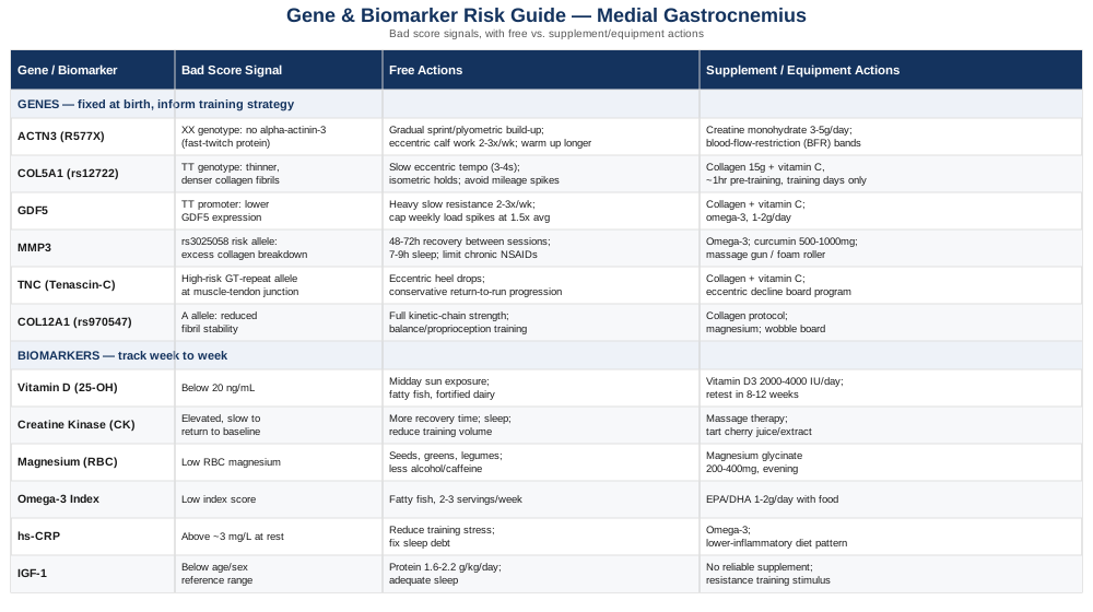
The Genetic Side of Medial Gastrocnemius Strains: What the Research Shows
There is no published genetic study that isolates the medial gastrocnemius alone — it is too narrow a target for most research funding. What exists instead is a broader, well-established field studying genetic variation in collagen genes, matrix remodeling enzymes, and muscle fiber composition, almost always in the context of hamstring strains, Achilles tendinopathy, and ACL ruptures. Because the medial gastrocnemius sits at a musculotendinous junction with biology very similar to these more-studied tissues — same collagen types, same remodeling enzymes, same load-bearing demands — this research is a reasonable, evidence-based proxy, even though it is not a perfect one. Researchers like Malcolm Collins and Stuart Raleigh built much of this field through South African and European cohort studies over the past two decades, and more recently, figures such as Ali Torkamani, a genomics researcher known for mining consumer genetic data for actionable health signals, and Gary Brecka, who popularized applying genetic and biomarker panels to athletic performance and injury risk, have pushed this kind of analysis into the mainstream. Their broader point is a useful frame here: raw genetic data, whether from a clinical panel or a consumer test like 23andMe, often contains information relevant to injury risk that never gets surfaced in a standard report.
ACTN3 (R577X) and Fast-Twitch Fiber Capacity
ACTN3 codes for alpha-actinin-3, a protein found almost exclusively in fast-twitch (type II) muscle fibers, where it helps anchor the contractile machinery during forceful, rapid contractions. About one in five people worldwide carry two copies of the non-functional "X" variant (the XX genotype), meaning they produce no alpha-actinin-3 at all and rely more heavily on slow-twitch fiber characteristics. In a study of professional football players, XX carriers had 2.66 times higher odds of injury and 2.13 times higher odds of severe injury compared to RR carriers, a finding published in the American Journal of Sports Medicine. For a muscle like the gastrocnemius, which is built for explosive, fast-twitch-dominant contraction during sprinting and jumping, this genotype may mean the tissue is being asked to do something it is structurally less prepared for.
If the gene is bad, the plan without supplements starts with respecting load progression rather than fighting the genotype directly. That means building sprint and plyometric exposure gradually across weeks, not days; prioritizing eccentric-focused calf work (slow, controlled lowering on calf raises, both straight-knee for the gastrocnemius and bent-knee for the soleus) two to three times per week; and extending warm-up time before any high-speed activity, since XX carriers appear to tolerate abrupt intensity spikes less well.
If the score is bad, the plan with supplements or equipment centers on creatine monohydrate, one of the most consistently studied supplements for supporting fast-twitch force output — 3 to 5 grams daily, taken continuously with no need to cycle off, since the safety data on long-term daily use is strong. Mild water retention and occasional GI discomfort are the main reported side effects, usually resolved by splitting the dose with food. Blood flow restriction training bands are a reasonable equipment addition, allowing strength gains at lower absolute loads, which may suit a fiber profile that is more injury-prone under maximal mechanical stress.
COL5A1 (rs12722) and Collagen Fibril Structure
COL5A1 codes for collagen type V, a minor but critical component that regulates the diameter of the much more abundant type I collagen fibrils. A systematic review and meta-analysis, published via the National Center for Biotechnology Information, found that the TT genotype at the rs12722 marker was associated with a 58% higher risk of musculoskeletal soft-tissue injury overall, an effect seen most consistently in Caucasian populations. The likely mechanism is that this genotype produces thinner, more densely packed collagen fibrils, which some other research links to greater flexibility but less tensile stiffness — a plausible trade-off for a tissue like the gastrocnemius-Achilles complex that needs to store and return elastic energy without over-stretching.
If the gene is bad, the plan without supplements leans on tempo-controlled training rather than pure volume: slow eccentric tempos (3 to 4 seconds on the lowering phase) during calf raises, isometric mid-range holds two to three times weekly to build tendon stiffness without added ballistic stress, and a hard rule against sudden mileage or intensity jumps — the classic "10% per week" guideline is a reasonable ceiling here.
If the score is bad, the plan with supplements or equipment uses a collagen-loading protocol with real supporting evidence: 15 grams of hydrolyzed collagen or gelatin combined with roughly 50 to 200 milligrams of vitamin C, taken about an hour before a training session, only on training days rather than daily. This timing matters — vitamin C is a required cofactor for collagen crosslinking, and the combination appears to meaningfully increase collagen synthesis markers only when consumed before mechanical loading. Side effects are minimal at these doses; people with a history of oxalate kidney stones should keep total vitamin C intake modest, since very high doses have been linked to oxalate formation in that specific population.
GDF5 and Tendon-Adjacent Tissue Development
Growth differentiation factor 5 plays a role in joint, ligament, and tendon development, and a specific promoter variant reduces GDF5 expression. In a study of a Chinese Han population, the TT genotype was significantly more common in people with non-contact ACL ruptures than in matched controls, as reported in research published via PubMed. Lower GDF5 expression is thought to translate into somewhat weaker or less well-organized connective tissue at load-bearing junctions, which plausibly extends to the myotendinous junction of the gastrocnemius, though direct calf-strain data specific to this gene does not yet exist.
If the gene is bad, the plan without supplements emphasizes heavy, slow resistance training for the calf-Achilles unit — three sets of 6 to 8 slow repetitions, two to three non-consecutive days per week — along with monitoring training load ratios so that a week's total workload never spikes more than roughly 1.5 times the trailing four-week average, a heuristic drawn from broader soft-tissue injury-prevention research.
If the score is bad, the plan with supplements or equipment repeats the collagen and vitamin C timing protocol described above, and adds a resistance band or decline board for eccentric calf-loading progressions. An omega-3 supplement (1 to 2 grams combined EPA/DHA daily, with food, no strict cycling required though periodic bloodwork every six months is reasonable) may support a less inflamed baseline tissue environment, which matters more for genotypes already working with a weaker structural starting point.
MMP3 and Collagen Remodeling Balance
Matrix metalloproteinase 3 is one of the primary enzymes responsible for breaking down and remodeling collagen and other extracellular matrix proteins — a necessary process for tissue adaptation, but one that needs to stay balanced. A meta-analysis in the NCBI PMC database found the rs3025058 variant associated with increased tendon-ligament injury risk, notably in Caucasian and Brazilian cohorts, while a different variant in the same gene (rs679620) was linked to reduced Achilles tendon rupture risk — a reminder that "one gene" rarely means "one effect."
If the gene is bad, the plan without supplements focuses on respecting recovery windows rather than trying to out-train the enzyme system: 48 to 72 hours between hard loading sessions on the same tissue, since collagen remodeling continues well after the workout ends, and prioritizing 7 to 9 hours of sleep, since much of connective tissue repair is concentrated during deep sleep phases. Chronic, high-dose NSAID use is worth minimizing where medically appropriate, since some evidence suggests it can blunt the same remodeling processes needed for adaptation.
If the score is bad, the plan with supplements or equipment includes omega-3 fatty acids at the same dose noted above, which have some evidence for moderating excessive matrix metalloproteinase activity as part of a broader anti-inflammatory effect. Curcumin (500 to 1000 milligrams daily with a black pepper extract for absorption, cycled 8 to 12 weeks on and 2 to 4 weeks off) has early evidence for influencing MMP expression, though the human data specific to soft-tissue injury is still thin — worth trying cautiously, and worth stopping if any GI irritation appears, especially if combined with blood-thinning medication. A foam roller or massage gun for regular soft-tissue work around the calf has minimal direct evidence for MMP modulation but is low-risk and may support local circulation.
TNC (Tenascin-C) and the Myotendinous Junction
Tenascin-C is an extracellular matrix protein concentrated at myotendinous junctions — precisely the transition zone where the medial gastrocnemius muscle belly meets the Achilles tendon, and where most calf strains and "tennis leg" injuries actually occur. A study in the American Journal of Sports Medicine found that certain GT-repeat lengths in the TNC gene were overrepresented in people with Achilles tendon injuries, conferring roughly six times the risk compared to protective alleles. Given the shared anatomy, this is arguably the single most directly relevant genetic finding in this entire list for a medial gastrocnemius strain specifically.
If the gene is bad, the plan without supplements centers on junction-specific loading: eccentric heel-drop exercises performed slowly and deliberately, a conservative return-to-running progression after any calf tightness (days, not sessions, of graded reintroduction), and caution around sudden changes in footwear, running surface, or terrain, all of which alter loading patterns at the myotendinous junction abruptly.
If the score is bad, the plan with supplements or equipment again uses the vitamin C and collagen protocol on training days, plus an eccentric decline board for a structured heel-drop program — twice daily for 12 weeks is the classic dosing from tendon-loading research, though this level of volume should be introduced under professional guidance rather than self-prescribed, since overloading an already vulnerable junction can worsen symptoms. Any sharp, localized pain during the protocol is a stop signal, not something to push through.
COL12A1 (rs970547) and Fibril-Associated Collagen
Collagen XII is a FACIT (fibril-associated collagen with interrupted triple helices) protein that helps organize and stabilize type I collagen fibrils under mechanical stress. A meta-analytic review available through PubMed Central found the rs970547 A allele associated with elevated ACL injury susceptibility in athletes, and by extension a plausibly relevant marker for other high-load soft-tissue injuries, including calf strains, though direct evidence in the gastrocnemius is not yet published.
If the gene is bad, the plan without supplements broadens training beyond isolated calf work to the full lower-limb kinetic chain — hip, knee, and ankle strength and control together, since collagen XII variants appear to affect overall tissue resilience under multi-joint load rather than one muscle in isolation. Balance and proprioception training, such as single-leg stance progressions on an unstable surface, three times weekly for 10 minutes, has reasonable supporting evidence for reducing lower-limb soft-tissue injury broadly.
If the score is bad, the plan with supplements or equipment includes the same collagen-loading protocol plus attention to magnesium status (see the biomarker section below), since magnesium supports the neuromuscular control that protects poorly stabilized tissue during unexpected loads. A wobble board or balance pad, used consistently rather than sporadically, is a low-cost equipment addition with a reasonable evidence base in general lower-limb injury prevention literature.
Epigenetic Factors: Why the Sequence Isn't the Whole Story
None of these six genes operate in isolation from behavior. Mechanical loading itself changes gene expression at these loci — a concept sometimes called mechanotransduction — and the vitamin C and collagen protocol referenced throughout this section works precisely because it interacts with that loading-dependent expression window, not because it changes anyone's underlying DNA sequence. Chronic under-recovery, poor sleep, and low vitamin C or vitamin D status can all suppress the expression of collagen genes regardless of genotype, meaning a "good" genetic profile can still underperform under poor conditions, and a "difficult" genetic profile can still perform reasonably well under good ones. This is the most actionable takeaway from the genetics research as a whole: the DNA sets the range of outcomes, but daily inputs — load, nutrition, sleep, and recovery timing — decide where within that range a person actually lands.
Biomarkers Worth Tracking Alongside Your Genetic Picture
Genetic risk is fixed at birth, but biomarkers move week to week, which makes them the better tool for answering the question that actually matters day-to-day: is this tissue recovering the way it should be right now? The six markers below are the ones with the clearest evidence base and the most practical relevance to calf strain risk and recovery.
Creatine Kinase (CK)
Creatine kinase leaks into the bloodstream when muscle fibers are damaged, making it one of the most direct available markers of muscle tissue breakdown. A literature review in PMC confirms CK typically peaks two to six days after a damaging bout of exercise, tracking closely with delayed-onset muscle soreness, though individual variability is high enough that trends matter more than any single reading.
How to measure it: a standard blood draw, $20 to $40 out of pocket if not bundled into a broader panel, with no special preparation beyond avoiding hard exercise for 48 hours prior.
If the score is bad, the plan without supplements is simply more recovery time before the next hard session on the same tissue, prioritizing sleep and reducing training volume until CK trends back toward your personal baseline — there is no way to force CK down faster through willpower alone.
If the score is bad, the plan with supplements or equipment includes massage therapy, which a systematic review found measurably lowers post-exercise CK alongside reducing soreness (detailed further in the complementary approaches section), and tart cherry juice or extract, which has modest supporting evidence for reducing exercise-induced muscle damage markers — typically 8 to 12 ounces of juice or an equivalent extract dose daily for the days surrounding a hard training block, with no known serious side effects beyond occasional GI upset.
Vitamin D (25-hydroxyvitamin D)
Vitamin D receptors are present directly in skeletal muscle tissue, and a review on athletes' vitamin D status, available via PubMed, notes that deficiency is associated with reduced muscle strength and function, with indoor-training athletes at particular risk.
How to measure it: a standard blood test, $30 to $50, widely available and often included in annual panels; most labs flag deficiency below 20 ng/mL, though many sports-medicine practitioners target 40 to 60 ng/mL for athletic populations.
If the score is bad, the plan without supplements starts with 15 to 30 minutes of midday sun exposure on bare skin several times weekly where climate allows, plus dietary sources like fatty fish and fortified dairy — though this alone is often insufficient to correct a real deficiency.
If the score is bad, the plan with supplements or equipment uses vitamin D3, typically 2000 to 4000 IU daily with a fat-containing meal, retested after 8 to 12 weeks rather than adjusted blindly; very high doses (above 10,000 IU daily sustained) can cause hypercalcemia over time, so ongoing supplementation should be guided by follow-up bloodwork rather than indefinite guessing.
Magnesium (RBC Magnesium)
Magnesium supports normal neuromuscular signaling, and a review in PMC notes that deficiency may distort neuromuscular function and that adequate magnesium status is positively associated with strength and exercise performance measures, though direct evidence linking magnesium specifically to muscle cramping remains limited.
How to measure it: serum magnesium is cheap ($15 to $25) but a poor proxy for total body status; red blood cell (RBC) magnesium, at $40 to $70, is a more informative option since most magnesium is stored intracellularly rather than in serum.
If the score is bad, the plan without supplements emphasizes magnesium-rich foods — pumpkin seeds, leafy greens, almonds, and legumes — alongside reducing excess alcohol and caffeine intake, both of which increase magnesium excretion.
If the score is bad, the plan with supplements or equipment uses magnesium glycinate, 200 to 400 milligrams in the evening, chosen over magnesium oxide for better absorption and less GI disturbance; loose stools are the main side effect at higher doses, resolved by reducing the amount rather than stopping entirely.
Omega-3 Index
A systematic review of randomized controlled trials, published in PMC, found that omega-3 supplementation may reduce post-exercise inflammation, mitigate exercise-induced muscle damage, and lower oxidative stress markers, though optimal dosing protocols are still being refined across studies.
How to measure it: a specialized blood-spot test (commercial options like OmegaQuant), $50 to $90, not typically covered by insurance and not part of a standard panel — a reasonable one-time or annual check rather than something to track weekly.
If the score is bad, the plan without supplements means increasing fatty fish intake (salmon, sardines, mackerel) to two to three servings weekly, which for many people is sufficient to shift the index meaningfully over two to three months.
If the score is bad, the plan with supplements or equipment uses a combined EPA/DHA supplement, 1 to 2 grams daily with food, taken continuously rather than cycled, since the benefit accrues gradually over months; mild fishy aftertaste or GI upset are the main side effects, and anyone on blood-thinning medication should discuss dosing with a physician first.
hs-CRP (High-Sensitivity C-Reactive Protein)
CRP is a general marker of systemic inflammation, and a systematic review in PMC found that intense, prolonged exercise raises CRP and related inflammatory markers for up to 28 hours afterward, while moderate exercise produces a comparatively smaller response — useful context for distinguishing normal training inflammation from a baseline that never settles.
How to measure it: a standard blood test, $15 to $30, commonly bundled into cardiometabolic panels; a persistently elevated resting hs-CRP (above roughly 3 mg/L with no acute illness) is worth discussing with a physician regardless of athletic context.
If the score is bad, the plan without supplements focuses on adequate recovery between hard sessions, reducing overall training stress temporarily, and addressing sleep debt, since chronic sleep restriction independently raises baseline CRP.
If the score is bad, the plan with supplements or equipment includes the same omega-3 protocol described above, alongside a lower-inflammatory-load diet pattern (more vegetables and fiber, fewer ultra-processed foods) — no strict cycling required, retested every three to six months to confirm the trend is actually moving.
IGF-1 (Insulin-Like Growth Factor 1)
IGF-1 plays a direct role in muscle repair, and a study published via PubMed found that localized IGF-1 expression accelerates recovery of injured skeletal muscle by down-regulating pro-inflammatory cytokines and limiting excess fibrosis — the kind of scar tissue that can make a calf strain re-injury more likely.
How to measure it: a blood test, $60 to $120, less commonly ordered in routine care but available through most labs or longevity-focused clinics; results should be interpreted relative to age- and sex-adjusted reference ranges rather than a single universal number.
If the score is bad, the plan without supplements centers on adequate total protein intake (roughly 1.6 to 2.2 grams per kilogram of bodyweight daily for anyone actively training) and sufficient sleep, since growth hormone — and downstream IGF-1 — release is concentrated in deep sleep stages.
If the score is bad, the plan with supplements or equipment has limited strong evidence beyond ensuring adequate dietary protein and resistance training stimulus itself, which is the primary driver of IGF-1 signaling; no supplement reliably and safely raises IGF-1 on its own, and any product claiming to should be treated with real skepticism.
The picture that emerges from combining a genetic panel with these six markers is more useful than either alone — genetics explains why certain training decisions matter more for you specifically, while the biomarkers confirm, in real time, whether the plan is actually working. From here, it's worth looking at how that recovery process unfolds physiologically, which is where a widely discussed podcast conversation on tendon and collagen biology adds a useful layer.
The Podcast Episode That Reframes Tendon and Calf Recovery
Andrew Huberman's conversation with Dr. Keith Baar, a physiologist whose lab has spent years studying collagen synthesis and connective tissue adaptation, is one of the more practically dense discussions available on tendon and muscle-tendon junction recovery — directly relevant to the medial gastrocnemius, since it sits exactly at that junction. The episode challenges a few assumptions many people carry from standard sports-medicine advice. Below are ten of its most useful, evidence-connected takeaways.
1. Tendons and Muscle-Tendon Junctions Heal on a Much Slower Clock Than Muscle
Muscle tissue has a rich blood supply and can meaningfully remodel in days; tendon and the collagen-dense junction tissue has far less blood flow and may take months to fully adapt to a new loading stimulus, which is one reason calf strains at this junction feel "mostly better" long before they are actually structurally sound.
2. Vitamin C Is Not Optional for Collagen Synthesis, It's a Cofactor
Without adequate vitamin C, the enzymes that crosslink collagen (prolyl and lysyl hydroxylase) cannot function properly, regardless of how much collagen protein is consumed — meaning collagen supplementation without vitamin C is likely far less effective.
3. Timing the Vitamin C and Collagen Intake Before Loading Matters
The supporting human study, published in the American Journal of Clinical Nutrition, found that consuming vitamin C-enriched gelatin about an hour before intermittent exercise roughly doubled a blood marker of collagen synthesis compared to placebo — the timing relative to loading appears to be what activates the effect, not just the intake itself.
4. Mechanical Loading Is the Primary Driver of Tendon Adaptation
A review on tendon physiology, available via PubMed, confirms that both acute and chronic mechanical loading increase tendon collagen synthesis and turnover, mediated in part through IGF-1 and TGF-beta signaling — reinforcing that passive treatments alone rarely rebuild tendon-adjacent tissue.
5. There May Be a Minimum Recovery Window Between Loading Bouts
Baar's research suggests connective tissue may need a period of roughly six hours between loading sessions for the synthesis response to fully register, which challenges the common habit of doing multiple same-day training sessions on the same tissue without any real spacing.
6. NSAIDs May Blunt the Adaptive Response to Loading
A recurring, more contrarian point in this discussion is that routine anti-inflammatory use around training sessions may interfere with the very remodeling signals that loading is supposed to trigger — worth discussing with a physician rather than defaulting to NSAIDs reflexively around minor calf tightness.
7. Copper Is an Underrated Cofactor in Collagen Crosslinking
Lysyl oxidase, an enzyme central to collagen crosslink strength, requires copper as a cofactor — a detail rarely mentioned in typical collagen-supplement marketing, and worth considering if dietary copper intake (shellfish, nuts, seeds, organ meats) is low.
8. Isometric Holds at Long Muscle Lengths May Help Injured Junction Tissue
Sustained isometric contractions, performed at a lengthened position for the calf-Achilles complex, are discussed as a way to load the tissue meaningfully while minimizing the ballistic stress that tends to aggravate an already irritated myotendinous junction.
9. Warm-Up Isn't Just About Feeling Loose, It Changes Tissue Properties
Raising tissue temperature before high-force activity genuinely changes the mechanical properties of tendon and muscle-tendon tissue, supporting the practical case for a longer, more deliberate warm-up before sprinting or explosive calf-dominant movement, rather than treating warm-up as optional.
10. Consistency Beats Intensity for Long-Term Connective Tissue Health
Because tendon and junction tissue adapts slowly, sporadic bursts of intense loading followed by long inactivity are described as a worse strategy than smaller, more consistent loading exposure spread evenly across weeks and months — a direct challenge to the common "weekend warrior" training pattern.
Taken together, this framework reinforces a theme that runs through the genetic and biomarker sections above: the tissue at the center of a medial gastrocnemius strain adapts slowly, depends on specific nutritional cofactors, and responds best to patient, consistent loading rather than aggressive correction. From there, several complementary, non-pharmacological approaches have real human evidence supporting a role in recovery and prevention.
Complementary Approaches Backed by Human Evidence
The approaches below were selected specifically because they have direct or closely related human evidence for muscle recovery, soft-tissue injury, or musculoskeletal rehabilitation outcomes — not because they are popular in general wellness circles.
Massage Therapy
Massage therapy is one of the more mechanistically plausible recovery tools for a calf strain, since it may support local circulation, reduce perceived muscle tension, and modestly influence circulating markers of muscle damage. For a muscle-tendon junction injury like this one, that combination of physical and subjective relief is part of why it remains a mainstay of sports physiotherapy programs.
A systematic review and meta-analysis in PMC found that massage therapy significantly reduced delayed-onset muscle soreness ratings and serum creatine kinase levels while improving muscle performance after strenuous exercise, with the largest effect observed around 48 hours post-exercise — closely matching the CK peak window described earlier in this article.
Realistically, this means scheduling a massage session roughly one to two days after a hard calf-loading session or a minor strain, rather than immediately afterward, and communicating clearly with the therapist about avoiding direct deep pressure on any acutely painful area during the first week of an actual strain injury.
Low-Level Laser Therapy (Photobiomodulation)
Photobiomodulation uses specific wavelengths of red or near-infrared light, theorized to support mitochondrial function in fatigued or damaged muscle tissue, which is why it has drawn interest as an adjunct for both performance recovery and soft-tissue healing.
A meta-analysis in PubMed found some effect favoring phototherapy in the 655 to 950 nanometer range for muscular performance and fatigue recovery after exercise, though the authors noted the underlying evidence quality ranged from very low to moderate, and protocols varied widely between studies.
In practice, this means photobiomodulation is reasonable to try as a low-risk adjunct to standard rehab — typically applied directly over the injured area for several minutes per session, several times weekly — but it should not replace progressive loading and should be treated as a modest supporting tool rather than a primary treatment.
EMG Biofeedback
Electromyographic (EMG) biofeedback provides real-time visual or auditory feedback on muscle activation, which can help retrain proper firing patterns in a calf that has been compensating around an injury or has developed guarding habits after a strain.
A review in PMC describes EMG biofeedback as the most widely investigated biofeedback modality, with evidence supporting its effectiveness across a range of musculoskeletal rehabilitation contexts.
For a medial gastrocnemius strain specifically, this typically means working with a physical therapist who has EMG equipment to help re-establish symmetrical, well-timed calf activation during walking and running gait before returning to full sprinting or jumping activity — a useful middle step between pain resolution and full sport return.
Motor Imagery (Guided Imagery)
Motor imagery involves mentally rehearsing a movement without physically performing it, a technique that appears to engage some of the same neural pathways as actual movement and may help maintain motor patterns during periods when physical loading has to be limited.
A systematic review and meta-analysis in PubMed found that adding motor imagery to standard rehabilitation produced significantly greater improvements in pain and range of motion than rehabilitation alone across a range of chronic musculoskeletal disorders.
For someone in the early, load-restricted phase of a calf strain, this could mean spending five to ten minutes daily mentally rehearsing normal, pain-free walking or running mechanics, as a way to preserve movement patterns and potentially ease the transition back into actual loaded rehab exercises.
Yoga
Yoga combines controlled stretching, strength-through-range-of-motion work, and breath regulation, all of which have plausible relevance to calf and ankle flexibility, general lower-limb resilience, and the kind of consistent, moderate loading that connective tissue research favors over sporadic intense bouts.
A review of prospective clinical studies in PubMed, covering more than 4,000 patients across 52 studies, found marked benefits from yoga for musculoskeletal disorders generally, with outcomes in yoga groups often comparable to or better than standard drug-based approaches, though the evidence specific to calf strains in isolation is limited.
Realistically, this means using yoga as a general lower-limb mobility and strength-endurance practice two to three times weekly during the later stages of calf strain recovery, focusing on poses that load the ankle and calf through a controlled range, while avoiding deep, forced calf stretches in the first weeks after an acute strain when tissue is still fragile.
Conclusion
A medial gastrocnemius strain sits at the intersection of collagen biology, muscle fiber composition, and recovery capacity — all of which have a measurable genetic and biochemical component, even if none of it is destiny. The six genes covered here shape how resilient your calf-Achilles junction tends to be under load, while the six biomarkers give you a real-time read on whether your current training, nutrition, and recovery habits are actually working in your favor. The podcast-derived framework and the complementary approaches both point toward the same underlying principle: slow, consistent, well-timed loading paired with the right nutritional cofactors tends to outperform aggressive correction or passive waiting.
None of this replaces a proper clinical evaluation, especially for anything beyond a mild strain, or for pain that isn't settling on the expected timeline. But it does give you a more specific set of questions to bring to that evaluation — which biomarkers are worth checking, which genetic variants might explain a slow or repeat recovery, and which recovery tools have actual evidence behind them. The next practical step is simple: pick one or two markers from this article that you haven't checked before, track your symptoms and training load for a few weeks, and bring both to a physical therapist or sports medicine physician who can put them in the context of your specific injury.
Musculoskeletal: Muscle Conditions Tendon & Ligament Conditions Sports Injuries