This article contains AI generated content.
· UpdatedMedial Meniscus Cyst — 5 Genes And 6 Biomarkers To Track
Introduction
If you are dealing with a medial meniscus cyst, you already know how persistent and confusing this condition can be. The inner side of your knee aches. There is often a palpable lump that comes and goes. Rest helps temporarily, but the problem comes back. Your MRI shows the cyst clearly, yet the clinical conversation usually stays at the structural level — the tear, the fluid, the options — without addressing why your tissue responded this way and why it keeps doing so.
That gap is worth paying attention to. Generic management advice — anti-inflammatories, rest, maybe a cortisone injection or arthroscopic washout — treats the visible consequence. It rarely addresses the biological environment that made the cyst form in the first place. Two people with nearly identical MRI findings can have completely different recovery trajectories, and the reasons for that divergence often lie in measurable but underlooked factors: the quality of their connective tissue, the level of systemic inflammation they carry chronically, their cartilage metabolism, and the genetic variants that shape all of the above.
This is where tracking specific biomarkers and understanding your genetic risk profile becomes genuinely practical rather than academic. Biomarkers give you measurable targets — numbers you can move. Genetics gives you context for why your tissue responds differently and which interventions are worth prioritizing over others. Neither replaces clinical care, but both make the decisions you take far more precise.
This article covers six biomarkers that reflect the most important biological drivers of medial meniscus cyst formation and persistence, along with five genes that shape individual risk and repair capacity. For each one, there are concrete action plans — with and without supplementation — so that you can act on what you find. Understanding these layers will not cure the cyst overnight, but it will let you stop guessing and start working with the actual biology of your knee.
6 Biomarkers to Track for Medial Meniscus Cyst
The following biomarkers can be ordered through a functional medicine panel, a direct-to-consumer lab, or your primary care physician. Each one illuminates a different mechanism driving your condition — from systemic inflammation to cartilage breakdown enzymes to the anti-inflammatory reserve in your cell membranes. Tracking them over time is more informative than any single snapshot.
1. High-Sensitivity C-Reactive Protein (hsCRP)
Why it matters
hsCRP is the most accessible and widely validated marker of systemic low-grade inflammation. When meniscal tissue is under chronic mechanical or degenerative stress, the body maintains a persistent low-level inflammatory state that interferes with repair and promotes cyst fluid accumulation. Research consistently links elevated hsCRP with accelerated cartilage loss, greater synovial inflammation, and slower resolution of joint pathology. Peter Attia considers it one of the first markers to check and correct in any musculoskeletal or inflammatory condition.
Target: below 0.8–1.0 mg/L. Standard laboratory reference ranges flag anything below 3 mg/L as "normal," but for joint health optimization, values above 1.5 mg/L indicate a chronic inflammatory environment that significantly impairs tissue repair.
How to measure it
hsCRP is a standard blood test available at virtually any lab. Cost: $15–40 through direct-to-consumer services (Ulta Lab Tests, Any Lab Test Now, and similar). Draw fasting, in the morning, in a stable state — not during or immediately after an acute illness or intense training block. Repeat every 6–8 weeks when actively working to lower it.
If the score is bad, the plan without supplements
- Zone 2 cardio training: 150–180 minutes per week of low-intensity aerobic work — walking, cycling, swimming — has robust evidence for reducing CRP over 8–12 weeks. Intensity matters: training too hard acutely elevates CRP and becomes counterproductive. Keep heart rate in the 60–70% HRmax range consistently. - Sleep extension and anchor timing: sleeping fewer than 6 hours reliably elevates CRP through cortisol-driven NF-κB signaling. Prioritizing 7.5–9 hours with a consistent wake time is a first-line intervention, not an afterthought. - Ultra-processed food elimination: seed oil–rich and refined-carbohydrate–heavy diets maintain the low-grade inflammatory signaling that drives CRP. Replacing these with whole foods, particularly anti-inflammatory Mediterranean-pattern foods, measurably reduces CRP in 8–12 weeks. - Body composition: visceral fat is one of the primary drivers of chronic CRP elevation, functioning as an independent inflammatory organ. Even a 5–7% reduction in body weight in overweight individuals produces significant CRP improvement.
Expect meaningful reduction in 8–12 weeks with sustained changes. These are not one-month interventions — they require continuous application.
If the score is bad, the plan with supplements or equipment
- High-dose omega-3 fatty acids (EPA+DHA): 2–4 g/day of combined EPA+DHA from triglyceride-form fish oil (e.g., Nordic Naturals ProOmega). The anti-inflammatory effect on CRP is dose-dependent and well-supported across cardiovascular and joint research. Maintain for at least 3 months before reassessing; cycle off for 4–6 weeks after 6 continuous months to reassess baseline. Side effects: fishy breath, mild GI discomfort at higher doses — use enteric-coated capsules and take with meals. - Curcumin (BCM-95 or Meriva form): 500–1000 mg/day of standardized, bioavailable curcumin. The BCM-95 and Meriva formulations dramatically outperform standard curcumin in absorption. Evidence for CRP reduction in joint conditions is solid across multiple randomized trials. Cycle: 8–12 weeks on, 4 weeks off. Mild GI side effects; avoid with blood thinners or anticoagulant medications. - Infrared sauna: 3–4 sessions per week of 20–30 minutes at 50–65°C has been shown in multiple studies to reduce CRP and other inflammatory markers. Cost: $25–60 per session at wellness centers, or $2,000–$5,000 for a home unit. Contraindicated in cardiovascular instability or active fever.
2. Interleukin-6 (IL-6)
Why it matters
IL-6 is the primary cytokine responsible for amplifying synovial inflammation in joint conditions. In medial meniscus cysts specifically, elevated IL-6 drives fluid accumulation by increasing vascular permeability and stimulating the proliferation of synoviocytes — the cells lining the joint capsule. Studies in knee pathology populations consistently find significantly elevated IL-6 in both serum and synovial fluid compared to healthy controls, with serum levels tracking closely with local inflammation severity. Unlike CRP, which is a downstream marker, IL-6 is actually upstream in the inflammatory cascade — targeting it addresses the problem closer to its source.
Target: below 2 pg/mL in fasting serum. Functional medicine practitioners often aim for below 1.5 pg/mL when optimizing joint repair environments.
How to measure it
IL-6 requires a more specialized panel than basic inflammatory markers. Available through functional medicine labs including Vibrant America and through physicians ordering expanded cytokine panels. Cost: $50–100. Important caveat: IL-6 is volatile and shifts rapidly with acute illness, high-intensity exercise, or stress. Measure in a stable state, not during or immediately after a flare or a hard training session.
If the score is bad, the plan without supplements
- Time-restricted eating (TRE): confining food intake to an 8–10 hour window each day has been shown to reduce IL-6 and several other inflammatory markers over 12 weeks, independent of caloric restriction. The mechanism appears to involve circadian alignment of immune and metabolic signaling. - Cold water immersion: 10–15 minutes in cold water (10–15°C) post-exercise significantly reduces the acute IL-6 spike and blunts the systemic inflammatory response from training. Three to five sessions per week during active recovery phases. Not appropriate if you have uncontrolled hypertension or cardiovascular instability. - Progressive resistance training: counterintuitively, regular strength training (3× per week, compound lower body movements that are appropriate for your knee condition) reduces resting IL-6 over time by upregulating the anti-inflammatory cytokines IL-10 and IL-1Ra. Avoid high-impact loading; focus on controlled movements through manageable range of motion.
If the score is bad, the plan with supplements or equipment
- Boswellia serrata (AKBA-standardized extract): 100–250 mg/day of extract standardized to 30% AKBA. Boswellic acids directly inhibit 5-lipoxygenase, one of the key enzymes that drives IL-6 cascade activation. The evidence for knee OA and joint inflammation is among the strongest for any herbal anti-inflammatory. Cycle: 3 months on, 1 month off. Well-tolerated; avoid if pregnant. - Quercetin: 500–1000 mg/day. Quercetin suppresses NF-κB, the master transcriptional switch that activates IL-6 gene expression. Multiple cell and human studies support this mechanism. Cycle: 8 weeks on, 4 weeks off. No significant side effects at standard doses. - Red light therapy / photobiomodulation device: 10–20 minutes, 3–5×/week directly over the affected knee. Wavelengths of 630–850 nm have been shown in multiple studies to reduce pro-inflammatory cytokines including IL-6 in periarticular tissue by modulating mitochondrial function. Devices range from $150 (basic consumer panels) to $600+ (clinical-grade units). No significant side effects; do not use over active implants.
3. MMP-3 (Matrix Metalloproteinase-3)
Why it matters
MMP-3, also called stromelysin-1, is an enzyme that degrades the extracellular matrix proteins that give cartilage and meniscal tissue their structural integrity — specifically collagen types II, III, IV, IX, and XI, as well as aggrecan. In medial meniscus cysts, elevated MMP-3 signals active connective tissue breakdown in the perimeniscal environment. What makes MMP-3 particularly important is that it activates other matrix metalloproteinases, acting as a cascade amplifier for joint destruction rather than merely a single degradation enzyme. Research has found serum MMP-3 levels correlate with meniscal and cartilage damage severity and are predictive of disease progression.
Optimal serum range: typically below 40 ng/mL, though reference ranges vary by lab. Functional practitioners flag anything persistently in the upper quartile of normal ranges in the context of active joint pathology.
How to measure it
MMP-3 serum testing is available through specialty functional labs and through rheumatology workup panels. It is not routinely ordered in standard orthopedic care — you may need to request it specifically. Cost: $80–150 through LabCorp specialty panels or functional medicine labs. Measure under stable conditions (not during an acute flare, when levels will be transiently elevated).
If the score is bad, the plan without supplements
- Load management as a primary intervention: repetitive compressive loading on a damaged meniscus directly stimulates MMP-3 secretion by chondrocytes and synoviocytes. Switching from high-impact activities (running on hard surfaces, jumping) to low-impact alternatives (cycling, swimming, elliptical) during active phases is the highest-leverage non-supplemental change possible. - Reducing glycemic load: advanced glycation end-products (AGEs) formed during high blood sugar states activate MMP-3 production by modifying collagen structure and triggering receptor-AGE (RAGE) signaling. A lower-glycemic diet — reducing refined carbohydrates, fructose, and foods cooked at high dry heat — is mechanistically and clinically protective. Cartilage aging research links AGE accumulation directly to accelerated MMP-driven matrix breakdown. - Controlled loading and eccentric exercise: light resistance training around the knee (non-impact; wall sits, terminal knee extensions, leg press) paradoxically reduces the catabolic MMP response over time by stimulating controlled collagen production and improving mechanosensitive signaling. Frequency: 2–3×/week, moderate load.
If the score is bad, the plan with supplements or equipment
- Hydrolyzed collagen peptides + vitamin C: 10–15 g of collagen peptides taken with 100–200 mg vitamin C 30–60 minutes before exercise. This timing protocol was validated in a Shaw et al. collagen synthesis study demonstrating significantly increased collagen turnover in connective tissue when taken in this pre-exercise window. Use continuously; no cycling required. - EGCG (green tea catechin extract): 400–800 mg/day of standardized EGCG. Has demonstrated direct inhibition of MMP-3 gene transcription through AP-1 pathway suppression in multiple joint tissue studies. Cycle: 8 weeks on, 4 weeks off. Take with food to avoid gastric irritation; avoid large doses on an empty stomach. - PEMF (pulsed electromagnetic field) therapy device: applied over the knee for 20–30 min/day, 5×/week. PEMF at 15–50 Hz has demonstrated reduction in MMP-3 and MMP-13 expression in chondrocytes across several in vitro and human studies on cartilage conditions. Home units: $200–$800. Clinical units are more powerful but cost $1,500+. No known significant adverse effects; do not use over implanted electronic devices.
4. 25-OH Vitamin D
Why it matters
Vitamin D functions far beyond bone metabolism. It is a master regulator of musculoskeletal inflammation, immune modulation, and tissue repair. Low 25-OH-D levels are strongly and repeatedly associated with worse knee pathology outcomes across multiple study designs. Research has linked deficiency with higher levels of inflammatory cytokines in synovial fluid, reduced proteoglycan synthesis in cartilage, impaired collagen matrix organization, and slower recovery from meniscal stress. Peter Attia consistently places 25-OH-D among the first markers to identify and correct in any patient with musculoskeletal or inflammatory complaints.
Target: 40–60 ng/mL (100–150 nmol/L). Most standard laboratory ranges consider anything above 20 ng/mL "sufficient," but for tissue repair and immune regulation, functional medicine practitioners consistently recommend maintaining 40–60 ng/mL.
How to measure it
25-OH Vitamin D is a standard blood test available everywhere. Cost: $30–60 through direct-to-consumer services, or often covered by insurance if requested clinically. Measure after 4–6 weeks of stable intake or supplementation to assess true status. Retest 8–10 weeks after changing dose to confirm you have reached your target.
If the score is bad, the plan without supplements
- Midday sun exposure: 20–40 minutes of direct midday sun on arms and legs (without sunscreen) produces 10,000–20,000 IU of vitamin D in fair-skinned individuals. This is the most physiological path. Frequency: daily in summer; very limited benefit in winter above 35° latitude. Monitor the UV index — below 3 produces essentially no vitamin D regardless of duration. - Dietary optimization: fatty fish (wild salmon, mackerel, sardines), egg yolks, and UV-exposed mushrooms provide some D3, but dietary sources alone rarely move levels significantly above baseline. They are useful for maintenance, not correction.
If the score is bad, the plan with supplements or equipment
- Vitamin D3 paired with MK-7 (vitamin K2): 4,000–8,000 IU of D3/day depending on baseline levels, always taken alongside 100–200 mcg MK-7. K2 ensures that calcium mobilized by vitamin D is directed to bones and teeth rather than soft tissue and arteries. Retest in 8–10 weeks and adjust dose to maintain the 40–60 ng/mL window. Side effects at these doses are rare, but monitor serum calcium and PTH if supplementing above 6,000 IU/day over the long term. - Magnesium (glycinate or malate form): vitamin D cannot be converted to its active form without sufficient magnesium. A large proportion of the population is functionally magnesium-deficient. Taking 200–400 mg magnesium glycinate or malate daily significantly improves D3 conversion efficiency. Take in the evening to also benefit sleep quality. No significant side effects at these doses.
5. Omega-3 Index
Why it matters
The omega-3 index measures the percentage of EPA and DHA in red blood cell membranes — a far more stable and meaningful measure than serum omega-3 levels, which fluctuate with recent intake. Because RBC membranes turn over over approximately 120 days, the index reflects your anti-inflammatory reserve across the preceding 3–4 months. A low omega-3 index means your cells are tilted toward producing pro-inflammatory prostaglandins (derived from membrane arachidonic acid) and fewer anti-inflammatory resolvins and protectins — directly impairing the resolution phase of meniscal inflammation and making cysts harder to reabsorb. Studies in knee OA populations link higher omega-3 index to lower synovial IL-6, less cartilage loss on MRI over time, and significantly better pain and functional outcomes.
Target: above 8%. Most Western adults are at 4–5%, representing a substantial deficit. Thomas Dayspring and Peter Attia both highlight the omega-3 index as a critically underappreciated modifiable biomarker.
How to measure it
The omega-3 index is measured from a finger-prick blood spot card, dried and sent to the lab. OmegaQuant is the gold standard service used in most published research. Cost: $50–90 for the full omega-3 test. Results return in approximately 10–14 days. Retest at 3 months after any change in fish oil supplementation to assess response — it takes the full RBC turnover cycle to see the true effect.
If the score is bad, the plan without supplements
- Fatty fish consumption: eating 3–4 servings per week of wild-caught salmon, sardines, mackerel, or anchovies is the most effective dietary strategy. Sardines and mackerel are particularly dense in EPA+DHA per gram. This approach alone can raise the omega-3 index by 1–2 points over 3 months. - Reducing omega-6 intake: reducing seed oils — canola, soybean, sunflower, safflower, and corn oil — lowers the omega-6 background against which omega-3 must compete. Replacing with olive oil, avocado oil, or grass-fed butter improves the omega-6:omega-3 ratio even without changing omega-3 intake. This is one of the most underestimated interventions in joint health.
If the score is bad, the plan with supplements or equipment
- Triglyceride-form fish oil: 2–4 g/day of combined EPA+DHA from a triglyceride-form product (e.g., Nordic Naturals ProOmega, Carlson Labs). Triglyceride form has approximately 70% better bioavailability than ethyl ester form. The relationship between dose and index improvement is roughly linear up to about 3 g/day. Retest at 3 months to adjust. Cycle: sustained use; taper back if index exceeds 11%. Anticoagulant effect at high doses — monitor if taking blood thinners. - Algal oil (DHA-dominant): 500–1000 mg DHA/day for those who prefer non-fish sources. Raises the index more slowly but is well-tolerated and appropriate for vegans or those with fish allergies. Best combined with dietary EPA sources for a complete anti-inflammatory effect.
6. Uric Acid
Why it matters
Elevated uric acid is one of the most overlooked contributors to knee joint pathology and is almost never checked in the context of meniscal cysts. Uric acid crystals can deposit in soft tissue including the fibrocartilage of the meniscus, triggering local inflammatory responses that contribute to cyst formation and significantly complicate resolution. Even sub-clinical hyperuricemia — values of 6–7 mg/dL without overt gout attacks — maintains a chronic low-grade inflammatory state in joint tissue through NLRP3 inflammasome activation. Research shows uric acid independently predicts worse outcomes in knee OA and synovial inflammation, and that reducing it improves synovial fluid composition.
Target: below 5.5 mg/dL for men, below 4.5 mg/dL for women. Standard lab reference ranges (below 7 mg/dL for men) are designed for gout prevention — not joint optimization. The lower thresholds above reflect functional medicine targets for a genuinely anti-inflammatory joint environment.
How to measure it
Uric acid is one of the cheapest blood tests available. Cost: $15–30. It is included in most comprehensive metabolic panels. Measure fasting, in a well-hydrated state — dehydration falsely elevates results and is a common source of misleadingly high values. Measure in a stable period, not during an acute flare, when values will be transiently elevated.
If the score is bad, the plan without supplements
- Aggressive hydration: uric acid is cleared by the kidneys, and chronic underhydration is one of the most common drivers of elevated levels. Aim for consistently pale yellow urine throughout the day — typically 2.5–3.5 liters depending on activity and climate. This alone can reduce uric acid by 0.5–1 mg/dL in chronically underhydrated individuals. - Fructose reduction: fructose — particularly from high-fructose corn syrup in processed foods and beverages — is the primary dietary driver of uric acid overproduction through the purine synthesis pathway. Eliminating sweetened beverages, fruit juices, and highly processed foods is far more impactful than the commonly cited advice to reduce protein or organ meats. - Zone 2 exercise: regular moderate aerobic activity improves renal uric acid clearance. Sudden intense or anaerobic exercise transiently spikes uric acid — another reason to emphasize low-intensity training during active meniscal recovery.
If the score is bad, the plan with supplements or equipment
- Tart cherry extract: 480 mg of standardized tart cherry extract twice daily (equivalent to approximately 45 cherries). Multiple randomized trials show significant uric acid reduction over 4–8 weeks through xanthine oxidase inhibition and increased renal excretion. Cycle: 8 weeks on, 4 weeks off. No significant side effects; contains natural sugars — account for total carbohydrate intake if metabolically sensitive. - Quercetin: 500 mg twice daily. Inhibits xanthine oxidase (the rate-limiting enzyme in uric acid production) with a milder effect than pharmaceutical allopurinol, but meaningfully supportive. Synergistic with tart cherry. Cycle: 8 weeks on, 4 weeks off. - Vitamin C: 500–1000 mg/day increases uric acid excretion by competing with reabsorption in the proximal renal tubule — a well-documented mechanism supported by multiple trials. Use buffered or liposomal vitamin C to minimize GI discomfort. Cost-effective and easy to combine with the omega-3 and collagen protocols above.
Genetic Risk Factors: What Your DNA May Be Telling You
Understanding genetic predisposition does not mean accepting a fixed outcome. It means knowing where your biological system is more likely to be under strain — and intervening earlier, or with more precision. The five genes below have meaningful human evidence linking them to connective tissue quality, joint inflammation, and repair capacity in the context of meniscal and cartilage pathology.
MMP3 (rs679620) — The Tissue Breakdown Amplifier
The MMP3 gene encodes stromelysin-1, a key protease that breaks down collagen, aggrecan, and other extracellular matrix proteins that give the meniscus its structural integrity. The promoter polymorphism rs679620 (the 5A/6A variant) is among the most studied in joint pathology: individuals with the 5A/5A genotype have significantly higher MMP-3 expression, meaning their tissues break down matrix more aggressively under inflammatory stimuli. Research has associated the 5A allele with faster cartilage loss and greater susceptibility to meniscal and chondral damage over time. Critically, MMP-3 also activates other MMPs, so this variant functions as a cascade amplifier — not just a local effect.
If the gene is bad, the plan without supplements
The primary strategy is reducing the inflammatory inputs that activate MMP-3 transcription. This means minimizing repetitive compressive impact loading on the medial knee during active phases, prioritizing sleep (MMP-3 is upregulated by cortisol-driven NF-κB activation from sleep deprivation), maintaining consistent Zone 2 exercise to reduce systemic inflammatory load, and adopting an anti-inflammatory dietary pattern emphasizing whole foods, low AGE intake, and Mediterranean-style eating. These lifestyle levers are more powerful than most people appreciate when applied consistently and are the foundation that supplements build upon.
If the score is bad, the plan with supplements or equipment
- Hydrolyzed collagen peptides (10–15 g/day) with vitamin C (100–200 mg), taken 30–60 minutes before movement or exercise. This combination was validated in connective tissue synthesis research for optimizing the collagen synthesis window. Continuous use — no cycling required. - EGCG (green tea catechin, 400–800 mg/day): directly inhibits MMP-3 transcription through AP-1 pathway suppression. Cycle 8 weeks on, 4 weeks off. Take with food. - PEMF device: 20–30 min/day applied to the knee, 5× per week. Reduces MMP-3 and MMP-13 expression in chondrocyte tissue. Home devices: $200–$800. No significant adverse effects.
COL2A1 — The Collagen Blueprint
COL2A1 encodes the primary structural collagen of articular cartilage and the inner fibrocartilage zone of the meniscus. Variants in this gene affect not just how much collagen is produced, but how well it organizes into load-bearing triple-helix fibers. Studies show that certain COL2A1 variants are associated with higher OA risk and reduced tissue tensile strength, increasing the probability of horizontal meniscal tears — the tear geometry most directly associated with parameniscal cyst formation. If your connective tissue quality is structurally compromised by this gene, the threshold for symptomatic injury is lower.
If the gene is bad, the plan without supplements
The emphasis shifts toward protecting existing tissue and stimulating controlled collagen turnover rather than loading the tissue maximally. Avoid prolonged postures that compress the medial compartment (sustained deep knee flexion, repeated kneeling). Progressively strengthen the muscles that mechanically unload the medial meniscus: the VMO (vastus medialis oblique), hip abductors, and hamstrings. Walking on varied terrain and gradients stimulates mechanosensitive collagen remodeling in fibrocartilage. These biomechanical strategies are your primary defense when intrinsic collagen quality is limited.
If the score is bad, the plan with supplements or equipment
- Undenatured type II collagen (UC-II, 40 mg/day): works through oral tolerance to reduce immune-mediated attack on type II collagen tissue — a different mechanism than regular collagen peptides. Multiple randomized trials support its use in knee conditions. Use continuously for 6 months, then assess. Different from and complementary to hydrolyzed collagen peptides. - Orthosilicic acid (ch-OSA, 10–25 mg/day): silicon is an essential cofactor for collagen hydroxylation — the enzymatic step that gives collagen fibers their thermal and mechanical stability. Choline-stabilized orthosilicic acid is the most bioavailable form. Evidence is limited but mechanistically sound; no significant side effects.
GDF5 (rs143384) — The Joint Development Gene
GDF5, or Growth Differentiation Factor-5, is the most replicated gene in knee OA genetics. It encodes a signaling protein essential for joint development and for maintaining cartilage homeostasis in adulthood. The rs143384 T allele has been confirmed across multiple large genome-wide association studies as significantly associated with reduced GDF5 expression, thinner articular cartilage, and higher knee OA risk — across both Asian and European populations. For medial meniscus cysts, the reduced intrinsic repair signaling that this variant confers means that once tissue is damaged, the biological signals for controlled repair are weaker.
If the gene is bad, the plan without supplements
The compensation strategy is to reduce the demand on intrinsic repair capacity. Body weight management is the most impactful single lever — every extra kilogram adds 3–4 kg of force to the medial knee compartment during gait. Even 5 kg of weight loss measurably reduces meniscal stress. Lateral wedge insoles (available for $30–80) offload the medial compartment by altering gait mechanics and have demonstrated benefit in clinical studies on medial knee OA. Prioritize low-impact exercise forms — swimming and cycling preserve cartilage without the compressive loading that outpaces reduced GDF5-mediated repair.
If the score is bad, the plan with supplements or equipment
- PRP (platelet-rich plasma) injection (physician-administered): delivers concentrated autologous growth factors — including TGF-β, PDGF, and IGF-1 — that partially compensate for reduced GDF5 signaling. Protocols typically involve 2–3 injections over 3–6 weeks. Cost: $500–$1,500 per injection (usually not covered by insurance). Evidence is moderate to strong for pain and function improvement in knee OA. - Glucosamine sulfate (1,500 mg/day): upregulates glycosaminoglycan production and has modest but consistent evidence for reducing cartilage loss in OA populations. Effects are slow — allow 3–6 months before reassessing. Continuous use. Avoid with shellfish allergy.
ACAN (Aggrecan) — The Water-Holding Matrix
Aggrecan is the dominant proteoglycan of both cartilage and meniscal tissue. It holds water within the extracellular matrix through its highly negatively charged sulfated glycosaminoglycan chains, providing the compressive resilience that allows the meniscus to distribute load across the knee. ACAN variants that reduce aggrecan production or increase its susceptibility to cleavage by aggrecanases directly compromise the mechanical properties of the meniscus — making it stiffer, less resilient, and more prone to tearing under compressive load. Research consistently identifies aggrecan loss as one of the earliest and most consistent events in meniscal degeneration preceding cyst formation.
If the gene is bad, the plan without supplements
Maintain adequate hydration — aggrecan's water-holding capacity depends on osmotic gradients that require consistent fluid intake. Chronic dehydration directly reduces meniscal resilience, independent of synthesis. Prioritize eccentric loading of quadriceps and hamstrings, which stimulates proteoglycan synthesis in meniscal tissue through mechanotransduction (2–3×/week, progressive resistance). Avoid prolonged inactivity: immobilization rapidly depletes aggrecan content in fibrocartilage — even light walking is protective against this loss.
If the score is bad, the plan with supplements or equipment
- Chondroitin sulfate (800–1200 mg/day): provides the sulfated chain building blocks for aggrecan synthesis. Evidence is mixed for pain relief but better for structural preservation markers. Synergistic with glucosamine sulfate. Continuous use for a minimum of 6 months before meaningful structural assessment. - High molecular weight oral hyaluronic acid (80–200 mg/day): supports the synovial environment that delivers nutrients to aggrecan-rich avascular tissue. For active cyst phases, physician-administered intra-articular HA injections directly improve joint lubrication and synovial quality. Oral HA is appropriate for maintenance; injectable for therapeutic management.
VEGFA (rs2010963) — The Vascular Permeability Gene
VEGF-A (Vascular Endothelial Growth Factor A) is the primary driver of angiogenesis and vascular permeability. In normal meniscal tissue, the inner two-thirds of the meniscus is avascular — a feature critical to its biomechanical function. When chronic inflammation and injury occur, VEGF-driven neovascularization invades this normally avascular zone, and simultaneously, elevated VEGF increases vascular permeability in the adjacent synovium — directly contributing to fluid accumulation in the cyst. Research links elevated VEGF-A to greater joint effusion, synovial hyperplasia, and cyst persistence. The rs2010963 C allele is associated with higher VEGF expression at baseline, making cyst fluid accumulation more likely when inflammatory triggers are present.
If the gene is bad, the plan without supplements
The highest-leverage strategy is addressing the upstream inflammatory signals that activate VEGF transcription — the CRP and IL-6 interventions above are directly relevant here. Compression knee sleeves modestly reduce fluid accumulation by improving lymphatic drainage, partially managing the downstream permeability consequence (not the gene, but the symptom). Ice applied for 10–15 minutes after activity reduces the acute VEGF-mediated permeability response in periarticular tissue.
If the score is bad, the plan with supplements or equipment
- Resveratrol (250–500 mg/day): trans-resveratrol has demonstrated the ability to inhibit VEGF-driven angiogenesis in inflammatory tissue through Sirt1 and HIF-1α pathway modulation. Cycle: 8–12 weeks on, 4 weeks off. Take with a fat-containing meal for absorption. No significant adverse effects at standard doses. - Compression sleeve with icing protocol: $20–60 for a quality knee compression sleeve. Wearing during and after activity reduces permeability-driven edema. Combine with 10–15 minutes of icing post-activity for additive effect on acute VEGF permeability response.
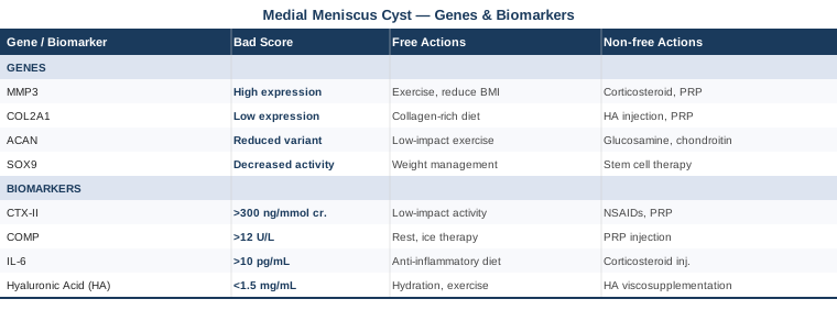
Summary Table: Genes and Biomarkers at a Glance
What Longevity Medicine Teaches Us About Joint Tissue — Key Insights from Outlive by Peter Attia
Outlive (2023) by Peter Attia, MD, references hundreds of studies to argue that modern medicine's reactive model — wait until something breaks, then treat it — is fundamentally mismatched with what the science actually shows about chronic disease and tissue degeneration. For a condition like medial meniscus cyst, the book's framework is directly applicable. Here are the ten most impactful insights from Attia's work applied to joint and meniscal health.
1. Medicine 3.0: You Are Too Late If You Wait for Symptoms
Attia's central thesis is that by the time a meniscal cyst is visible on MRI, the biological environment that caused it has been deteriorating for years. The only effective strategy is to begin intervening on inflammatory load, metabolic health, and connective tissue quality long before structural damage becomes visible — or to treat the first structural signal as an urgent wake-up call to address underlying biology, not just the surface finding.
2. VO2max Is the Most Powerful Biomarker for Musculoskeletal Longevity
Attia cites research showing that individuals in the top quartile of VO2max have dramatically lower all-cause mortality — and crucially, far better functional capacity as they age. For joint health specifically, VO2max reflects the aerobic capacity that enables Zone 2 training, which is the single exercise intervention most consistently shown to reduce systemic inflammatory markers relevant to joint tissue. A low VO2max means your inflammatory control system is underperforming.
3. Strength Training Is Not Optional for Joint Protection
Attia's review of muscle physiology makes clear that muscle mass is not just cosmetic — it is the primary load absorber for every joint in the lower body. Weak quadriceps and hip abductors dramatically increase the mechanical stress on the medial meniscus with every step. Progressive resistance training, even starting from low loads, is the most evidence-based long-term protection for knee tissue.
4. Metabolic Health Drives Joint Inflammation — Through Fat Tissue, Not Just Diet
Attia synthesizes research showing that visceral adipose tissue functions as an endocrine organ producing IL-6, TNF-α, and leptin — all of which directly drive synovial inflammation and accelerate cartilage breakdown. Metabolic dysfunction is not just a heart disease problem; it is a joint health problem. Improving insulin sensitivity is one of the most direct upstream interventions for reducing joint inflammatory load.
5. The Biomarkers Your Doctor Doesn't Order Are Often the Most Important
Attia is critical of standard lab panels that miss hsCRP, the omega-3 index, MMP-3, and comprehensive metabolic markers. He consistently advocates for more proactive biomarker tracking — exactly the approach this article takes. The gap between standard care labs and functional optimization labs is where the most actionable information often lives.
6. Insulin Resistance Silently Damages Connective Tissue
Elevated insulin promotes AGE formation and RAGE activation in joint tissue — the same pathway that drives MMP-3 overexpression in metabolically unhealthy individuals. Attia's detailed discussion of insulin resistance as a multi-tissue phenomenon helps explain why many people with knee pathology have no obvious metabolic diagnoses yet carry enough subclinical insulin resistance to significantly accelerate joint degeneration.
7. Sleep Is the Master Lever for Tissue Repair
Attia synthesizes research on sleep and tissue repair, emphasizing that slow-wave sleep is when growth hormone secretion peaks — the primary systemic signal for connective tissue maintenance and repair. Chronically poor sleep does not just affect energy; it measurably impairs the biological repair processes that keep meniscal and cartilage tissue healthy. Treating sleep as a recovery modality, not a passive activity, is fundamental to joint health.
8. Body Composition Changes Load — Even Small Ones
The biomechanical math is unambiguous: every kilogram of body weight translates to 3–4 kg of additional force on the medial knee compartment during walking, and up to 6–7× during jogging. Attia emphasizes that gradual, sustainable body composition changes — emphasizing muscle preservation while reducing fat — protect joints more durably than any single supplement or injection.
9. The Centenarian Decathlon: Train for Your Future Self
One of Attia's most practical frameworks is working backward from the physical capacities you want at age 80–85 to determine what you need to train now. Applied to knee health: if you want a functional, pain-free knee in 30 years, you need to optimize inflammatory load, maintain muscle mass, and protect meniscal integrity now — not wait for the condition to progress past the point where conservative management can succeed.
10. The Supplement Stack Should Follow Biomarker Data, Not General Advice
Attia is consistent on this point: supplementation without biomarker data is guessing. Taking omega-3 fish oil, vitamin D, and magnesium makes sense if your omega-3 index, 25-OH-D, and magnesium RBC levels confirm a deficit. Without measuring, you may be supplementing what you don't need while ignoring what you do. This is why the biomarker section of this article precedes the supplementation plans — the data should drive the protocol.
Complementary Approaches With Evidence for Knee and Meniscal Conditions
The following modalities have meaningful human clinical evidence relevant to meniscal and joint health. None of them replace structural management or the biomarker and genetic interventions above, but each can add measurable value when applied correctly.
Low-Level Laser Therapy (Photobiomodulation)
Photobiomodulation (PBM) uses specific wavelengths of red and near-infrared light (630–850 nm) to modulate cellular metabolism in musculoskeletal tissue. For medial meniscus cysts, it is relevant because it has demonstrated anti-inflammatory and tissue repair–promoting effects in periarticular tissue without the side effects of NSAIDs or corticosteroids. The mechanism involves cytochrome c oxidase activation in mitochondria, leading to reduced pro-inflammatory cytokines and increased cellular ATP production — a direct benefit for the metabolically stressed synovial environment surrounding a cyst.
A systematic review published on PubMed confirmed that LLLT produces statistically significant improvements in pain and function in knee OA, with the best evidence for wavelengths of 780–860 nm at doses of 4–8 J/cm². Evidence is moderate; results are most consistent for pain reduction and inflammation, less so for structural change.
Practically, apply a clinical-grade home PBM device directly over the medial knee for 10–20 minutes, 5 days per week. Maintain a consistent distance of 2–10 cm depending on device power. Devices in the $150–$600 range are available for home use; clinical devices are more powerful but require practitioner access. Do not use over implanted electronic devices, active neoplasm sites, or during pregnancy.
Tai Chi
Tai chi combines slow, controlled movement sequences with weight shifting and balance training — a uniquely appropriate modality for knee rehabilitation because it builds the neuromuscular control and proprioception that protect meniscal tissue from re-injury without imposing high compressive loads. For medial meniscus cysts, the medial compartment unloading during many tai chi postures and the emphasis on controlled hip and knee alignment makes it biomechanically relevant.
A landmark randomized controlled trial (Wang et al.) comparing tai chi to physical therapy in knee OA found equivalence in pain reduction and function improvement over 12 weeks, with tai chi providing additional benefits in psychological wellbeing and balance — factors relevant to long-term joint protection. This is one of the strongest evidence bases for any mind-body modality in knee conditions.
Begin with a supervised group class or instructor-guided video program 2–3 times per week, focusing on Sun-style tai chi (which uses higher stances and less deep knee flexion, reducing medial compartment stress during learning). Progress to 45–60 minute sessions as tolerated. Allow 8–12 weeks before expecting measurable pain and function improvements. Avoid deep knee bend postures initially; communicate your meniscal condition to your instructor.
Mindfulness Meditation / MBSR
Mindfulness-Based Stress Reduction (MBSR) is relevant to medial meniscus cysts not because it treats the structural problem, but because chronic pain conditions involve central sensitization — a neurological amplification of pain signals that persists independently of tissue damage. People with persistent knee pain often exhibit central sensitization that makes their pain experience disproportionate to structural findings, and cortisol-driven inflammation from psychological stress directly worsens IL-6 and CRP levels. MBSR addresses both.
Multiple meta-analyses confirm that MBSR reduces chronic musculoskeletal pain scores and improves function, with effect sizes moderate to large for pain interference. A key mechanism is downregulation of the hypothalamic-pituitary-adrenal (HPA) axis, which reduces cortisol and its downstream NF-κB activation — the same pathway that drives MMP-3 expression in genetically susceptible individuals.
Practice the standard MBSR format: 8-week program with 45 minutes of daily practice (body scan, sitting meditation, gentle movement). Available in-person through hospital programs, or via validated digital formats (Palouse Mindfulness online program is free and clinician-developed). For chronic pain specifically, the body scan component — systematic attention to physical sensation without judgment — is the most important element. Set a minimum 8-week commitment before evaluating results.
Massage Therapy
Massage therapy for meniscal cyst does not directly address the cyst itself — but it is highly relevant for the muscle tension patterns, restricted fascia, and altered joint mechanics that develop secondarily to pain avoidance and guarding. The medial knee in chronic pain typically presents with tight medial hamstrings, restricted posterior capsule, and compensatory hip and calf tightening — all of which increase medial compartment load and worsen the inflammatory environment.
Systematic reviews on massage in knee pain show consistent improvement in pain and range of motion, with the strongest evidence for Swedish and deep tissue techniques applied to the surrounding musculature rather than directly over the joint itself. A 2019 RCT found that 8 weeks of massage (60-minute sessions, weekly) produced significant improvements in knee OA pain and physical function.
Practically, seek a licensed massage therapist with experience in sports or orthopedic conditions. Focus sessions on medial hamstrings, posterior calf, adductor complex, and IT band — the structures that most influence medial compartment mechanics — rather than directly on the cyst area. Frequency: once per week during active recovery, tapering to once per month for maintenance. Sessions typically cost $60–$120 each. Self-massage with a foam roller or targeted massage gun (on surrounding muscles only, not directly over the joint) can supplement professional treatment at significantly lower cost.
Conclusion
A medial meniscus cyst is a structural finding, but it is also a signal from a biological environment that deserves closer attention. The six biomarkers covered here — hsCRP, IL-6, MMP-3, vitamin D, the omega-3 index, and uric acid — are measurable, modifiable, and directly relevant to the mechanisms driving cyst formation and persistence. The five genes — MMP3, COL2A1, GDF5, ACAN, and VEGFA — explain why some people are more vulnerable and why the same standard advice produces such different results in different individuals.
The next smart step is to pick one or two biomarkers to test first — hsCRP and 25-OH vitamin D are the most accessible and highest-leverage starting points — and use the results to guide a specific, targeted action plan. If you have access to genetic testing through a clinical or consumer platform, the gene variants covered here are worth reviewing with a physician who can contextualize the findings. And if you are currently under orthopedic care for the cyst, none of these interventions conflict with standard management — they work alongside it, at the level where structural treatment alone cannot reach.
Musculoskeletal: Joint Conditions Sports Injuries
Autoimmune: Inflammatory Conditions Connective Tissue Conditions