This article contains AI generated content.
· UpdatedMedial Patellofemoral Ligament Tear — 5 Genes and 6 Biomarkers to Track
Introduction
A medial patellofemoral ligament tear rarely happens in isolation from the rest of your biology. The ligament ruptures when the patella dislocates laterally — during a pivot, a collision, or something as routine as a misstep off a curb — and what follows depends on far more than your surgical technique or how consistently you show up to physical therapy. Two patients can receive identical reconstructions, follow identical protocols, and arrive at six months in entirely different states. One returns to sport; the other still has instability, persistent swelling, and the unsettling feeling that healing has stalled.
That gap is not random. Much of it is biochemical, and much of that biochemistry is measurable. The speed of collagen synthesis, the degree of chronic low-grade inflammation running in the background, the hormonal environment your tissue is remodeling inside, and the genetic architecture of your connective tissue all shape the recovery trajectory in ways that standard orthopedic protocols never address. This is not a criticism of orthopedic care — it simply operates at a different level of resolution than the molecular one.
Generic post-injury advice — eat well, sleep enough, do your exercises — is accurate but imprecise. It describes the right direction without telling you where you actually stand. If your vitamin D is low, your IGF-1 is blunted, and your body is running in a low-grade inflammatory state, the standard eight-month MPFL recovery timeline is a fantasy. Knowing your numbers changes the quality of every decision you make during rehab.
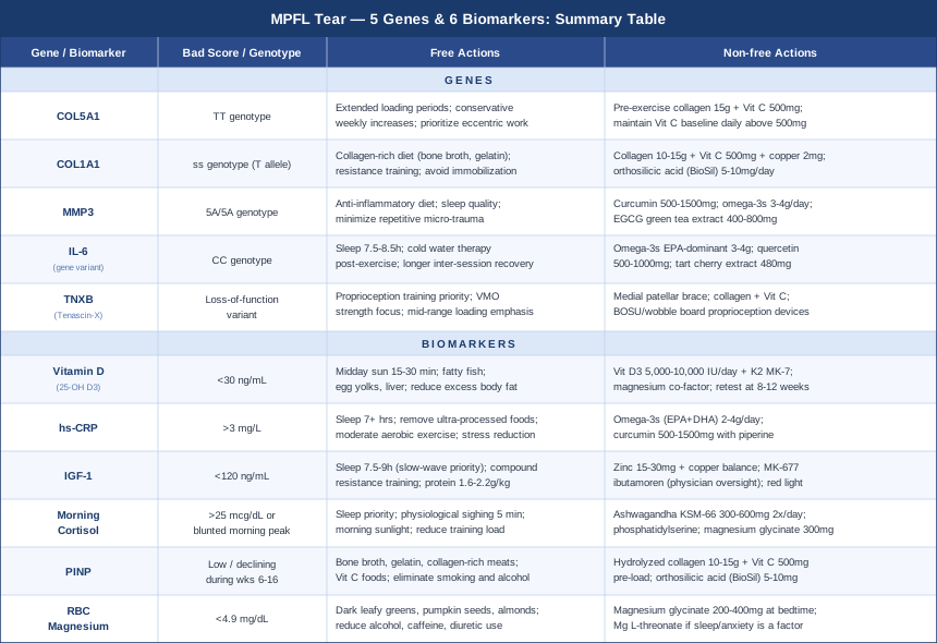
This article approaches the problem from two complementary angles. The first — and most actionable for most people — is a set of six measurable biomarkers that directly reflect your body's capacity to repair connective tissue. All are accessible through standard blood work or low-cost add-ons. The second covers five gene variants with well-documented or emerging links to soft tissue injury susceptibility and healing rate. Together, these frameworks offer a sharper, more individualized picture of what your body needs — and where it may already be limiting your progress.
6 Biomarkers That Reflect Your MPFL Healing Capacity
Post-injury blood work is typically ordered to rule out infection or check general organ function. None of the standard panels assess what your connective tissue repair machinery is actually doing. The six markers below are different — they reflect the specific biological processes that drive ligament remodeling, collagen synthesis, and inflammatory resolution. Most can be added inexpensively to a routine panel. What they show, collectively, is the biochemical environment your body is working in while trying to rebuild.
1. Vitamin D (25-OH Vitamin D)
Vitamin D functions as a steroid hormone, and its receptors are present in skeletal muscle, cartilage, and ligament fibroblasts — the very cells responsible for producing new MPFL tissue. It upregulates TGF-β signaling in fibroblasts, driving collagen type I synthesis, and simultaneously modulates the NF-κB inflammatory pathway to keep chronic inflammation in check. Low vitamin D does not just affect bone; it measurably blunts the cellular machinery that rebuilds soft tissue. Several prospective cohort studies have found that athletes with levels below 30 ng/mL experience higher soft tissue injury rates and longer recovery windows than those maintaining levels between 40 and 60 ng/mL.
The practical relevance for MPFL recovery is direct. A person with a level of 18 ng/mL entering a post-surgical rehab program is not operating the same repair biology as someone at 52 ng/mL. No amount of physical therapy compensates for a fibroblast environment that is vitamin D deficient.
How to Measure It
Test: Serum 25-hydroxyvitamin D (25-OH D3). Standard through any blood lab. Cost: $30–$70 without insurance; frequently covered with a physician's note. Target for soft tissue repair: 40–60 ng/mL. Below 30 ng/mL is deficient; 30–40 ng/mL is suboptimal in the context of active ligament healing.
If the Score Is Low: Plan Without Supplements
Midday sun exposure: Direct sun on large skin surface areas (arms, legs, torso) for 15–30 minutes around solar noon, depending on latitude and skin tone. This can raise levels by 5–15 ng/mL over 8–12 weeks in suitable climates. Food: Fatty fish (salmon, mackerel, sardines), egg yolks, and liver contribute meaningfully. Food alone rarely corrects a significant deficit, but it reduces the gap. Body composition: Adipose tissue sequesters vitamin D; reducing excess body fat improves circulating levels over time.
If the Score Is Low: Plan with Supplements or Equipment
Vitamin D3 (cholecalciferol), not D2: For levels below 20 ng/mL, 5,000–10,000 IU daily for 8–12 weeks is a common repletion protocol, with follow-up testing before adjusting. For levels between 20 and 35 ng/mL, 3,000–5,000 IU daily is typically sufficient. Essential co-factors: Always pair with vitamin K2 (100–200 mcg MK-7 form) to correctly route calcium metabolism. Magnesium is required for vitamin D conversion — address it simultaneously. Frequency and cycling: Daily dosing; retest at 8–12 weeks. Maintain the dose that keeps you in the 40–60 ng/mL range without cycling. Side effects: Hypercalcemia is possible at sustained doses above 10,000 IU/day without monitoring. Stay within evidence-supported ranges and retest.
2. High-Sensitivity CRP (hs-CRP)
Chronic low-grade inflammation is one of the most underappreciated barriers to connective tissue recovery. Ligament repair requires an organized inflammatory environment — acute resolution followed by clean matrix remodeling. When systemic inflammation is persistently elevated, even mildly, the balance tips toward degradation. Pro-inflammatory cytokines like TNF-α and IL-6 upregulate matrix metalloproteinases (MMPs), enzymes that break down collagen scaffolding before new fibers can form. The repair process is essentially competing against its own demolition crew.
High-sensitivity CRP is the most accessible proxy for this state. It is not a direct measure of local tissue inflammation, but it reflects the hepatic response to systemic inflammatory signaling and is inexpensive, standardized, and clinically meaningful. A persistently elevated hs-CRP — above 1–3 mg/L outside the acute post-injury phase — signals that the biological environment is working against organized repair.
How to Measure It
Test: High-sensitivity CRP. Standard add-on to any metabolic panel. Cost: $15–$40. Interpretation: Below 1 mg/L is optimal; 1–3 mg/L is mildly elevated; above 3 mg/L warrants investigation. Note: hs-CRP will naturally be elevated in the first 4–8 weeks after surgery or acute injury — this test is most useful once the acute inflammatory phase has resolved.
If the Score Is High: Plan Without Supplements
Sleep: Chronic sleep restriction (under 7 hours) is one of the most reliable drivers of elevated CRP. Addressing sleep quality is the single highest-leverage free action available. Diet: Remove ultra-processed foods, refined seed oils, and high-glycemic staples. Increase omega-3-rich fish, colorful vegetables, and polyphenol-dense fruits. Moderate aerobic activity: Sustained moderate-intensity exercise (not intense training, which acutely spikes CRP) is a well-documented chronic inflammation suppressor. Appropriate even during MPFL recovery through low-impact modalities. Stress management: Chronic psychological stress drives cortisol dysregulation, which feeds hs-CRP. Breathwork, social connection, and reducing sustained cognitive load all contribute.
If the Score Is High: Plan with Supplements or Equipment
Omega-3 fatty acids (EPA+DHA): 2–4 g/day of combined EPA+DHA from pharmaceutical-grade fish oil. This is among the most evidence-supported anti-inflammatory interventions available. Measurable effects on hs-CRP appear at 6–8 weeks. Curcumin (with piperine or phospholipid delivery): 500–1500 mg/day of standardized curcumin extract. Works through NF-κB inhibition — the same pathway that drives MMP upregulation. Cycling: Omega-3s can be maintained indefinitely. Curcumin: 8–12 week cycles with breaks, or maintained at lower doses. Side effects: High-dose fish oil may mildly reduce platelet aggregation — note this in perioperative contexts. Curcumin rarely causes GI discomfort at standard doses.
3. IGF-1 (Insulin-Like Growth Factor 1)
IGF-1 is the downstream effector of growth hormone and the primary anabolic signal driving tissue repair in ligaments, tendons, and muscle. Ligament fibroblasts express IGF-1 receptors, and IGF-1 signaling directly stimulates collagen type I and III synthesis, fibroblast proliferation, and new extracellular matrix formation. After MPFL reconstruction, the adequacy of IGF-1 signaling is a primary determinant of how quickly quality tissue rebuilds.
IGF-1 declines with age, poor sleep, chronic caloric restriction, elevated cortisol, and sedentary behavior. Many patients presenting with MPFL tears — particularly those who are older, overtrained, or chronically sleep-deprived — are already operating with suboptimal IGF-1 before the injury. Peter Attia and other clinicians who prioritize tissue quality and longevity routinely include IGF-1 in their panels precisely because it sits at the intersection of repair and aging biology.
How to Measure It
Test: Serum IGF-1. Cost: $50–$120. Optimal range: Context-dependent by age, but generally 150–250 ng/mL in adults is associated with effective tissue repair. Below 120 ng/mL in someone under 60 warrants attention. Lab reference ranges are age-adjusted — what matters is where you sit in the healthy upper quartile for your age bracket, not merely "within range."
If the Score Is Low: Plan Without Supplements
Sleep optimization: Growth hormone — and therefore IGF-1 — is released primarily during slow-wave sleep. Prioritizing 7.5–9 hours of quality sleep (cool, dark room; consistent schedule; minimal alcohol before bed) is the most powerful lever for raising IGF-1 without any supplement. Resistance training: Multi-joint compound movements acutely spike growth hormone and raise baseline IGF-1 over time. Upper-body resistance work during MPFL recovery preserves this stimulus while the lower extremity heals. Protein distribution: Adequate dietary protein (1.6–2.2 g/kg body weight) distributed across meals supports IGF-1 signaling. Note: aggressive intermittent fasting predictably reduces IGF-1 — relevant if you practice it. Zinc adequacy: Zinc deficiency blunts GH-IGF-1 axis function; dietary sources include oysters, red meat, and pumpkin seeds.
If the Score Is Low: Plan with Supplements or Equipment
Zinc (if deficient): 15–30 mg elemental zinc daily with food; balance with 1–2 mg copper for long-term use. MK-677 (ibutamoren): A GH secretagogue that reliably raises IGF-1 by 40–70% in clinical trials. Not an approved pharmaceutical in most jurisdictions; requires physician oversight. Red light therapy / photobiomodulation: Applied over large muscle groups, some trials report increases in local GH-related signaling as a secondary effect — useful as an adjunct. Cycling: MK-677: 8–12 weeks on, 4 weeks off. Monitor fasting glucose during use. Side effects: MK-677 increases appetite and may cause transient water retention; insulin sensitivity should be monitored, especially in predisposed individuals.
4. Morning Cortisol
Cortisol is catabolic — it breaks down tissue to liberate fuel — which makes chronically elevated cortisol directly antagonistic to ligament repair. The normal pattern sees cortisol rise sharply in the 30–45 minutes after waking (the cortisol awakening response), then decline through the day. When cortisol is persistently high due to sustained psychological stress, poor sleep, overtraining, or aggressive caloric restriction, it suppresses collagen synthesis, reduces fibroblast activity, and accelerates the MMP-driven matrix degradation that undoes organized repair.
What makes this clinically relevant is the reality of how many MPFL patients recover: under genuine psychosocial pressure, fearful of re-injury, often with disrupted sleep. Testing morning cortisol gives a quantitative read on whether catabolic pressure is actively competing with the anabolic repair process — and by how much.
How to Measure It
Test: Morning serum cortisol (blood draw between 7–9 AM) or a 4-point salivary cortisol panel (showing the full diurnal curve). Serum cost: $30–$60. Salivary panel: $100–$250 through functional medicine labs. Optimal: Morning serum cortisol 10–20 mcg/dL. Consistently above 25 mcg/dL, or a blunted morning peak with elevated evening values, both indicate dysregulation worth addressing.
If the Score Is Dysregulated: Plan Without Supplements
Sleep: The most powerful cortisol modulator available. Chronic sleep debt directly and reliably elevates cortisol — there is no supplement workaround for this. Physiological sighing: A double inhale through the nose followed by a long exhale through the mouth, practiced for 5 minutes daily, reliably reduces sympathetic tone. A 2023 Stanford randomized controlled trial found this specific pattern outperformed mindfulness-based techniques for acute stress reduction. Morning sunlight: Sun exposure within 60 minutes of waking calibrates the cortisol awakening response appropriately. Reducing training load: If overtraining is the driver, reducing volume or intensity lowers cumulative allostatic load meaningfully.
If the Score Is Dysregulated: Plan with Supplements or Equipment
Ashwagandha (KSM-66 standardized extract): The most rigorously studied adaptogen for cortisol management. 300–600 mg twice daily with food. A published randomized controlled trial (Chandrasekhar et al., 2012) demonstrated a 27.9% reduction in serum cortisol compared to placebo over 60 days. Phosphatidylserine: 400–800 mg/day specifically blunts cortisol response to exercise stress — useful when overtraining is the primary driver. Magnesium glycinate: Magnesium deficiency directly elevates cortisol; supplementation normalizes it. 300–400 mg before bed. Cycling: Ashwagandha: 8–12 week cycles, 4-week break. Phosphatidylserine: continuous use at standard doses is well-tolerated. Side effects: Ashwagandha may cause GI discomfort in a minority; avoid in pregnancy. Phosphatidylserine has an excellent safety profile.
5. PINP (Procollagen Type I N-Propeptide)
PINP is a direct byproduct of collagen type I synthesis — it is cleaved from the procollagen molecule as new collagen fibers form and released into the bloodstream. Measuring serum PINP provides a near-real-time, quantitative signal of how actively your body is building collagen. This makes it mechanistically specific to MPFL healing in a way that no general inflammation marker can be, since the MPFL is primarily a type I collagen structure.
PINP is most commonly used in osteoporosis management, where it tracks bone collagen formation in response to treatment. Its relevance to soft tissue repair is equally direct and significantly underused. A low PINP during a phase of active expected repair suggests the collagen synthesis machinery is underperforming — possibly due to vitamin C insufficiency, low vitamin D, inadequate protein intake, suppressed IGF-1, or chronic inflammation blocking fibroblast activity.
How to Measure It
Test: Serum PINP (also written P1NP). Available through specialty and hospital labs; ask specifically, as it is not on standard panels. Cost: $60–$150 depending on the lab. Interpretation: Adult reference ranges vary by sex and lab (approximately 15–70 mcg/L in healthy adults). The trend over time is more informative than a single reading — you want to see levels rise during active remodeling (roughly weeks 6–16 post-injury) and normalize subsequently.
If the Score Is Low: Plan Without Supplements
Collagen-rich dietary protein: Collagen synthesis requires glycine, proline, and hydroxyproline in abundance — the specific amino acid profile found in bone broth, skin-on slow-cooked meat, and gelatin. These supply structural amino acids that standard protein powders do not. Vitamin C from food: Vitamin C is an essential co-factor for prolyl and lysyl hydroxylase — the enzymes that crosslink collagen chains into mechanically functional fibers. Bell peppers, citrus, kiwi, and berries are high-density sources. Remove collagen suppressors: Smoking, chronic alcohol intake, and glucocorticoid use all reliably suppress collagen synthesis. Each must be addressed directly.
If the Score Is Low: Plan with Supplements or Equipment
Hydrolyzed collagen peptides + vitamin C protocol: 10–15 g of hydrolyzed collagen taken with 500 mg vitamin C, 30–60 minutes before loading exercise. This timing is supported by research from Keith Baar's lab (Shaw et al., 2017) showing that this combination doubles collagen synthesis markers in tendons compared to placebo. Orthosilicic acid (BioSil): Silicon supports collagen crosslinking quality. 5–10 mg of stabilized orthosilicic acid daily has been shown in a small RCT to increase PINP. Frequency: Daily. Cycling: Collagen peptides and vitamin C require no cycling — indefinitely safe at these doses. Side effects: Very well-tolerated. Vitamin C above 2 g/day may cause loose stools in sensitive individuals.
6. RBC Magnesium
Standard serum magnesium — the version that appears on most blood panels — is a poor indicator of actual magnesium status. The body maintains serum levels within a tight range by drawing from intracellular stores, meaning serum magnesium can be entirely normal while intracellular reserves are significantly depleted. RBC (red blood cell) magnesium provides a far more accurate picture of true functional status.
Why does this matter for MPFL recovery? Magnesium participates in over 300 enzymatic reactions, including ATP production (the energy currency of cellular repair), protein synthesis regulation, and inflammatory signaling. Magnesium insufficiency increases systemic inflammation — elevating both hs-CRP and IL-6 — impairs protein synthesis, reduces sleep quality, and destabilizes cortisol regulation. A meaningful fraction of athletes and high-stress individuals are functionally magnesium insufficient, often without knowing it because their serum level appears normal.
How to Measure It
Test: RBC magnesium — not serum magnesium. Specify this explicitly when ordering. Cost: $40–$80 through specialty labs; available via LabCorp and Quest in the US, and through GP request in most other countries. Optimal: 5.5–6.5 mg/dL. Below 4.9 mg/dL is functionally low regardless of serum values.
If the Score Is Low: Plan Without Supplements
Dietary sources: Dark leafy greens (spinach, Swiss chard), pumpkin seeds, almonds, dark chocolate, and whole grains are the highest-density food sources. A deliberate effort to include these daily can raise RBC magnesium meaningfully over 8–12 weeks. Reduce depletors: Alcohol, high caffeine intake, excessive sweating, and diuretic medications all significantly increase magnesium excretion and should be reduced or managed. Mineral water: Some still mineral waters have high magnesium content and contribute to intake over time.
If the Score Is Low: Plan with Supplements or Equipment
Magnesium glycinate: 200–400 mg elemental magnesium before bed. The glycinate form is best-tolerated (minimal GI effect) and well-absorbed. Avoid magnesium oxide — poorly absorbed despite appearing high on labels. Magnesium malate: Useful for daytime use and energy metabolism support. Magnesium L-threonate: Penetrates the blood-brain barrier more effectively — useful if sleep disruption or anxiety is a significant factor in recovery. Cycling: No cycling required. Long-term daily use is safe and beneficial in insufficient individuals. Side effects: High doses of oxide or citrate forms cause loose stools; glycinate is consistently better tolerated. Magnesium toxicity is only possible in renal impairment.
The biomarker picture is most useful when read alongside the genetic layer — because what your genes predict, your biomarkers often confirm.
The Genetic Architecture of Soft Tissue Injury: 5 Variants That Matter
Genetics does not determine outcomes, but it shifts baseline risk and alters how the body responds to both injury and recovery interventions. Certain gene variants reliably change the quality of collagen produced, the efficiency of matrix remodeling, or the intensity of inflammatory responses — all of which shape how an MPFL heals. Understanding this layer transforms vague lifestyle recommendations into mechanistically justified decisions.
The five variants below are the best-characterized in the context of ligament and soft tissue injury. Most are accessible through consumer genetic panels (23andMe, AncestryDNA) with third-party analysis tools, or through clinical genetic testing when ordered by a physician.
COL5A1 — The Collagen Fibril Organizer
COL5A1 encodes the alpha-1 chain of collagen type V, which regulates the diameter and uniformity of collagen type I fibrils. Smaller, more regular fibrils produce mechanically superior connective tissue; COL5A1 variants that reduce its function result in larger, less organized fibrils that are more prone to tearing under load. The BstUI RFLP (rs12722) is the most studied polymorphism, with the TT genotype associated with Achilles tendinopathy, ACL injury, and broader soft tissue vulnerability across multiple independent cohorts.
If the Genotype Is Unfavorable: Plan Without Supplements
Extended progressive loading: COL5A1 unfavorable genotypes require longer connective tissue adaptation periods. Double conservative return-to-sport estimates. The connective tissue lags further behind muscular recovery in this genotype, and the gap is measurable. Eccentric exercise priority: Controlled eccentric loading (slow lengthening contractions) is the gold standard for driving collagen fibril remodeling and is especially critical when baseline fibril organization is compromised. Conservative weekly load increases: The 10% volume increase rule is especially relevant — rapid load increases impose forces that outstrip the adaptation capacity of already-inferior fibrils.
If the Genotype Is Unfavorable: Plan with Supplements or Equipment
Pre-exercise collagen protocol: 15 g hydrolyzed collagen with 500 mg vitamin C, taken 30–60 minutes before loading activity, 5–6 days per week. This timing seeds the tissue with building blocks during the mechanical loading window when synthesis is upregulated. Vitamin C baseline: Maintain daily intake above 500 mg to support prolyl hydroxylase-dependent crosslinking. Cycling: Daily, with training; sustainable long-term without breaks. Side effects: None significant at these doses.
COL1A1 — The Structural Backbone
COL1A1 encodes the primary α-1 chain of type I collagen — the predominant structural protein in virtually all major ligaments, including the MPFL. The Sp1 G/T polymorphism (rs1800012) affects transcription of the gene; the "s" allele (T variant) alters collagen fiber quality, and the homozygous ss genotype is associated across multiple studies with ACL rupture risk, reduced bone mineral density, and general soft tissue vulnerability. This is not a rare variant — it appears in a meaningful minority of the population and is consistently linked to connective tissue injury outcomes.
If the Genotype Is Unfavorable: Plan Without Supplements
Collagen-rich dietary protein: Glycine and proline — the building blocks that COL1A1 encodes — should be supplied liberally through bone broth, slow-cooked connective tissue-rich meats, and gelatin. Avoid prolonged immobilization: Immobilization rapidly degrades collagen quality and density. Even within MPFL recovery constraints, early gentle range of motion maintains tissue turnover and slows net collagen loss. Consistent resistance training: Resistance training upregulates COL1A1 gene expression in tendons — one of the few ways to partially compensate for a less favorable starting point in this gene.
If the Genotype Is Unfavorable: Plan with Supplements or Equipment
Collagen synthesis stack: 10–15 g hydrolyzed collagen + 500 mg vitamin C + 2 mg copper daily. Copper is a co-factor for lysyl oxidase, the enzyme that crosslinks collagen fibers into mature, mechanically strong structures — directly targeting the structural weakness associated with COL1A1 variants. Orthosilicic acid: BioSil or equivalent at 5–10 mg daily supports collagen crosslinking quality. Cycling: Continuous; all components safe for long-term use. Side effects: Copper supplementation above 3 mg/day long-term may compete with zinc; ensure adequate zinc intake (15–25 mg daily).
MMP3 — The Matrix Remodeler
Matrix metalloproteinase 3 (MMP-3) is an enzyme that degrades extracellular matrix proteins — collagen, fibronectin, and proteoglycans. In acute, controlled bursts it is essential for clearing damaged tissue and enabling repair. Chronically upregulated, it destroys the repair scaffold before new tissue can consolidate. The 5A/6A promoter polymorphism (rs3025058) directly affects MMP-3 transcription: the 5A allele produces higher MMP-3 activity, and the 5A/5A genotype is associated with more aggressive connective tissue degradation post-injury, harder-to-maintain surgical repairs, and greater cartilage vulnerability in osteoarthritis models.
If the Genotype Is Unfavorable: Plan Without Supplements
Anti-inflammatory lifestyle as primary strategy: MMP-3 expression is driven by pro-inflammatory cytokines — specifically IL-1β and TNF-α. Any practice that consistently reduces chronic inflammation (sleep, low-glycemic diet, stress reduction) directly reduces the transcriptional signal that activates excess MMP-3. Minimize repetitive micro-trauma: Each minor trauma event re-triggers inflammatory cascades that upregulate MMP-3. Protective bracing, proper footwear with adequate cushioning, and surface awareness matter more in this genotype.
If the Genotype Is Unfavorable: Plan with Supplements or Equipment
Curcumin (NF-κB inhibitor): One of the most evidence-backed MMP-3 modulators. 500–1500 mg standardized extract daily. Omega-3 fatty acids (EPA): EPA reduces the pro-inflammatory cytokines that drive MMP-3 transcription. 3–4 g EPA+DHA daily. EGCG (green tea extract): A well-characterized MMP inhibitor in multiple human and in-vitro studies. 400–800 mg standardized extract daily. Cycling: Curcumin and EGCG: 8 weeks on, 4 weeks off. Omega-3s: continuous. Side effects: EGCG above 800 mg/day may cause GI discomfort or mild transient liver enzyme elevation in sensitive individuals — stay within standard ranges.
IL-6 — The Inflammatory Amplifier
IL-6 plays a dual role in tissue biology: in short, acute pulses it is pro-regenerative and necessary for initiating repair. Chronically elevated, it sustains a low-grade inflammatory state that actively inhibits late-phase ligament remodeling. The -174 G/C promoter polymorphism (rs1800795) affects how much IL-6 is transcribed in response to stimuli. The CC genotype produces higher baseline and stimulated IL-6 levels. In an MPFL recovery context, this means injury-related inflammation may persist longer, resolve less cleanly, and interfere with the late remodeling phase that determines final ligament quality and tensile strength.
If the Genotype Is Unfavorable: Plan Without Supplements
Sleep as non-negotiable: Even partial sleep restriction markedly elevates IL-6 in healthy subjects. For CC carriers, this effect is amplified. Targeting 7.5–8.5 hours consistently is not optional. Cold water therapy post-exercise: Brief cold water immersion following loading sessions acutely reduces inflammatory cytokine activity and may help CC genotype carriers manage excessive post-load IL-6 responses that would otherwise delay recovery. Longer recovery between sessions: High-intensity training drives acute IL-6 sharply; CC genotype carriers benefit from longer inter-session recovery compared to average recommendations.
If the Genotype Is Unfavorable: Plan with Supplements or Equipment
Omega-3s (EPA-dominant): EPA is one of the most evidence-backed IL-6 modulators. 3–4 g EPA+DHA daily. Quercetin: A flavonoid with demonstrated IL-6 inhibition properties. 500–1000 mg/day, often combined with bromelain for improved absorption and synergistic effect. Tart cherry extract: Shown in multiple human trials to reduce post-exercise IL-6 and inflammatory marker profiles. 480 mg concentrate or equivalent daily. Cycling: Quercetin: 8-week cycles; tart cherry: continuous use is generally safe. Side effects: Quercetin at very high doses (above 2 g/day) may modestly inhibit thyroid peroxidase — stay within standard dose ranges.
TNXB — The Hidden Hypermobility Factor
Tenascin-X, encoded by TNXB, is an extracellular matrix glycoprotein that organizes collagen fiber assembly and regulates mechanical tissue tension. Haploinsufficiency or loss-of-function variants in TNXB produce a connective tissue phenotype closely resembling hypermobile Ehlers-Danlos syndrome: joint hypermobility, skin extensibility, and impaired ligamentous mechanical integrity. TNXB deficiency disrupts the regular lateral orientation of collagen fibrils, reducing the tensile capacity of ligaments across the body. Individuals with TNXB variants may experience recurrent patellar instability even after technically successful MPFL reconstruction — not because the surgery failed, but because the reconstructed ligament cannot develop normal tension in a TNXB-deficient tissue environment.
If the Genotype Is Unfavorable: Plan Without Supplements
Proprioception training as a priority, not an afterthought: In TNXB-associated hypermobility, mechanosensory feedback from joints is impaired alongside structural laxity. Single-leg balance training, wobble board work, and progressive neuromuscular re-education are not optional extras — they are the primary compensatory strategy. VMO strength priority: The VMO (vastus medialis oblique) is the most important active patellar stabilizer. When passive ligamentous stabilization is compromised, dynamic muscular stability must carry more of the load. Mid-range loading emphasis: Hypermobile connective tissue is most vulnerable at end-range positions. Train through mid-range and build progressively toward end-range only as strength and proprioceptive control are established.
If the Genotype Is Unfavorable: Plan with Supplements or Equipment
Medial patellar stabilizing brace: A well-fitted brace with medial buttress offloads the MPFL during activity — especially critical in TNXB-compromised tissue where the reconstructed ligament faces increased laxity demands. Collagen + vitamin C protocol: Same as described above — TNXB-deficient tissue derives particular benefit from optimizing crosslinking quality since fibril organization is the structural weakness. Proprioceptive training devices: BOSU balance trainers, wobble boards, and resistance-band feedback training progressively rebuild neuromuscular joint stabilization in hypermobile joints. Cycling: Equipment use as needed; collagen protocol continuous. Side effects: None specific to these interventions.
What the Huberman Lab Gets Right About Connective Tissue Healing — 10 Things Worth Knowing
The Huberman Lab podcast episode on connective tissue biology — particularly the conversation with Dr. Keith Baar (University of California, Davis), one of the world's leading researchers on tendon and ligament physiology — presents a picture of connective tissue healing that diverges significantly from standard clinical guidance. Baar's work directly challenges the idea that rest is the optimal healing strategy, and his research on collagen synthesis timing, loading windows, and supplementation protocols has shifted how elite sports medicine practitioners approach soft tissue injuries. Ten things from that body of work that most patients are never told:
1. Ligaments and Tendons Are Poorly Vascularized — and That Changes Everything
Unlike muscle, which is richly supplied with blood vessels that deliver nutrients and remove metabolic waste rapidly, tendons and ligaments are largely avascular. They receive nutrients through diffusion, not direct blood flow. This is why connective tissue heals more slowly than muscle, why nutritional interventions must be timed precisely around mechanical loading (when local blood flow temporarily increases), and why systemic interventions like vitamin D or omega-3 status matter so much — they represent the molecular environment that avascular tissue must work with.
2. There Is a 6-Hour Collagen Synthesis Window After Loading
Mechanical loading triggers a burst of collagen gene expression in fibroblasts that peaks approximately 4–6 hours post-load and returns to baseline within 24 hours. This means that every loading session opens a brief anabolic window for connective tissue — but only if the necessary substrates (amino acids, vitamin C, growth factors) are present. Miss the window, and the mechanical stimulus produces no net synthesis.
3. Gelatin + Vitamin C Before Exercise Is One of the Few Supplement Combinations With Direct Mechanistic Support
Consuming 15 g of gelatin (or hydrolyzed collagen) with 500 mg of vitamin C approximately 30–60 minutes before a loading session significantly increases circulating hydroxyproline and pro-collagen type I — the building blocks and markers of collagen synthesis. In the Shaw et al. 2017 RCT, this protocol doubled collagen synthesis markers in tendon tissue compared to placebo. The timing is the mechanism: substrate delivery coincides with mechanically-triggered synthesis upregulation.
4. Intermittent Loading Is Vastly Superior to Continuous Loading for Connective Tissue
Continuous loading stresses connective tissue past its recovery capacity. Intermittent loading — short bouts of exercise separated by recovery periods — produces dramatically better collagen synthesis outcomes. The practical implication for MPFL rehab: multiple short sessions per day (10–15 minutes each) may produce better tissue-level outcomes than one 45-minute session, particularly in the earlier phases of remodeling.
5. Isometric Holds Reduce Pain While Stimulating Repair Simultaneously
Sustained isometric contractions at moderate intensity (around 70% of maximum, held for 30–45 seconds) have been shown to produce powerful analgesic effects in patellar and Achilles tendon conditions, with pain reduction persisting 45 minutes post-exercise. Crucially, these isometric holds also stimulate collagen synthesis without the compressive or shear forces of dynamic loading. For early MPFL recovery, appropriately targeted isometrics are among the most evidence-aligned tools available.
6. You Cannot Out-Pace Muscle Adaptation With Connective Tissue — But You Can Close the Gap
Muscle can roughly double its cross-sectional area in 12 weeks with aggressive training. Connective tissue adapts in the range of 10–15% over the same period. This mismatch is where re-injury happens: muscle gets stronger, the patient feels confident, loads increase — and the connective tissue is not ready. Understanding this biological gap changes the logic of return-to-sport decisions. The gap can be narrowed (not eliminated) with optimal nutrition, loading protocol design, and the collagen synthesis timing strategies above.
7. Ice Immediately After Exercise May Blunt Connective Tissue Adaptation
Applying ice immediately after a connective tissue loading session vasoconstricts the already poor local blood supply, reduces the post-exercise inflammatory signal that drives adaptation, and may impair the collagen synthesis response. Baar's position — and increasingly the consensus in sports science — is that cryotherapy immediately post-exercise is counterproductive for adaptation goals. For acute injury with significant swelling, the calculus is different; but for routine post-exercise recovery during rehab, ice should be used with caution.
8. Sleep Is a Structural Healing Variable, Not Just Recovery
Growth hormone, released primarily during slow-wave sleep, drives IGF-1 signaling that directly stimulates collagen synthesis in ligament fibroblasts. A night of disrupted sleep is not merely a subjective inconvenience — it is a measurable reduction in the hormonal signal that drives MPFL repair. Optimizing sleep is a structural intervention, not a lifestyle suggestion.
9. Blood Flow Restriction Training Changes the Calculus for Injured Joints
BFR training — using cuffs or wraps to restrict venous return during low-load exercise — enables meaningful hypertrophic and strength stimulus at loads too low to stress healing connective tissue. In MPFL recovery contexts, BFR allows the surrounding musculature (VMO, quadriceps) to be loaded productively while the ligament is still in early remodeling, preventing the muscle atrophy that typically accompanies conservative rehab.
10. The Biological Timeline for Ligament Remodeling Is Longer Than Most Return-to-Sport Protocols
Collagen type III (weaker, laid down in early repair) begins transitioning to type I (stronger) approximately 6 weeks post-injury, but this remodeling continues for 12–18 months. Most return-to-sport timelines are based on functional milestones — strength ratios, hop tests — not on tissue maturity. Patients returned to full sport at 6 months on the basis of functional tests are returning with connective tissue that is still in active remodeling. Knowing this does not mean waiting 18 months, but it does justify continued low-load collagen synthesis support, progressive loading, and prophylactic bracing well past functional clearance.
Complementary Approaches with Clinical Relevance for MPFL Injuries
Several complementary modalities have meaningful human evidence for soft tissue healing, pain management, and neuromuscular rehabilitation in knee injuries. The three below were selected for their evidence specificity and practical applicability to MPFL recovery — not as replacements for medical care, but as well-supported additions to a structured rehab program.
Low-Level Laser Therapy / Photobiomodulation
Photobiomodulation (PBM) uses specific wavelengths of red and near-infrared light (typically 630–1000 nm) to stimulate mitochondrial cytochrome c oxidase, increasing ATP production and reducing oxidative stress in target tissue. In the context of soft tissue injuries, PBM has been shown to accelerate fibroblast proliferation, increase collagen synthesis, and reduce post-injury inflammation — all mechanisms directly relevant to MPFL healing. The low vascular supply of ligaments makes any treatment that boosts local cellular energy availability particularly meaningful.
A 2014 systematic review and meta-analysis published in the Brazilian Journal of Physical Therapy examined PBM for musculoskeletal conditions and found significant improvements in pain and tissue healing markers across multiple RCTs, particularly in tendon and ligament injuries. Device parameters matter considerably — treatment dose (J/cm²), wavelength, and contact versus non-contact application all affect outcomes. Professional-grade devices (10–500 mW) show the most consistent results; consumer devices exist but vary widely in delivered dose.
For realistic application to MPFL recovery: PBM sessions are typically 5–15 minutes over the medial knee, 3–5 times weekly. It is best used as an adjunct during the active remodeling phase (weeks 4–16), not as a substitute for mechanical loading. Contraindications include direct irradiation over active malignancy or photosensitizing medications. The evidence is promising but not definitive for ligament-specific outcomes; most studies examine tendons rather than MPFL specifically.
Biofeedback
Biofeedback provides real-time physiological data — in the orthopedic context, typically electromyography (EMG) — to help patients consciously modulate muscle activation patterns. For MPFL injuries, where recurrent patellar instability is driven by imbalanced quadriceps activation (VMO inhibited relative to VL — vastus lateralis), EMG biofeedback offers a mechanistically precise rehabilitation tool. Patients can see when the VMO is firing, learn to preferentially recruit it, and correct the activation imbalance that allows lateral patellar tracking to persist.
A randomized controlled trial by Dursun et al. (2001) compared biofeedback-assisted VMO training to conventional physical therapy in patellofemoral pain and found significantly greater VMO activation and functional improvement in the biofeedback group. The mechanism is straightforward: proprioceptive feedback from an injured knee is temporarily disrupted, and biofeedback provides an external signal to replace the impaired internal one while neural retraining occurs.
In practice, biofeedback is administered by a physiotherapist using surface EMG electrodes placed over the VMO and VL. Sessions of 30–45 minutes, 2–3 times weekly during the active rehab phase, are common protocols. Home biofeedback devices have become more accessible (under $200 for basic units) and allow daily practice between clinic sessions. For MPFL recovery specifically, it addresses the neuromuscular driver of patellar instability at the level of muscle activation — something that standard strengthening exercises cannot target with the same precision.
Massage Therapy
Manual therapy applied to the soft tissue structures surrounding the knee serves multiple functions relevant to MPFL recovery: reducing post-surgical scar tissue adhesions, improving local blood flow to hypovascular ligament tissue, reducing protective muscle guarding that restricts rehabilitation movement, and modulating local pain through gate-control mechanisms. Specifically, myofascial release and cross-fiber friction massage techniques have been used in the management of knee ligament injuries to maintain tissue extensibility during remodeling.
A systematic review examining manual therapy in knee rehabilitation found evidence of improved pain and function outcomes when soft tissue mobilization was added to exercise-based rehabilitation, with the greatest effects seen in the early-to-mid rehabilitation phase. Evidence for MPFL specifically is limited, as most studies examine the knee broadly; extrapolation from ACL and patellofemoral pain research is necessary.
Practically, massage therapy for MPFL recovery is best administered by a licensed massage therapist or physiotherapist with experience in post-surgical soft tissue management. Sessions of 30–60 minutes, 1–2 times weekly during the active rehab phase, focusing on quadriceps, IT band, and medial retinaculum tissue, can meaningfully support rehabilitation progress. Self-massage tools (foam rollers, massage balls) provide a lower-cost adjunct for daily maintenance between professional sessions. It is appropriate to begin gentle effleurage around — not over — the surgical site once wound closure is confirmed, progressing to deeper work as healing advances.
Conclusion
An MPFL tear is a real structural injury that requires real structural repair — surgical, rehabilitative, and biological. The gap between the average outcome and the best possible outcome is largely filled by precision: knowing where your vitamin D, hs-CRP, IGF-1, cortisol, PINP, and RBC magnesium actually sit, and knowing whether your COL5A1, COL1A1, MMP3, IL-6, or TNXB genotype is creating silent headwinds. Neither the biomarkers nor the genes determine your fate. They describe your starting conditions, which you can then act on.
The next useful step is a targeted blood panel. Order hs-CRP, vitamin D, IGF-1, morning cortisol, and RBC magnesium — most can be requested through your GP or a direct-to-consumer lab. If you have consumer genetic data, run it through a third-party connective tissue panel. Bring the results to your orthopedic or sports medicine physician. The conversation you can have with actual numbers in hand is a fundamentally different one from the generic protocol discussion. That specificity is where real progress starts.