This article contains AI generated content.
· UpdatedMedial Retinacular Tear: 6 Biomarkers And 5 Genes To Track
Introduction
A medial retinacular tear rarely announces itself with a single dramatic moment. More often, it follows a lateral patellar dislocation, a sudden rotational load, or years of cumulative stress on the inner side of the knee — and then you are left managing swelling, instability, and the unsettling feeling that your kneecap might shift again without warning. Standard care tells you to rest, brace, ice, and follow through with physical therapy. Most people recover. But for a meaningful subset, healing is slower than expected, re-injury frustrates every attempt at return, and nobody has stopped to ask why.
That is the gap this article addresses. Generic rehabilitation protocols for connective tissue injuries are designed for the statistical average. The problem is that no individual is exactly average. Your inflammatory response, your collagen quality, your capacity to remodel fibrous tissue after damage — all of these are shaped by variables that standard clinical exams never measure. Some of those variables are readable in your blood right now, through a handful of targeted tests. Others are written into your DNA and have influenced your connective tissue biology since birth.
Two layers of biology matter most here: your biomarkers (what your blood chemistry reveals today about your healing environment) and your genetics (what inherited tendencies mean for collagen structure, inflammation regulation, and tissue repair capacity). Neither is destiny. But both provide information that most patients recovering from a medial retinacular tear never receive — and that can meaningfully shift how you approach recovery and prevention.
This article walks through both layers in depth. The first section covers six biomarkers worth tracking during and after injury: what each reveals, how to measure it affordably, and what to do when it reads out of range. The second section covers five key genes associated with connective tissue vulnerability and healing capacity, with practical steps for each variant. Subsequent sections draw on the recovery science discussed in Andrew Huberman's work, and review three complementary modalities with genuine clinical support for soft tissue healing. Better information leads to better decisions — and for a condition as frustratingly variable as this one, that matters more than most people realize.
Summary
This article examines the biology beneath the surface of a medial retinacular tear — beyond the standard rest-and-rehab protocol. You will find six clinically meaningful biomarkers that can reveal your healing environment right now, including two that most orthopedic panels never order. You will also find five genetic variants that determine how your body builds and remodels connective tissue, with a practical intervention plan for each unfavorable genotype — with and without supplements. Following that, ten of the most impactful insights from Andrew Huberman's connective tissue and recovery science are distilled into immediately applicable protocols. Finally, three complementary modalities with real clinical support for soft tissue healing close the article. If your recovery has been slower than expected, or if you want to do everything possible to prevent re-injury, the answers are likely already measurable.
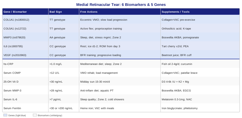
The biomarker section below is the most directly actionable entry point — these are things you can test, track, and address within weeks.
6 Biomarkers to Track for Medial Retinacular Tear Recovery
The medial retinaculum is a fibrous stabilizing band on the inner aspect of the knee that resists lateral displacement of the patella. When it tears — most commonly during lateral patellar dislocation — healing unfolds across three overlapping phases: acute inflammation, fibroblast-driven tissue proliferation, and matrix remodeling. Each phase depends on a distinct set of molecular signals, and measuring those signals through specific biomarkers gives you a functional window into how well the process is progressing and where it may be stalling.
The six biomarkers below were selected for their relevance to connective tissue repair, their availability through commercial or specialty labs, and the strength of clinical evidence linking them to musculoskeletal recovery outcomes.
1. High-Sensitivity C-Reactive Protein (hs-CRP)
Why it matters for medial retinacular healing
CRP is a liver-produced acute-phase protein that rises in response to inflammatory signaling. After an acute retinacular tear, a short-term CRP elevation is biologically appropriate — it signals that the immune system has initiated repair. The problem emerges when CRP remains persistently elevated weeks or months later, indicating that chronic low-grade inflammation has replaced the normal repair cascade. This smoldering inflammatory state degrades the extracellular matrix through sustained enzyme activation, inhibits fibroblast function, and impairs collagen maturation. In practical terms: patients with chronic elevated hs-CRP often plateau in physical therapy with unexplained stiffness and slow strength return.
How to measure it
The high-sensitivity assay (hs-CRP, not standard CRP) is required because standard CRP misses the low-grade range that matters most in chronic injury. A basic hs-CRP blood test is widely available through standard labs and typically costs $10–$40. It can be added to routine metabolic panels. Target for connective tissue healing: below 1.0 mg/L. Values between 1–3 mg/L signal moderate background inflammation; above 3 mg/L warrants investigation. The most useful measurement windows are at baseline shortly after injury, at six weeks, and at three months.
If the score is high — the plan without supplements
The most powerful lifestyle-level anti-inflammatory intervention is dietary pattern change: eliminating refined seed oils, processed grains, and added sugars while increasing whole-food sources of omega-3 fatty acids such as fatty fish, sardines, and mackerel. A Mediterranean-style pattern has been shown across multiple meta-analyses to reduce hs-CRP by 20–30% over 8–12 weeks. Consistent sleep of 7–9 hours per night reduces inflammatory cytokine output measurably within weeks. Zone 2 aerobic exercise — low intensity, conversational pace — performed 30–45 minutes, 3–4 times per week, normalizes the IL-6 pathway and produces sustained CRP reduction over 6–8 weeks. Extreme caloric restriction, counterintuitively, elevates CRP and should be avoided during healing.
If the score is high — the plan with supplements or equipment
Fish oil (EPA+DHA): 2–4 g of combined EPA+DHA daily. The anti-inflammatory effect is supported by multiple meta-analyses. No cycling needed; continuous use is appropriate. Side effects include mild GI upset at high doses and minor blood-thinning — take with the largest meal of the day.
Curcumin with piperine or in liposomal/phospholipid form: 500–1000 mg/day of a bioavailable formulation (Meriva, BCM-95, or with black pepper extract). Cycle 8 weeks on, 2 weeks off. Side effects: GI distress at high doses; avoid combining with anticoagulants without medical oversight.
2. Serum COMP (Cartilage Oligomeric Matrix Protein)
Why it matters for medial retinacular healing
COMP is a non-collagenous glycoprotein that forms part of the extracellular matrix in cartilage, tendons, and ligamentous tissue — including the medial retinaculum. When connective tissue is mechanically stressed or actively degrading, COMP is released into the bloodstream. Studies in knee ligament injury and tendinopathy show that serum COMP rises sharply after acute connective tissue damage, then should decline progressively as repair completes. A COMP level that remains elevated at six to twelve weeks post-injury suggests ongoing tissue breakdown — a sign that the remodeling phase is not completing normally. Very low COMP during a chronic injury may indicate insufficient matrix activity and stalled biological healing.
How to measure it
Serum COMP is available through specialty labs and some academic medical center panels, typically at a cost of $80–$180. It is not a standard orthopedic order, so you will likely need to request it through a sports medicine or integrative medicine physician. Normal reference ranges fall generally below 12 U/L in healthy non-injured adults. The most informative measurement windows are 1–2 weeks post-injury for a baseline, and again at 6 and 12 weeks to assess trajectory.
If the score remains persistently elevated — the plan without supplements
Persistent COMP elevation almost always points to ongoing mechanical stress on the retinaculum — driven by poor patellar tracking, quadriceps-to-hamstring imbalance, or premature return to loading activity. The primary intervention is structured progressive load management: a rehabilitation program centered on VMO (vastus medialis oblique) strengthening, hip abductor activation, and neuromuscular retraining of patellar stability. Reducing impact loading — avoiding running, jumping, and deep knee flexion — while the remodeling phase completes is critical. Gait and alignment assessment for excessive knee valgus is frequently underutilized and directly relevant.
If the score remains persistently elevated — the plan with supplements or equipment
Collagen peptides with Vitamin C pre-exercise: 15 g of hydrolyzed collagen combined with 50–100 mg Vitamin C, taken 30–60 minutes before rehabilitation sessions. Shaw et al. demonstrated that this timing protocol enhances collagen synthesis markers in periarticular connective tissue when substrate is available during peak anabolic demand. Daily use during active rehabilitation; the safety profile supports continuous use.
Patellar stabilizing brace (J-brace or dynamic patellar stabilizer): applied during loading activities to reduce mechanical stress on the medial retinaculum while strengthening progresses. Not a permanent strategy, but directly reduces repetitive micro-stress during the vulnerable remodeling phase.
3. 25-Hydroxyvitamin D (25-OH Vitamin D)
Why it matters for medial retinacular healing
Vitamin D deficiency is one of the most commonly overlooked modulators of musculoskeletal healing. Vitamin D receptors are expressed on fibroblasts — the cells responsible for producing and remodeling collagen in fibrous tissue like the medial retinaculum. Deficiency impairs fibroblast proliferation, reduces type I collagen gene expression, and increases the risk of ligament laxity and connective tissue fragility. Multiple studies have linked low vitamin D status to prolonged soft tissue recovery times and elevated re-injury risk in athletes and general populations alike.
How to measure it
Serum 25-OH Vitamin D is widely available and typically costs $30–$60. It is included in many preventive health panels. Optimal range for musculoskeletal healing: 40–70 ng/mL (100–175 nmol/L). Values below 30 ng/mL represent deficiency; below 20 ng/mL is severe deficiency requiring prompt correction. Retest 8–12 weeks after beginning supplementation. Late winter and early spring testing captures the most accurate baseline for most temperate climates.
If the score is low — the plan without supplements
Direct midday sunlight on exposed skin (arms and legs, no sunscreen on the exposed areas) for 15–30 minutes, 4–5 days per week, remains the most efficient natural source. This is significantly climate and season dependent. Dietary sources — fatty fish, egg yolks, fortified foods — contribute but are rarely sufficient to correct significant deficiency on their own. These are worth maximizing as a complement to sunlight, particularly for individuals at high latitudes.
If the score is low — the plan with supplements or equipment
Vitamin D3: 4,000–6,000 IU per day for 3 months, then retest. For severe deficiency (below 20 ng/mL), a short-term supervised loading protocol may be appropriate. Always pair with Vitamin K2 as MK-7 at 100–200 mcg/day — K2 ensures that the calcium mobilized by Vitamin D is directed appropriately into bone rather than soft tissue. Side effects: toxicity is a real risk at very high prolonged doses; retesting at 3 months prevents over-supplementation.
Magnesium glycinate: 200–400 mg/day. Magnesium is an essential cofactor for converting Vitamin D to its active hormonal form. Deficiency in magnesium blocks this conversion, which is why some patients supplement D3 aggressively and see little improvement in 25-OH D levels. This is a frequently missed co-factor in standard clinical practice.
4. Matrix Metalloproteinase-3 (MMP-3)
Why it matters for medial retinacular healing
Matrix metalloproteinases are a family of enzymes that degrade extracellular matrix components including collagen, fibronectin, and proteoglycans. MMP-3 (stromelysin-1) is particularly active in retinacular and ligamentous tissue remodeling. After an acute tear, a rise in MMP-3 is appropriate: it clears damaged matrix so new tissue can form. But chronically elevated MMP-3 — driven by persistent inflammation, mechanical overload, or genetic predisposition — continues to degrade freshly forming collagen fibers before they can mature and cross-link. Research on knee ligament healing has demonstrated that excessive MMP-3 activity correlates with structurally weaker scar tissue and incomplete functional recovery.
How to measure it
Serum MMP-3 is available through specialty labs and some integrative medicine panels, typically costing $100–$200. It is not a routine clinical order. General reference range: under 28 ng/mL in healthy adults, though the optimal ceiling for active tissue healing is less precisely defined in the literature. The most informative measurement window is 4–10 weeks post-injury, when chronic remodeling patterns have become apparent.
If the score is high — the plan without supplements
Chronically elevated MMP-3 is primarily driven by systemic inflammation, so the anti-inflammatory dietary and sleep strategies described under hs-CRP are equally relevant here. Additionally, mechanical offloading during the remodeling phase directly reduces the inflammatory signaling that drives MMP-3 production. Aquatic therapy — pool walking or pool-based rehabilitation — is particularly well-suited here because it maintains range of motion and muscle activation while eliminating the impact loading that sustains MMP-3 release.
If the score is high — the plan with supplements or equipment
Boswellia serrata extract standardized for AKBA: 100–200 mg of AKBA per day from a 65–75% AKBA standardized extract. Boswellia directly inhibits MMP-3 and other leukotriene-pathway enzymes involved in matrix degradation. Cycle: 12 weeks on, 4 weeks off. Side effects include mild GI effects at higher doses; potential interaction with anticoagulant medications.
Green tea extract (EGCG): 400–800 mg/day standardized to 45–50% EGCG. EGCG has demonstrated MMP inhibition in human connective tissue studies. Take with food to avoid GI effects. Cycle 8 weeks on, 2 weeks off. Avoid on an empty stomach at high doses due to a liver safety signal observed in rare cases with concentrated extracts.
5. Interleukin-6 (IL-6)
Why it matters for medial retinacular healing
IL-6 is a cytokine with a well-documented dual role in injury. During the acute phase — the first 24–72 hours — a spike in IL-6 is pro-healing: it recruits immune cells, activates fibroblasts, and initiates the anabolic repair signaling cascade. But when IL-6 remains chronically elevated — which occurs in individuals with adipose tissue dysregulation, poor sleep, systemic stress, or specific IL6 gene variants — it shifts into a pro-catabolic role that promotes muscle wasting and matrix degradation. Tracking IL-6 helps distinguish between a healthy acute response and a maladaptive inflammatory state that is slowing the remodeling phase.
How to measure it
Serum IL-6 is available through standard labs, typically costing $50–$120. It is highly sensitive to same-day stressors — acute infection, poor sleep the previous night, vigorous exercise within 48 hours — so measurements must be taken in a rested baseline state. No fasting required. Normal range in healthy adults: under 7 pg/mL. Values above 10 pg/mL in a non-acute injury phase warrant attention. Retest at 6-week intervals while interventions are in progress.
If the score is persistently high — the plan without supplements
Chronic IL-6 elevation is tightly linked to poor sleep quality and excess adipose tissue. Addressing sleep continuity — eliminating alcohol, maintaining consistent sleep and wake times, managing sleep apnea if present — can reduce IL-6 measurably within 4–6 weeks. Low-intensity aerobic exercise (zone 2) acutely raises IL-6 during the session but produces a progressive post-exercise reduction over weeks — an important distinction from the chronic resting elevation. Cold exposure through cold showers (3–5 minutes) or brief cold water immersion at approximately 15°C has emerging evidence for reducing systemic inflammatory cytokines including IL-6, particularly when applied in the morning rather than immediately after training.
If the score is persistently high — the plan with supplements or equipment
Melatonin (where sleep disruption is present): 0.3–1 mg taken 30–60 minutes before bed. Lower doses are typically more effective than high doses for sleep initiation, and melatonin directly reduces IL-6 through both sleep improvement and independent anti-inflammatory mechanisms. Continuous use is supported; some prefer a 5-nights-on, 2-nights-off rhythm.
N-Acetyl Cysteine (NAC): 600–1200 mg per day in divided doses. NAC restores glutathione levels and reduces oxidative stress, which is a primary driver of chronic cytokine elevation. Cycle 8 weeks on, 2–4 weeks off to avoid glutathione pathway downregulation. Side effects: GI discomfort at higher doses; a distinctive sulfur smell in breath and urine at high doses.
6. Serum Ferritin
Why it matters for medial retinacular healing
Ferritin occupies two distinct clinical positions depending on where it falls on the measurement spectrum. As an iron storage marker, it reflects whether the body has sufficient iron for tissue repair — including hemoglobin synthesis, cellular energy production, and the oxygen-dependent enzymatic steps of collagen cross-linking. Too low (below 30 ng/mL), and fibroblast function is measurably compromised, healing slows, and fatigue masks the actual biological deficit. Too high (above 150–200 ng/mL in the absence of known iron loading), and ferritin may be functioning as an inflammatory acute-phase reactant rather than a storage marker, indicating significant systemic inflammation or early iron overload. Peter Attia's longevity-focused clinical work repeatedly highlights ferritin as a frequently under-evaluated marker in recovering and athletic populations, sitting at the intersection of energy metabolism and inflammation.
How to measure it
Serum ferritin is typically included in standard iron panels and costs $15–$40. Optimal range for tissue healing: 50–100 ng/mL for women, 75–150 ng/mL for men. Always interpret alongside total iron and transferrin saturation for full context. Note that ferritin is an acute-phase reactant — it elevates during active inflammation independent of actual iron status — so a high reading must be cross-referenced with hs-CRP to determine whether it reflects true iron excess or inflammatory elevation.
If the score is low — the plan without supplements
Dietary iron optimization centers on heme iron sources — red meat, organ meats, shellfish — which are absorbed at 25–35% efficiency compared to 3–8% for non-heme sources like legumes and spinach. Non-heme iron absorption can be meaningfully improved by combining it with Vitamin C-rich foods in the same meal. Avoid consuming calcium-rich foods, coffee, or black tea within 2 hours of iron-rich meals, as these substantially inhibit absorption. This dietary adjustment alone can raise ferritin by 15–30 ng/mL over 8–12 weeks in mild deficiency.
If the score is low — the plan with supplements or equipment
Iron bisglycinate: 25–36 mg elemental iron every other day. Alternate-day dosing improves net absorption by allowing hepcidin levels to normalize between doses — a pharmacokinetic approach supported by recent absorption studies. Take with 250 mg Vitamin C and away from other mineral supplements. Retest ferritin at 8 weeks. Side effects include constipation (significantly reduced with bisglycinate vs. sulfate forms) and dark stools. Do not supplement without confirmed deficiency.
For elevated ferritin above 200 ng/mL: rule out hereditary hemochromatosis with HFE gene testing. Verify with transferrin saturation greater than 45%. Phlebotomy remains the most effective intervention for hereditary iron overload and is managed by a physician. Moderate reduction in red meat intake helps in milder non-genetic elevation.
The six biomarkers above map your body's current healing environment. The following section adds a deeper, more permanent layer — the genetic predispositions that explain why some individuals' biomarkers drift out of optimal range more readily than others.
5 Key Genes That Influence Medial Retinacular Healing
Genetics does not determine your recovery outcome, but it does set the baseline conditions under which your connective tissue operates. Understanding your genetic tendencies — particularly in collagen architecture, inflammatory signaling, and matrix enzyme activity — allows you to address structural weaknesses proactively rather than discovering them through repeated injury or unexpectedly slow healing.
Genetic testing for the variants below is available through direct-to-consumer platforms such as 23andMe, with analysis through tools like SelfDecode, StrateGene, or Genetic Genie, or through clinicians using more comprehensive panels. The variants discussed here are among the most studied in human populations in the context of connective tissue injury, recovery, and structural vulnerability.
Gene 1: COL1A1 (rs1800012)
What it affects
COL1A1 encodes the alpha-1 chain of Type I collagen — the dominant structural protein in ligaments, tendons, and the fibrous retinacular tissue of the knee. The rs1800012 polymorphism (specifically the TT genotype) is associated with reduced mechanical stiffness of collagen fibers and increased structural vulnerability in ligamentous tissue. Studies in athletic populations have linked this variant to higher rates of ACL rupture, Achilles tendon rupture, and related soft tissue injuries — all conditions mechanically analogous to medial retinacular tears in terms of collagen architecture demands.
If the gene is unfavorable — the plan without supplements
The compensation strategy centers on protecting the tissue from peak mechanical loads while building neuromuscular protection capacity. Eccentric strengthening of the VMO and hip abductors — performed slowly with controlled loading rather than ballistic movement — has the strongest evidence for improving tissue-level protection in collagen-vulnerable individuals. Plyometric and high-impact activities should be progressed very gradually, with no more than a 10% increase in load per week and longer-than-average deload periods. Footwear and orthotic assessment is relevant: excessive pronation increases medial patellar stress and places repeated strain on a retinaculum that is already structurally less stiff.
If the gene is unfavorable — the plan with supplements or equipment
Collagen peptides with Vitamin C pre-exercise: the same protocol described under the COMP biomarker — 15 g hydrolyzed collagen combined with 50 mg Vitamin C, 45–60 minutes before loading sessions. This directly stimulates Type I collagen synthesis in the tissues under mechanical demand. Daily use during active rehabilitation; the safety profile supports long-term maintenance.
Prolyl hydroxylase cofactors — specifically Vitamin C and adequate iron stores — are required for the enzyme that cross-links collagen fibers. Ensuring both are in the optimal range (not just Vitamin C alone, but also ferritin above 50 ng/mL) directly addresses the downstream structural consequence of this variant, as cross-link density is a primary determinant of collagen stiffness.
Gene 2: COL5A1 (rs12722)
What it affects
COL5A1 encodes the alpha-1 chain of Type V collagen, which regulates the diameter and uniformity of Type I collagen fibrils. Smaller, more uniform fibril diameter produces greater tensile strength in ligamentous tissue. The TT genotype of rs12722 has been repeatedly associated with elevated risk of tendon and ligament injury in multiple athletic cohorts. For medial retinacular tissue specifically, disrupted fibril diameter regulation means that healed tissue may be structurally weaker than the original and more susceptible to re-tear under comparable loads.
If the gene is unfavorable — the plan without supplements
Excessive passive stretching carries a specific risk for this genotype: prolonged passive holds may reduce fibril integrity in already-vulnerable collagen. Instead, prioritize active flexibility — controlled movement through full range under mild load, as in yoga-based active-stretch work or slow eccentric movements — over passive prolonged holds. This maintains mobility without compromising fibril integrity. Proprioception training (single-leg balance, wobble board, eyes-closed balance work) for 10–15 minutes daily is particularly valuable, because COL5A1 variants affect mechanoreceptor function within ligaments as well as structural tensile strength.
If the gene is unfavorable — the plan with supplements or equipment
Bioavailable orthosilicic acid (silica): 6–10 mg/day of orthosilicic acid (as in BioSil or similar stabilized forms). Silica is a structural cofactor for collagen fibril formation and has shown connective tissue density improvements in preliminary human studies. Continuous supplementation appears safe at these doses; no established cycling protocol.
Kinesiology tape (McConnell patellar taping technique): during the return-to-activity phase, medial glide taping directly reduces retinacular mechanical stress. Not a supplement, but an important equipment-level tool specifically for individuals with structural collagen vulnerability who are returning to loading activity before full tissue maturation.
Gene 3: MMP3 (rs679620)
What it affects
This polymorphism in the MMP3 gene promoter region influences basal expression level of MMP-3 (stromelysin-1). The AA genotype is associated with higher MMP-3 production and more aggressive matrix degradation activity. As discussed in the biomarker section, excessive MMP-3 undermines collagen cross-linking and produces weaker, less organized scar tissue in healing ligamentous structures. Individuals with the AA genotype are more likely to show the elevated serum MMP-3 levels described above and will benefit disproportionately from protocols that reduce MMP-3 activity — both through lifestyle and targeted supplementation.
If the gene is unfavorable — the plan without supplements
The lifestyle protocol centers on minimizing the three primary drivers of MMP-3 overexpression: ultra-processed food, sleep debt, and chronic psychosocial stress. These three factors produce the sustained inflammatory cytokine environment that promotes MMP-3 gene transcription. A structured daily stress modulation practice — even 10–15 minutes of diaphragmatic breathing or progressive relaxation — produces measurable reductions in inflammatory cytokine expression within weeks. Moderate aerobic exercise (zone 2) reduces resting MMP-3 activity over time; high-intensity, high-volume training may transiently elevate it, making load management particularly important for this genotype.
If the gene is unfavorable — the plan with supplements or equipment
Boswellia serrata (AKBA): as detailed under the MMP-3 biomarker section — 100–200 mg AKBA from a standardized extract, 12 weeks on / 4 weeks off. This genotype makes Boswellia particularly high-priority.
Pomegranate extract (punicalagins): 500 mg/day of a standardized pomegranate extract. Polyphenols from pomegranate have demonstrated direct MMP inhibition in human tissue studies. Cycle 8 weeks on, 2 weeks off. Side effects are minimal at typical doses and the compound is generally well-tolerated.
Gene 4: IL6 (rs1800795)
What it affects
The rs1800795 variant in the IL6 gene promoter region influences baseline interleukin-6 expression. The CC genotype has been associated with higher resting IL-6 levels and a more amplified inflammatory response to tissue injury. For connective tissue healing, this translates to faster initial inflammation — which may actually accelerate the early repair phase — but with a significantly higher risk of failing to resolve appropriately and transitioning into the chronic inflammatory state that impairs remodeling. Research linking this variant to musculoskeletal recovery outcomes continues to develop, but the mechanistic basis is well-supported in the broader IL-6 biology literature.
If the gene is unfavorable — the plan without supplements
For CC genotype individuals, the critical window is the first 2–5 days post-injury. While some acute inflammation is biologically necessary, excessive early inflammation driven by mechanical stress, sleep deprivation, or dietary inflammatory load may set a dysregulated remodeling trajectory that is difficult to reverse. Practical first-line steps: immediate relative rest, intermittent ice application (10 minutes on, 20 off in the first 48 hours), elevation, and early gentle range-of-motion movement beginning at days 3–5 to mobilize synovial fluid and prevent chronic swelling pooling. This is consistent with current sports medicine evidence favoring active early mobilization over prolonged immobilization.
If the gene is unfavorable — the plan with supplements or equipment
Tart cherry concentrate: 30 mL twice daily during the acute and early remodeling phases. Anthocyanins in tart cherry have demonstrated IL-6 reduction in multiple small randomized trials involving exercise-induced inflammation. No significant cycling protocol is needed; continuous use is safe.
Palmitoylethanolamide (PEA): 300–600 mg twice daily. PEA is an endogenous fatty acid amide with documented anti-inflammatory, analgesic, and IL-6 modulating effects. The safety profile is favorable and well-established. Cycle: 8 continuous weeks, then reassess with an IL-6 retest if initial level was significantly elevated.
Gene 5: VEGF (rs2010963)
What it affects
Vascular Endothelial Growth Factor is the primary driver of angiogenesis — the formation of new blood vessels — which is essential for delivering oxygen and nutrients to healing connective tissue. The medial retinaculum is relatively avascular compared to skeletal muscle, making adequate VEGF signaling particularly important for capillary ingrowth into the repair tissue. The CC genotype of rs2010963 is associated with lower VEGF expression, contributing to slower vascularization of the healing retinaculum, reduced collagen deposition rates, and measurably longer recovery timelines. Early human studies in soft tissue injury suggest this variant contributes meaningfully to the wide variation in recovery duration seen between individuals with similar-severity tears.
If the gene is unfavorable — the plan without supplements
Angiogenesis in healing tissue is most powerfully stimulated by mechanical loading and localized hypoxic signaling — the same signals that make progressive exercise so central to tendon and ligament recovery. Blood Flow Restriction (BFR) training — applying a calibrated cuff or band to produce mild limb occlusion during low-load exercise — creates localized hypoxia that powerfully upregulates VEGF expression in the targeted tissue. Several studies support BFR for tendon and ligament healing specifically in populations with low angiogenic signaling capacity. Protocol: 20% of 1RM, 4 sets (30/15/15/15 repetitions) with the cuff applied at 60–80% limb occlusion pressure, 3 times per week during rehabilitation.
If the gene is unfavorable — the plan with supplements or equipment
Inorganic nitrates from dietary sources or beetroot juice: 500 mL of concentrated beetroot juice or approximately 6 mmol of dietary nitrate equivalent daily. Nitric oxide — produced from dietary nitrates — is a key upstream activator of VEGF signaling and endothelial vasodilation. No cycling required. Side effects include benign pink or red discoloration of urine and stool.
BFR training equipment (dedicated cuff system): for individuals with the CC VEGF genotype, BFR training is arguably more impactful than any supplement intervention for stimulating vascularization in hypoavascular connective tissue. Clinical-grade pneumatic cuffs (e.g., Delfi or B-Strong systems) provide calibrated occlusion; lower-cost elastic cuffs can be used with physiotherapist guidance on appropriate tension.
The genetic and biomarker data above tell the biological story of an individual recovery. The following section translates some of the most practically useful connective tissue science into directly applicable protocols.
What Andrew Huberman's Research on Connective Tissue Recovery Reveals
Across multiple Huberman Lab podcast episodes covering inflammation, sleep science, musculoskeletal adaptation, and recovery biology, a consistent body of evidence-based tools emerges for those managing connective tissue injury. These are not opinions — they represent consistent themes from peer-reviewed literature that Huberman cites explicitly, which makes them particularly useful for applying to a condition like medial retinacular tear, where the standard clinical guidance often stops at rest and physical therapy.
1. Collagen Synthesis Has a Precise Timing Window
Huberman has extensively covered the work of Keith Baar's lab showing that collagen synthesis in tendons and ligaments peaks approximately one hour after loading exercise and requires circulating substrate at that precise moment. The protocol — 15 g hydrolyzed collagen with 50 mg Vitamin C, 30–60 minutes before a rehabilitation session — is not about flooding the system; it is about having supply present at peak demand. Without the substrate, the synthesis stimulus is wasted.
2. Sleep Is the Primary Recovery Tool — Not a Recovery Supplement
Growth hormone, released in the first portion of the night during deep slow-wave sleep, is the dominant anabolic signal for connective tissue repair. Disrupting sleep by even 90 minutes measurably reduces GH pulse amplitude. No supplement compensates for this deficit. Huberman's non-negotiable sleep protocols — consistent wake time, complete darkness in the bedroom, minimal blue light exposure after sunset, room temperature of 65–67°F (18–19°C) — are directly relevant to anyone healing a retinacular tear where GH-dependent fibroblast activity is critical.
3. Cold Immersion Timing Can Either Help or Harm Recovery
Cold water immersion applied within 4 hours of exercise blunts the inflammatory signal that drives tissue adaptation and repair. Huberman is explicit: do not apply ice or cold immersion immediately after rehabilitation sessions if the goal is tissue adaptation. The inflammatory window post-exercise is biologically meaningful for repair. Cold exposure is most beneficial in the morning, 6+ hours after the last training session, where it provides systemic anti-inflammatory benefit without suppressing the local repair signal.
4. Zone 2 Exercise Is Anti-Inflammatory Medicine
Low-intensity aerobic work — sustainable conversational pace — drives mitochondrial biogenesis and progressively reduces chronic IL-6 and CRP levels over weeks of consistent practice. Huberman recommends 150–200 minutes per week as a baseline. For someone healing a medial retinacular tear, pool walking or stationary cycling at zone 2 intensity simultaneously provides the systemic anti-inflammatory benefit and the gentle joint loading needed to stimulate VEGF-driven vascularization and collagen synthesis without overloading the healing structure.
5. Morning Sunlight Sets the Biology of Repair
Consistent morning light exposure — 10–20 minutes outdoors within 30–60 minutes of waking — sets circadian timing for cortisol, melatonin, growth hormone, and immune regulation throughout the day. Irregular circadian rhythm is independently associated with higher inflammatory cytokines and reduced growth hormone amplitude. This zero-cost, zero-equipment protocol directly influences the hormonal environment in which connective tissue repair occurs, and Huberman highlights it consistently as one of the highest-leverage behaviors available.
6. The NSAID Trade-Off That Most Patients Are Not Told About
Huberman has discussed the accumulating evidence that chronic NSAID use during the acute injury phase may blunt the healing signal. Prostaglandins suppressed by NSAIDs are part of the initial repair cascade for connective tissue, not merely bystanders to pain. For retinacular tears, the question is whether short-term pain reduction justifies a potential cost to repair tissue quality. This is a clinical conversation worth having with a sports medicine physician, but most patients are not aware that the trade-off exists.
7. Deliberate Heat Exposure Enhances Tissue Perfusion
Regular sauna use — 15–20 minutes at 80–100°C, 3–4 times per week — improves cardiovascular function, heat shock protein expression, and tissue perfusion including in the relatively avascular connective tissue structures. For individuals with the low-VEGF CC genotype, sauna sessions may provide a supplementary vascularization stimulus. Huberman recommends avoiding sauna immediately after intense training; morning or early afternoon sessions are preferable.
8. Omega-3s Resolve Inflammation Rather Than Simply Suppressing It
Rather than blocking inflammation (as NSAIDs do), omega-3 fatty acids produce specialized pro-resolving mediators — resolvins and protectins — that actively promote the transition from acute inflammation to tissue repair. Huberman distinguishes this mechanism clearly: fish oil does not merely reduce inflammation, it promotes resolution. At 2–3 g/day of EPA+DHA, this is one of the most evidence-supported supplements for accelerating the transition from the inflammatory phase to productive remodeling.
9. Stress Chronically Suppresses Connective Tissue Repair
Elevated cortisol from chronic psychological stress directly suppresses fibroblast activity, reduces collagen synthesis rates, and prolongs the inflammatory phase of healing. The physiological sigh — a double inhale through the nose followed by an extended exhale through the mouth — produces the fastest acute reduction in sympathetic activation of any known breathing technique, according to the Stanford research Huberman cites. Performed 1–5 times when stress is acute, it has immediate relevance for anyone managing the frustration and anxiety of an injury affecting mobility and daily function.
10. Creatine Supports Tissue Repair Beyond Muscle
Huberman has covered creatine's roles extending well beyond muscle energy buffering — including in connective tissue cellular metabolism and in reducing inflammatory markers during recovery. At 3–5 g/day as a maintenance dose, creatine monohydrate is among the safest and most extensively researched supplements in sports science. No cycling is required, and a loading phase is optional. Its relevance to medial retinacular recovery is indirect but meaningful: supporting the energy demands of fibroblast activity and satellite cell function during the remodeling phase.
With the biological tools established, the following section covers hands-on modalities that directly address the local tissue environment.
Complementary Approaches With Evidence for Soft Tissue Healing
Low-Level Laser Therapy (Photobiomodulation)
Low-level laser therapy (LLLT) — also called photobiomodulation — applies specific wavelengths of red and near-infrared light (typically 630–850 nm) to penetrate tissue and stimulate cellular energy production through mitochondrial photoreceptors. For medial retinacular tears, its relevance is direct: LLLT has been shown to accelerate fibroblast proliferation, reduce MMP activity in healing tissue, and increase collagen synthesis rates in soft tissue injury models. A systematic review in Photomedicine and Laser Surgery found significant positive effects on tendon and ligamentous repair across human trials. The evidence is most robust for near-infrared wavelengths in superficial fibrous structures — which is precisely the depth of the medial retinaculum.
A practical protocol: a device delivering 30–100 mW at 810 nm (near-infrared) applied to the medial knee for 60–90 seconds per point, at 2–3 points over the medial retinaculum, 3–4 sessions per week for 4–6 weeks during the remodeling phase. Clinical LLLT devices provide more consistent dosing; FDA-cleared home near-infrared panels (Joovv, Mito Red, and similar) achieve therapeutic photon dosing for superficial tissue at adequate proximity. Evidence for specific home devices at this anatomical site is limited, but the physics of near-infrared penetration at 810 nm supports its reach to retinacular depth.
LLLT is most appropriately introduced 1–2 weeks after the acute phase, once swelling has substantially resolved. It pairs well with rehabilitation exercise by reducing post-session local inflammatory activity without suppressing the systemic healing signal that drives adaptation. The primary caution: avoid applying directly over areas of active infection, open wounds, or areas with uncertain diagnosis. For individuals with the high MMP-3 genotype or persistently elevated MMP-3 biomarker, LLLT's direct MMP-modulating effect makes it a particularly logical tool.
Massage Therapy
Soft tissue massage — specifically deep tissue work, myofascial release, and cross-friction technique — has a meaningful evidence base in connective tissue injury recovery. For the medial retinaculum, massage addresses several distinct mechanisms: reducing adhesion formation between the healing scar and adjacent periarticular structures, improving lymphatic drainage of residual swelling, and reducing protective muscle guarding in the VMO and medial quadriceps that can alter patellar mechanics and apply continued stress to the healing site. A review of soft tissue injury rehabilitation found moderate evidence for pain reduction and improved functional outcomes when massage was integrated with standard rehabilitation programs rather than used in isolation.
The technique with the most specific application to medial retinacular scar tissue is transverse cross-friction massage — firm, perpendicular pressure applied across the fibers of the healing scar, 5–7 minutes per session. This technique, originally systematized by James Cyriax, is designed to align newly forming collagen fibers longitudinally and prevent the formation of restrictive cross-fiber adhesions that limit patellar mobility and cause chronic aching. Typically performed by a sports massage therapist or physiotherapist with specific training in connective tissue techniques. Weekly sessions during the remodeling phase (weeks 3–12 post-injury) represent the standard clinical protocol; twice-weekly sessions are appropriate early in this phase where adhesion formation is more active.
Timing matters. Deep direct massage over the injury site should not begin until after the acute inflammatory phase — the first 5–7 days — when direct tissue pressure would worsen swelling and potentially disrupt the initial clot and early vascular ingrowth. During the first week, massage of the surrounding musculature (quadriceps, hip flexors, IT band) is appropriate and beneficial. Transition to targeted cross-friction at the medial retinaculum starting week 1–2, guided by the resolution of acute swelling and tenderness to direct pressure.
Mindfulness Meditation and MBSR
Mindfulness-Based Stress Reduction may appear peripheral to a structural knee injury, but its biological relevance is direct and mechanistic. Chronic psychological stress — the frustration of impaired mobility, the anxiety of re-injury risk, the monotony of rehabilitation — elevates cortisol, which suppresses fibroblast activity and prolongs the inflammatory phase of healing. Several randomized controlled trials have demonstrated that 8-week MBSR programs produce measurable reductions in cortisol, hs-CRP, and IL-6 in populations with chronic musculoskeletal conditions and pain — exactly the biomarker profile that slows retinacular healing.
The most evidence-backed protocol is Jon Kabat-Zinn's original 8-week MBSR structure: 2.5 hours per week of guided instruction plus 30–45 minutes of daily home practice. This is available through certified in-person instructors or the Palouse Mindfulness online curriculum, which follows the original MBSR format and is freely available. The recommended formal practice is seated or lying body scan meditation for 30–45 minutes daily. For individuals healing a knee injury who may find seated postures uncomfortable, body scan practice performed lying down is equally supported by the evidence and directly relevant given its focus on non-judgmental awareness of physical sensation — a particularly useful skill for managing recovery anxiety.
For those with the IL6 CC genotype or persistently elevated hs-CRP, the stress-inflammation pathway is especially active, making MBSR a physiologically meaningful adjunct to the biomarker-targeted protocols described earlier — not a soft add-on, but a measurable modifier of the molecular environment in which the repair is occurring.
Conclusion
A medial retinacular tear is a structural injury, but your recovery from it is shaped by biology that varies substantially between individuals. The six biomarkers covered here — hs-CRP, COMP, 25-OH Vitamin D, MMP-3, IL-6, and ferritin — give you a real-time, functional picture of your healing environment. The five genetic variants — COL1A1, COL5A1, MMP3, IL6, and VEGF — give you a baseline map of where your connective tissue biology may need targeted support. Neither replaces clinical care. Both allow you to have a more specific and informed conversation with the practitioners managing your recovery.
The most practical next step depends on where you are right now. If you have not had a recent blood panel, start with hs-CRP and 25-OH Vitamin D — both are inexpensive, widely available, and immediately actionable. If standard labs have been unremarkable but recovery has stalled, consider requesting COMP and MMP-3 through a sports medicine or integrative medicine physician. If you are interested in the longer-term picture of connective tissue risk and prevention, a genetic panel through 23andMe analyzed via SelfDecode or StrateGene will identify your collagen and inflammation gene variants reliably. The goal is not optimization for its own sake. It is to stop working in the dark — and to give your tissue the environment it actually needs to repair well.
Musculoskeletal: Joint Conditions Tendon & Ligament Conditions Sports Injuries
Autoimmune: Inflammatory Conditions Connective Tissue Conditions