This article contains AI generated content.
Medial Retinaculum Enthesopathy Genes And Biomarkers - 6 Genes And 6 Biomarkers To Track
Introduction
If you are reading this, you have probably already heard the phrase "medial retinaculum enthesopathy" from a radiology report or an orthopedic consult, most likely after a bout of anterior knee pain, a patellar subluxation, or months of inner-knee discomfort that never quite settled. The standard advice that follows - rest, ice, a knee sleeve, some quad sets, maybe a round of physical therapy - is not wrong. It is just incomplete. It treats the enthesis, the small but mechanically demanding zone where the medial retinaculum fibers insert into bone, as if it were the same in every knee and every patient.
It isn't. Two people with an identical MRI finding can heal at completely different speeds, redevelop symptoms under different loads, and respond to the exact same rehab program in opposite ways. Some of that difference comes down to training history and biomechanics. But a meaningful part of it comes down to the biology of the connective tissue itself - how well it builds and maintains collagen, how it responds to mechanical load, and what is currently happening in your blood chemistry that either supports or undermines tissue repair. Generic advice rarely accounts for any of that, which is exactly why it so often produces generic, inconsistent results.
This article takes a more specific approach. It looks at the genes that current research ties to collagen quality, tendon and ligament resilience, and enthesis biology - the same biological terrain the medial retinaculum lives in - and explains, honestly, where the human evidence is strong and where it is still early. It also looks at the blood biomarkers that reflect your tissue's current repair capacity right now, independent of what your genes say is possible.
None of this amounts to a guaranteed fix, and anyone promising one is overselling. But better information changes decisions. Knowing which genetic variants you carry can reframe how patient you need to be with a rehab timeline and which levers are actually worth pulling. Knowing your biomarkers can tell you whether your body is currently primed to repair tissue or working against itself. Add to that a look at the nutrient-timing research that is quietly changing how sports scientists think about tendon and ligament rehab, plus a short review of complementary approaches with real supporting evidence, and you have a more complete map than "rest and hope."
Summary
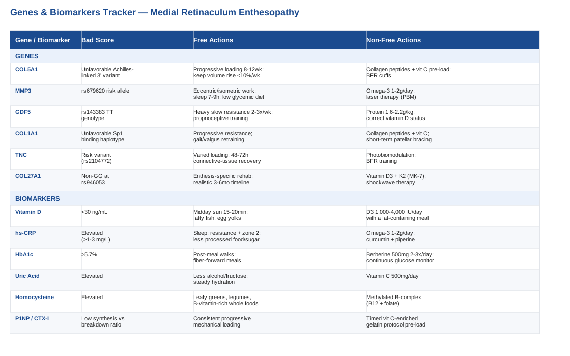
Medial retinaculum enthesopathy sits at the intersection of mechanics and biology, and this article works through both sides of that equation. On the genetics side, you will find six genes - COL5A1, MMP3, GDF5, COL1A1, TNC, and COL27A1 - that current human research links to collagen fibril structure, matrix remodeling, and the cartilage-to-bone transition zones that define an enthesis, along with a realistic plan, with and without supplements or equipment, for each one. On the biomarker side, you will find six blood markers, from vitamin D to collagen turnover proteins, that reveal whether your tissue is currently in a repair-friendly state, including cost ranges and what moves the needle.
Beyond genes and blood work, you will find a breakdown of the tendon-and-ligament research from physiologist Keith Baar, whose vitamin C and gelatin loading protocol has quietly challenged the old rest-first approach to connective tissue injuries, and a short, evidence-checked look at complementary approaches - including biofeedback, laser therapy, tai chi, and massage - worth considering alongside standard rehab. The goal throughout is not a miracle fix, but a clearer, more personalized way to think about a stubborn and often misunderstood injury.
The Genetic Blueprint Behind Medial Retinaculum Enthesopathy
No published study has looked at genetic variants specifically in people diagnosed with medial retinaculum enthesopathy - it is too narrow and too recently named a diagnosis for its own genetics literature to exist yet. What does exist is a substantial and growing body of human research on the genetics of tendon, ligament, and enthesis biology more broadly: Achilles tendinopathy, patellar tendinopathy, ACL rupture, and rotator cuff healing. Because entheses share the same core biology everywhere in the body - collagen fibril assembly, matrix remodeling enzymes, and mechanotransduction signaling - this research is a reasonable, if indirect, guide to what may be happening at the medial retinaculum's insertion points. Where the evidence is early or extrapolated rather than direct, that is noted below.
If you want to explore your own raw genetic data for these variants, researchers like Ali Torkamani, whose work at Scripps Research focuses on translating genomic data into actionable health insight, and Gary Brecka, who has popularized functional genetic panels covering methylation and collagen-related genes, are reasonable starting points for understanding how consumer genetic testing can be interpreted. Most mainstream consumer panels do not report on tendon-specific SNPs like the ones below directly, so you may need to pull your raw data file and run it through a third-party interpretation tool to see these specific markers.
COL5A1: The Fibril Diameter Regulator
COL5A1 encodes the alpha-1 chain of type V collagen, a minor collagen that nonetheless plays an outsized role: it sits at the core of newly forming collagen fibrils and helps set their final diameter during assembly. Thinner, more numerous fibrils tend to make tissue more compliant; thicker, fewer fibrils tend to make it stiffer. A well-studied polymorphism in the COL5A1 3' region has been linked to chronic Achilles tendinopathy risk, with one allele appearing to play a protective role against chronic symptoms.
The COL5A1 gene and Achilles tendon pathology (September et al.) found this association in a South African cohort, and it has since been examined in several other populations with mixed but generally supportive results. This is Achilles-specific evidence, not retinaculum-specific, so it should be read as suggestive of shared collagen biology rather than a direct diagnosis of your knee.
If The Gene Variant Is Unfavorable: The Plan Without Supplements
Because COL5A1 affects structural assembly rather than something a pill can directly correct, the actionable lever is mechanical. Progressive tendon and ligament loading - isometric holds advancing to heavy, slow resistance work over 8 to 12 weeks - stimulates the same remodeling pathways regardless of genotype. Pair this with quadriceps and hip strengthening to reduce the load transmitted through the medial retinaculum, and keep training volume increases below roughly 10 percent per week to avoid outpacing the tissue's adaptive capacity.
If The Gene Variant Is Unfavorable: The Plan With Supplements Or Equipment
Timed nutrition around loading sessions is the most directly studied option here (the full protocol is detailed in the Keith Baar section below): roughly 15 grams of hydrolyzed gelatin or collagen peptides plus 50 to 100 milligrams of vitamin C, taken about an hour before rehab loading, 3 to 4 times per week, cycled in 8 to 12 week blocks aligned with rehab phases. Blood flow restriction (BFR) cuffs can allow effective loading at lower absolute weights, which is useful early in rehab. Side effects are minimal - occasional mild GI upset from gelatin at higher doses, and vitamin C above 2 grams daily can cause GI discomfort or raise kidney stone risk in predisposed individuals, so staying within these ranges matters.
MMP3: The Remodeling Enzyme
MMP3 (stromelysin-1) is a matrix metalloproteinase that breaks down and remodels extracellular matrix components, making it a central player in the constant balance between tissue breakdown and repair. A functional variant at rs679620 has been associated with increased Achilles tendinopathy risk, and appears to interact with COL5A1 genotype to compound that risk further.
Variants within the MMP3 gene are associated with Achilles tendinopathy: possible interaction with the COL5A1 gene (Raleigh et al.) is the primary human study behind this association, again in Achilles tissue rather than the retinaculum specifically.
If The Gene Variant Is Unfavorable: The Plan Without Supplements
Excess or poorly regulated MMP activity is often driven by unresolved local inflammation and repetitive microtrauma rather than genetics alone. Eccentric and isometric loading protocols have been shown to help normalize tendon matrix turnover, and basic recovery hygiene - 7 to 9 hours of sleep, avoiding smoking, and limiting high-glycemic food intake - reduces the oxidative and glycation stress that can push MMP activity out of balance.
If The Gene Variant Is Unfavorable: The Plan With Supplements Or Equipment
Omega-3 fatty acids (1 to 2 grams combined EPA/DHA daily, taken continuously rather than cycled, since omega-3 status is a steady-state marker) have some evidence for modulating inflammatory and matrix-remodeling signaling; recheck status with an omega-3 index test every 3 to 4 months. Photobiomodulation (low-level laser therapy) has early evidence for influencing tendon matrix gene activity and is discussed further in the complementary approaches section. Doses of omega-3 above 3 grams daily can increase bleeding risk, so check with a physician if you take blood thinners.
GDF5: The Development And Loading Signal
Growth/differentiation factor 5 belongs to the BMP/TGF-beta superfamily and is essential for tendon, ligament, and joint formation during development; in animal models, GDF5 deficiency produces structurally weaker, thinner Achilles tendons with delayed healing. In humans, the TT genotype at the 5'UTR variant rs143383 has been linked to roughly double the risk of Achilles tendon pathology.
Components of the transforming growth factor-beta family and the pathogenesis of human Achilles tendon pathology (Posthumus et al.) is the key genetic association study here. There is also an interesting epigenetic layer: CpG methylation regulates allelic expression of GDF5 by modulating SP1 and SP3 binding at this same SNP, and the degree of methylation differs between knee and hip cartilage. In plain terms, this means the same "risk" variant may express itself differently depending on the joint and, potentially, on local factors like mechanical loading and inflammation that influence methylation. This mechanistic work is cartilage-focused and early, not retinaculum-specific, so treat it as a promising direction rather than settled science.
If The Gene Variant Is Unfavorable: The Plan Without Supplements
GDF5 signaling itself responds to mechanical load, so a structured, progressive loading program is the primary non-supplement lever: heavy, slow resistance training for the quadriceps-patellar-retinaculum unit, 2 to 3 times per week in 12-week blocks, increasing load by about 5 to 10 percent every 1 to 2 weeks. If you also have generalized joint hypermobility (worth checking with a simple Beighton score), add proprioceptive and neuromuscular control training, since laxity tends to compound risk when combined with GDF5-related variants.
If The Gene Variant Is Unfavorable: The Plan With Supplements Or Equipment
No supplement is known to directly upregulate GDF5 expression in humans. The most defensible adjacent levers are adequate protein intake (roughly 1.6 to 2.2 grams per kilogram of bodyweight daily) and correcting any vitamin D insufficiency, since related bone-tendon signaling pathways are vitamin D responsive (see the biomarker section for testing and dosing). Blood flow restriction training or a decline board for patellar-adjacent loading can provide effective mechanical stimulus at lower absolute loads for those managing joint laxity. Avoid aggressive end-range stretching if you are hypermobile, as it can worsen laxity rather than help it.
COL1A1: The Structural Workhorse
COL1A1 encodes the dominant chain of type I collagen, the primary structural protein in ligaments, tendons, and the dense connective tissue of the retinaculum itself. A well-studied Sp1-binding-site polymorphism affects how much collagen I is transcribed. Interestingly, a specific COL1A1 haplotype (the G-T combination of the -1997G/T and +1245G/T variants) has been found protective against ACL injury in athletes.
Gene variants within the COL1A1 gene are associated with reduced anterior cruciate ligament injury in professional soccer players (Collins et al.) is the primary study, with broader soft-tissue rupture associations described in The COL1A1 gene and acute soft tissue ruptures.
If The Gene Variant Is Unfavorable: The Plan Without Supplements
Collagen I genetics can't be edited, but load-driven remodeling improves tissue tolerance regardless of genotype. Focus on progressive resistance training for knee extensors and hip stabilizers, since better proximal control reduces the mechanical stress transmitted to the medial retinaculum, and use gait and movement retraining to correct dynamic knee valgus during cutting or landing tasks. Base return-to-sport decisions on objective criteria, such as a limb symmetry index above 90 percent, rather than a fixed timeline.
If The Gene Variant Is Unfavorable: The Plan With Supplements Or Equipment
The same timed collagen-peptide and vitamin C loading protocol described under COL5A1 applies here (roughly 15 grams collagen plus 50 milligrams vitamin C, an hour before loading, 3 to 5 times weekly during active rehab, cycling off during maintenance phases). Short-term patellar tracking braces or kinesiology taping, used for weeks rather than months during high-load return-to-sport phases, can offload the medial retinaculum while strength training catches up. Avoid prolonged continuous bracing, since it risks quadriceps deconditioning if not paired with active rehab.
TNC (Tenascin-C): The Mechanical Alarm Signal
Tenascin-C is an extracellular matrix glycoprotein that rises sharply in mechanically stressed and injured connective tissue, functioning as part of the signaling cascade that orchestrates matrix remodeling and healing after load or injury. Certain TNC variants have been linked to impaired healing and higher degenerative tear risk in the Achilles tendon and rotator cuff, and a recent case-control study found a TNC variant overrepresented in athletes with tendinopathy more broadly.
The Association of the COL27A1 rs946053 and TNC rs2104772 with Tendinopathies (Croatian athlete cohort) is a recent study worth flagging as early-stage evidence rather than an established finding, while a novel tenascin-C haplotype modifying failure-to-heal risk after rotator cuff repair adds supporting context from a different tendon.
If The Gene Variant Is Unfavorable: The Plan Without Supplements
Since TNC expression is itself driven by mechanical loading, the practical principle is to vary load rather than avoid it, while respecting recovery windows of 48 to 72 hours between loading sessions for connective tissue - longer than the 24 to 48 hours typical for muscle recovery - to avoid interrupting the TNC-mediated repair window.
If The Gene Variant Is Unfavorable: The Plan With Supplements Or Equipment
No human supplement directly targets TNC expression at this time. The most biologically plausible equipment-based options are photobiomodulation and blood flow restriction training, both discussed elsewhere in this article, which provide joint-friendly mechanical and cellular stimuli without a specific dosing protocol unique to TNC genotype. There are no additional side effects beyond the standard precautions for those modalities.
COL27A1: The Cartilage-To-Bone Transition Gene
COL27A1 encodes a fibrillar collagen expressed specifically at the transition zone between cartilage and bone - precisely the anatomical territory relevant to any enthesopathy, since an enthesis is defined by a graded transition from soft tissue through fibrocartilage into bone. A protective genotype (GG at rs946053) was overrepresented among healthy controls in the same recent Croatian athlete study referenced above, suggesting the opposite genotype may leave this transition zone less structurally organized.
This is genuinely early evidence - a single case-control study, not yet replicated in ligament-insertion-specific cohorts - so it should be treated as a promising lead rather than a settled risk marker.
If The Gene Variant Is Unfavorable: The Plan Without Supplements
Because this gene concerns the enthesis transition zone specifically, direct, tissue-specific loading through the range of motion that stresses the medial retinaculum's attachment matters more here than general quadriceps work alone. Work with a sports physiotherapist on enthesis-specific rehab, and set realistic timelines: fibrocartilage at the enthesis remodels more slowly than mid-substance tendon, often needing 3 to 6 months for meaningful adaptation rather than 6 to 8 weeks.
If The Gene Variant Is Unfavorable: The Plan With Supplements Or Equipment
Vitamin D and vitamin K2 co-supplementation has theoretical relevance to mineralization at the fibrocartilage-bone junction, extrapolated from general bone and cartilage biology rather than tested at the retinaculum directly. A common approach is vitamin D3 at 1,000 to 2,000 IU daily with K2 (MK-7) at 100 micrograms daily, taken with a fat-containing meal and reassessed via a vitamin D blood test every 3 to 6 months. For chronic, recalcitrant cases, extracorporeal shockwave therapy has documented evidence in other enthesopathy-type conditions, such as patellar tendon enthesopathy and plantar fasciitis, and is worth discussing with a sports medicine physician. Sustained vitamin D intake above 4,000 IU daily risks hypercalcemia and should be monitored with blood testing.
Genetics set the ceiling on how your tissue is built, but they say nothing about what is happening in your body right now. That is where blood biomarkers come in, offering a real-time picture that complements the genetic one.
Six Biomarkers That Reflect Your Current Tissue-Repair Capacity
Where genetic testing shows long-term structural tendencies, these biomarkers show current biochemical conditions - whether your body is in a state that supports or works against collagen repair right now. Each one is affordable to test, several are already on routine panels, and each has at least one lever you can pull if the number is off.
Vitamin D (25-Hydroxyvitamin D)
Vitamin D receptors are present on tendon and ligament fibroblasts, and vitamin D plays a role in regulating collagen synthesis and local inflammation. A large retrospective analysis found meaningfully higher rates of distal biceps tendinopathy among vitamin D-deficient patients compared to matched controls, described in Association of Vitamin D Deficiency With Distal Biceps Injury.
How To Measure It
A standard serum 25-OH-D blood draw costs roughly 30 to 50 dollars out of pocket, or is often bundled into routine panels. Most researchers target 30 to 50 ng/mL, though the exact optimal range is still debated.If The Score Is Bad: The Plan Without Supplements
Aim for 15 to 20 minutes of midday sun exposure on bare skin several times a week, adjusted for season and latitude, plus dietary sources like fatty fish and egg yolks.If The Score Is Bad: The Plan With Supplements Or Equipment
D3 at 1,000 to 4,000 IU daily with a fat-containing meal, retested in 8 to 12 weeks, then adjusted to a maintenance dose. Sustained intake above 4,000 IU daily should be physician-supervised due to hypercalcemia risk.High-Sensitivity CRP (hs-CRP)
This marker reflects systemic low-grade inflammation, which can keep local tissue in a chronic irritation state rather than a repair state, slowing enthesis healing.
How To Measure It
A standard blood test costing 15 to 30 dollars, commonly included in cardiometabolic panels of the kind clinicians like Peter Attia track routinely.If The Score Is Bad: The Plan Without Supplements
Prioritize 7 to 9 hours of sleep, combine resistance training with zone 2 cardio, reduce ultra-processed food and refined sugar intake, and address any untreated infections or dental issues that can quietly keep CRP elevated.If The Score Is Bad: The Plan With Supplements Or Equipment
Omega-3s (1 to 2 grams EPA/DHA daily) and curcumin with piperine (500 to 1,000 milligrams daily, cycled in 8 to 12 week blocks with breaks) both have supportive evidence for lowering inflammatory markers. Curcumin can cause mild GI upset and may interact with anticoagulants.HbA1c Or Fasting Glucose
Chronic high blood sugar promotes advanced glycation end-products that abnormally cross-link collagen, stiffening tissue and impairing normal healing. Diabetes is a recognized risk factor for tendinopathy broadly, as reviewed in Tendinopathy in diabetes mellitus patients - epidemiology, pathogenesis, and management.
How To Measure It
A standard blood draw, 10 to 20 dollars, part of most routine metabolic panels. Target HbA1c below 5.7 percent.If The Score Is Bad: The Plan Without Supplements
Resistance training combined with short walks after meals, fiber-forward meals, and minimizing liquid sugar calories all meaningfully lower glucose load over time.If The Score Is Bad: The Plan With Supplements Or Equipment
Berberine (500 milligrams, 2 to 3 times daily, cycled in 8 to 12 week blocks) has glucose-lowering evidence but can cause GI side effects and should not be combined with diabetes medications without medical guidance. A continuous glucose monitor (roughly 50 to 100 dollars per month) gives direct feedback on which foods and training sessions spike your glucose.Uric Acid
Elevated uric acid is associated with crystal deposition and inflammatory changes in tendon and enthesis tissue in some populations, and reflects broader metabolic and purine load.
How To Measure It
A standard blood test, 10 to 15 dollars, frequently included on metabolic panels.If The Score Is Bad: The Plan Without Supplements
Reduce alcohol, particularly beer, and high-fructose intake, and maintain steady hydration throughout the day.If The Score Is Bad: The Plan With Supplements Or Equipment
Vitamin C at a modest 500 milligrams daily has some uric-acid-lowering evidence and is safe to take continuously at this dose. Avoid multi-gram doses long-term due to kidney stone risk in predisposed individuals.Homocysteine (With B12 And Folate)
Elevated homocysteine is associated with abnormal collagen cross-linking chemistry, and B12 and folate are required cofactors for normal connective tissue metabolism.
How To Measure It
A blood test costing 20 to 40 dollars, less routine than the markers above, so ask for it specifically.If The Score Is Bad: The Plan Without Supplements
Increase intake of leafy greens, legumes, and other B-vitamin-rich whole foods.If The Score Is Bad: The Plan With Supplements Or Equipment
A methylated B-complex (B12 as methylcobalamin, 500 to 1,000 micrograms, plus folate at 400 to 800 micrograms daily), reassessed after 3 months. Side effects are minimal.Collagen Turnover Markers: P1NP And CTX-I
These markers give a direct blood readout of whole-body collagen synthesis (P1NP) versus breakdown (CTX-I) - the same class of marker used to demonstrate real increases in collagen synthesis in the vitamin C and gelatin research discussed next. They mostly reflect bone collagen turnover rather than tendon-specific turnover, so treat them as a useful proxy rather than a retinaculum-specific readout.
How To Measure It
A specialty lab order, typically 75 to 150 dollars per marker, not routinely offered by primary care and usually accessed through sports medicine, functional medicine practices, or direct-to-consumer lab services.If The Score Is Bad: The Plan Without Supplements
Consistent, progressive mechanical loading is the strongest known driver of collagen synthesis signal, more so than any single nutrient.If The Score Is Bad: The Plan With Supplements Or Equipment
The timed vitamin C-enriched collagen protocol described next is the specific, studied way to shift this marker. Retest after a full 8 to 12 week training block rather than week to week, since day-to-day variability is high.With the biology and the blood work covered, it helps to see how one research program has pulled these threads together into a single, practical protocol - and challenged some fairly entrenched rehab habits along the way.
What Keith Baar's Tendon Research Changes About Standard Rehab Advice
Keith Baar, a muscle and connective tissue physiologist at UC Davis, has spent years studying how tendons and ligaments adapt differently from muscle, and his work - shared across podcasts including Tim Ferriss's show, Sigma Nutrition, and IHMC's STEM-Talk - has quietly reshaped how sports scientists think about rehabbing tissue like the medial retinaculum. Below are ten of the most useful ideas from that body of research.
1. Connective Tissue Adapts On Its Own Schedule, Not Muscle's
Tendon and ligament tissue is far less metabolically active than muscle and turns over much more slowly. Rehab timelines built around muscle-recovery expectations - "you'll be fine in six weeks" - routinely underestimate how long an enthesis actually needs to remodel.2. Timing The Nutrient Matters More Than The Dose Alone
Baar's research found that consuming vitamin C-enriched gelatin roughly one hour before a loading session, not just at some point during the day, is what allows the amino acids to be available exactly when the tissue is mechanically signaling for repair.3. The Landmark Study Doubled A Real Collagen Synthesis Marker
In a randomized, double-blinded, crossover study, subjects who took 15 grams of vitamin C-enriched gelatin an hour before six minutes of jump rope showed double the blood marker of new collagen I formation compared with placebo, as reported in Vitamin C-enriched gelatin supplementation before intermittent activity augments collagen synthesis (Shaw et al., American Journal of Clinical Nutrition, 2017).4. Load Can Function As An Anti-Inflammatory
Contrary to the instinct to rest and avoid load after a connective tissue injury, Baar's work argues that controlled mechanical loading itself helps resolve inflammation and drive remodeling, rather than simply waiting it out - a direct challenge to a purely rest-and-ice-first mindset.5. Isometrics Can Reduce Pain Without Waiting For Inflammation To Fully Resolve
Isometric holds have been shown to reduce tendon and enthesis pain relatively quickly, making them a practical early-stage tool for a sore medial retinaculum before heavier loading is appropriate.6. Vitamin C Is A Non-Negotiable Cofactor, Not An Optional Extra
Vitamin C is required for the prolyl and lysyl hydroxylase enzymes that stabilize the collagen triple helix. Even marginal, sub-clinical deficiency - well short of scurvy - may quietly cap how well new collagen is formed.7. Oral Gelatin Genuinely Reaches Circulation As Usable Building Blocks
A common skepticism is that ingested collagen or gelatin is "just digested like any other protein and does nothing special." The data show that timed gelatin intake measurably raises blood markers of collagen synthesis, challenging that assumption.8. Genetics Sets The Ceiling, Loading And Nutrition Determine How Close You Get To It
This is the practical synthesis of the genetics section above: a favorable COL5A1 or COL1A1 genotype means little without the mechanical and nutritional inputs that actually drive remodeling, and an unfavorable genotype is not a fixed sentence either.9. Recovery Windows And Hormonal Status Shape Repair Capacity
Sleep quality and stress hormone levels influence growth hormone and IGF-1 signaling, both of which affect how efficiently connective tissue rebuilds - a reminder that nutrient timing works best layered on top of, not instead of, basic recovery habits.10. Tissue-Specific Rehab Beats Generic Rehab
Perhaps the broadest takeaway is that connective tissue responds best to loading that mimics the specific demands it will face again, reinforcing the case for enthesis-specific rehab over generic strengthening alone.Alongside targeted loading and nutrient timing, a handful of complementary approaches have real, if sometimes limited, human evidence worth knowing about.
Complementary Approaches Worth Considering
These are not substitutes for progressive loading or medical care, but each has genuine human evidence in closely related conditions - patellofemoral pain, tendinopathy, or knee osteoarthritis - that makes it reasonable to consider alongside standard rehab for medial retinaculum enthesopathy.
Biofeedback
Biofeedback, specifically EMG biofeedback, uses real-time electrical readouts of muscle activity to help retrain how the vastus medialis and vastus lateralis fire relative to each other. Since imbalanced quadriceps activation is a recognized contributor to patellar tracking problems and the mechanical stress placed on the medial retinaculum, this is directly relevant to the condition.
A controlled trial found that an 8-week exercise program combined with EMG biofeedback produced a significantly better VMO-to-VL activation ratio than exercise alone in patients with patellofemoral pain syndrome, described in Biofeedback exercise improved the EMG activity ratio of the medial and lateral vasti muscles in subjects with patellofemoral pain syndrome.
In practice, this means working with a physiotherapist who has EMG biofeedback equipment during the strengthening phase of rehab, typically over 6 to 8 weeks, rather than trying to self-diagnose activation imbalances. It is a reasonable add-on for anyone whose retinaculum symptoms seem tied to poor patellar tracking rather than a single traumatic event.
Low-Level Laser Therapy (Photobiomodulation)
Low-level laser therapy applies specific wavelengths of red or near-infrared light to tissue, with proposed effects on cellular energy production and local inflammatory signaling. Because tendinopathy and enthesopathy share overlapping tissue biology, this modality has been studied specifically in tendon conditions relevant to the medial retinaculum's tissue type.
A systematic review with meta-analysis found LLLT effective for reducing pain in tendinopathy when used at recommended dosages, summarized in Low level laser treatment of tendinopathy: a systematic review with meta-analysis (Tumilty et al.), though the review is honest that dosage parameters vary widely across studies and overall evidence quality remains moderate.
Realistically, this means seeking out a clinic using published dosage parameters (typically multiple sessions per week over several weeks) rather than a single one-off treatment, and treating it as a pain-modulation adjunct rather than a primary fix for the underlying mechanical problem.
Tai Chi
Tai chi combines slow, controlled movement with weight shifting and balance work, building the kind of proprioceptive and neuromuscular control that supports better knee joint mechanics - relevant to a condition often linked to patellar tracking and dynamic knee alignment.
A well-conducted randomized trial found tai chi produced pain and function improvements in knee osteoarthritis comparable to standard physical therapy, reported in Comparative Effectiveness of Tai Chi Versus Physical Therapy for Knee Osteoarthritis (Wang et al., Annals of Internal Medicine). This evidence is specific to osteoarthritis rather than enthesopathy, so it should be read as supportive of general knee joint mechanics rather than a direct treatment for retinaculum tissue itself.
A practical approach is a beginner tai chi class 2 to 3 times per week for 8 to 12 weeks, used as a low-load complement to strength-based rehab rather than a replacement for it, particularly useful once acute pain has settled and the focus shifts to long-term knee control.
Massage Therapy
Deep transverse friction massage applies focused pressure across the fibers of a tendon or ligament, with the proposed goal of stimulating local blood flow and breaking down disorganized scar tissue. It has been studied specifically in tendinitis and related overuse conditions.
A systematic review found that deep transverse friction massage combined with other physiotherapy modalities did not show a consistent additional benefit over those modalities alone for pain or function, as reported in Deep transverse friction massage for treating tendinitis (Cochrane review). This is a case where the evidence is genuinely mixed, and expectations should be set accordingly.
Given that honest limitation, massage is reasonable as a short-term comfort measure or adjunct - a few sessions per week during a flare - but should not be relied on as a primary treatment in place of progressive loading and the mechanical corrections discussed earlier.
Conclusion
Medial retinaculum enthesopathy sits where mechanics and biology meet, and understanding both sides gives you more to work with than generic rest-and-brace advice ever will. The genetics reviewed here - COL5A1, MMP3, GDF5, COL1A1, TNC, and COL27A1 - point to collagen structure and enthesis remodeling as the biological terrain worth understanding, even though the evidence is largely extrapolated from broader tendon and ligament research rather than the retinaculum specifically. The biomarkers offer something genetics can't: a current, changeable readout of whether your body is primed to repair tissue right now. And the loading and nutrient-timing research from Keith Baar's work ties both together into something genuinely actionable, backed by real human data rather than guesswork.
None of this replaces a proper clinical work-up if your symptoms are ongoing or worsening. The next useful step is a concrete one: track how your symptoms respond to a structured, progressive loading program over several weeks, ask your physician about testing the biomarkers covered here if you haven't already, and bring this information into your next conversation with a sports medicine physician or physiotherapist so your rehab plan reflects your actual biology, not just a generic protocol.
Musculoskeletal: Bone Conditions Joint Conditions
Autoimmune: Connective Tissue Conditions