This article contains AI generated content.
· UpdatedMedial Tibial Stress Syndrome Genes And Biomarkers — 6 Genes And 7 Biomarkers To Track
Introduction
If you have ever dealt with medial tibial stress syndrome, you already know the frustrating cycle: you scale back, let the shin settle, ease cautiously back into training, and the pain returns within weeks. You are doing everything the physiotherapist recommended. So is your training partner, who recovered in half the time. That gap in response is not a matter of effort or willpower. It reflects differences in bone biology, collagen architecture, inflammatory baseline, and hormonal status — factors that the standard rest-and-stretch prescription cannot address because it does not see them.
The clinical explanation for MTSS — repetitive tibial bending stress causing periosteal inflammation — is accurate but incomplete. It describes the mechanism of injury without explaining why some tibiae adapt and others do not. Whether your bone is actively remodeling well, whether your vitamin D and collagen substrates are sufficient, whether chronic cortisol elevation is suppressing the bone formation response your periosteum needs to thicken — none of that shows up in a standard assessment. And without it, management stays generic.
This article takes a more precise approach. Rather than repeating the usual advice about mileage limits and ice, it looks at what your blood work and, if relevant, your genetics can tell you about why you are susceptible — and what you can actually do about it. Biomarkers give you a real-time physiological picture. Genetic variants give you a longer-horizon view of structural tendencies. Neither replaces clinical care, but both raise the quality of decisions you make about training, supplementation, and recovery.
Better information does not promise a faster outcome. But it changes what you target and how confidently you target it. This article covers seven measurable biomarkers that directly shape MTSS risk and recovery, six genetic variants with meaningful relevance to bone and connective tissue biology, a synthesis of the most impactful insights from training and bone adaptation research, and four complementary approaches with genuine clinical evidence for this condition.
Summary
What this article covers in full: Seven biomarkers — vitamin D, bone turnover markers (CTX-I and P1NP), hsCRP, ferritin, morning cortisol, sex hormones, and RBC magnesium — that explain hidden drivers of MTSS recurrence and slow recovery. For each one: what optimal looks like, how to test affordably, and a specific plan without and with supplements. Six genetic variants — COL1A1, COL5A1, VDR, ACTN3, IL-6, and RUNX2 — that influence collagen integrity, bone formation speed, and inflammation regulation, with targeted compensatory protocols for each. Plus: ten evidence-based insights from bone adaptation science that challenge the standard rest-and-wait approach, and four complementary therapies with real clinical support. If your shin pain keeps returning despite doing everything right, the biology covered here may explain why — and it is measurable and addressable.
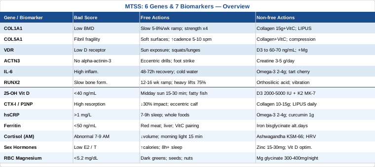
7 Biomarkers to Track for Medial Tibial Stress Syndrome
Biomarkers do not diagnose MTSS — imaging and clinical examination do that. But they reveal the physiological terrain your tibia is working within: how efficiently your bone is remodeling, how inflamed your systemic baseline is, how adequately fueled your musculoskeletal repair machinery actually is. Tracking the right panel can explain why certain runners remain stuck despite structural rehabilitation, guide supplementation decisions with actual data, and prevent recurrence more reliably than any single training modification. The seven below are ranked by actionability and evidence quality.
1. 25-OH Vitamin D
Why it matters: Vitamin D is not simply a calcium cofactor. Its active form binds the VDR receptor and regulates genes involved in osteoblast differentiation, periosteal bone formation, and type II muscle fiber recruitment. Low vitamin D status has been repeatedly associated with higher rates of bone stress injuries in athletes and military populations. A randomized controlled trial by Lappe et al. (2008, Journal of Bone and Mineral Research) found that female navy recruits supplemented with calcium and vitamin D had significantly lower incidence of stress fractures during basic training compared to placebo — a benchmark study in stress injury prevention (Lappe et al., 2008).
How to measure it
A standard 25-OH vitamin D serum test is available at any laboratory. Out-of-pocket cost typically ranges from $30–$80 in the US; it is frequently covered under insurance when ordered for musculoskeletal conditions. The functional optimal range for bone health and athletic adaptation is 40–60 ng/mL (100–150 nmol/L). Many labs flag deficiency only below 20 ng/mL — a threshold that misses functional insufficiency in active individuals. Test in late winter or early spring to capture your seasonal low.
If the score is low, the plan without supplements
Prioritize 15–30 minutes of midday sun exposure on large skin surface areas (arms, legs, upper back) four to five days per week. This approach is most effective at latitudes within 35°N–35°S during late spring through early fall. Dietary sources — fatty fish (salmon, mackerel, sardines), egg yolks, and beef liver — contribute meaningfully but rarely correct clinical insufficiency on their own. Pair sun exposure with progressive resistance training, which independently upregulates bone formation signaling via mechanical stimulation of osteoblasts.
If the score is low, the plan with supplements or equipment
Supplement with vitamin D3 (not D2), typically 2,000–5,000 IU daily depending on baseline deficiency and sun exposure, taken with your largest meal for fat-soluble absorption. Always co-supplement with vitamin K2 (MK-7 form, 100–200 mcg/day) to direct calcium into bone matrix rather than soft tissue and arterial walls. Retest at 90 days and adjust dose accordingly. No meaningful cycling is necessary at standard doses; ongoing maintenance is appropriate for athletes in low-sun climates. Toxicity risk appears only at sustained intakes consistently above 10,000 IU/day without monitoring.
2. Bone Turnover Markers: CTX-I and P1NP
Why they matter: Bone is constantly being broken down and rebuilt. CTX-I (C-terminal telopeptide of type I collagen) reflects osteoclast activity — the resorption side. P1NP (procollagen type 1 N-terminal propeptide) reflects osteoblast activity — the formation side. In MTSS, the periosteum endures repetitive tibial micro-bending that initially triggers a resorption signal before adaptive formation catches up. If resorption significantly outpaces formation — which an elevated CTX-I relative to P1NP reveals — the bone is in net negative structural territory and is vulnerable. Most MTSS sufferers have this assessed never.
How to measure it
CTX-I requires a fasting morning blood draw (values fluctuate significantly with food intake and time of day). P1NP can be drawn any time. Both are available through endocrinology or sports medicine referrals; out-of-pocket cost is $60–$150 each. The ratio matters more than absolute values. An elevated CTX-I alongside low or low-normal P1NP indicates inadequate formation relative to resorption — a physiological pattern that makes the tibia structurally more fragile under repeated loading.
If the score is bad, the plan without supplements
Reduce training impact volume by 20–30% for 4–6 weeks, specifically eliminating high-repetition-impact activities (stairs, downhill running, plyometrics). Replace with low-impact cross-training: pool running, cycling, or elliptical. Progressive resistance training — particularly calf raises (including eccentric-phase-focused), tibialis anterior strengthening, and single-leg press — stimulates osteoblast activity without high periosteal impact. Adequate caloric intake is non-negotiable: bone formation requires energy surplus, and even mild chronic caloric restriction suppresses the formation side of the equation more than most athletes realize.
If the score is bad, the plan with supplements or equipment
Collagen peptides (10–15g/day with 50mg vitamin C, taken 30–60 minutes before a loading session) supply hydroxyproline and proline, the structural amino acids for type I collagen. A study by Shaw et al. published in the American Journal of Clinical Nutrition found that vitamin C-enriched gelatin supplementation before intermittent activity significantly increased collagen synthesis markers, providing a rationale for pre-exercise timing (Shaw et al., 2017). Calcium (500–1,000 mg/day from combined food and supplement, spread across meals rather than taken at once) supports bone matrix mineralization. Low-intensity pulsed ultrasound (LIPUS) devices used 20 minutes daily over the affected tibial region have clinical evidence for accelerating bone healing. Recheck bone turnover markers at 90 days.
3. High-Sensitivity CRP (hsCRP)
Why it matters: Inflammation is the correct healing response — acutely. But chronically elevated systemic inflammation, reflected in hsCRP, creates a biological environment that impairs tissue repair, sensitizes pain pathways, and may suppress osteoblast activity. Athletes managing high training stress alongside poor sleep, suboptimal nutrition, or gut dysbiosis often face this compounding scenario: they generate the stimulus for bone adaptation but the recovery environment actively works against it. hsCRP is a simple, inexpensive proxy for this systemic state.
How to measure it
Request high-sensitivity CRP (not standard CRP — they differ in detection threshold). It is available at most labs, costs $15–$40, and is frequently included in cardiovascular risk panels. Optimal range: below 1 mg/L. Values between 1–3 mg/L reflect moderate systemic inflammation; above 3 mg/L is high. Critically, acute illness or a hard training session will transiently spike CRP — test during a true rest week, not the day after a tempo run.
If the score is bad, the plan without supplements
Sleep is the most powerful anti-inflammatory intervention available without a prescription: 7–9 hours in a cool, dark environment with consistent sleep and wake times. Reduce training load until inflammation markers normalize. Shift the dietary pattern toward whole foods: oily fish, colorful vegetables, olive oil, berries, and nuts. Reduce ultra-processed foods, refined sugar, and alcohol — all of which elevate hsCRP. Address gut health: chronic intestinal permeability is a significant driver of systemic inflammatory tone that most training-focused approaches ignore entirely.
If the score is bad, the plan with supplements or equipment
Omega-3 fatty acids (2–4g EPA+DHA per day from fish oil or algae oil) have the strongest evidence for reducing hsCRP in athletes and active individuals across multiple RCTs. Curcumin in a high-bioavailability form (phytosome or combined with piperine, 1,000 mg/day) has demonstrated anti-inflammatory effects in controlled trials — use for 8–12 weeks, then reassess; indefinite continuous use is not necessary. Magnesium glycinate (300–400 mg at night) supports sleep quality and has mild anti-inflammatory properties. Cold water immersion (10–15°C, 10–15 minutes, 3–4 times per week) may reduce hsCRP and accelerate systemic recovery, though it is best avoided immediately after strength training sessions to preserve the anabolic adaptation signal.
4. Serum Ferritin
Why it matters: Ferritin is the body's primary iron storage protein. Iron is essential for oxygen delivery, mitochondrial energy production, and collagen synthesis — it serves as a cofactor for prolyl hydroxylase, the enzyme that cross-links collagen fibrils. Runners with low ferritin, even within "normal" lab ranges, consistently report exaggerated fatigue, elevated perceived effort at sub-threshold speeds, and slower recovery. This training quality impairment directly undermines the gradual adaptation that MTSS rehabilitation requires. Functional iron deficiency — low ferritin with normal hemoglobin — is common and routinely missed by standard panels.
How to measure it
Serum ferritin is standard and costs $20–$60. The functional threshold for athletes differs substantially from general population reference ranges: many sports medicine practitioners working within the evidence-based performance framework (including those aligned with Peter Attia's approach) consider values below 50 ng/mL as functionally insufficient for distance runners, even when lab references cite 12–150 ng/mL as normal. For a complete picture, request a full iron panel: ferritin + serum iron + transferrin saturation + TIBC.
If the score is low, the plan without supplements
Increase dietary heme iron: red meat (particularly beef and lamb), organ meat (liver is the most iron-dense food available), and shellfish (oysters, clams). Pair iron-rich meals with vitamin C sources — citrus, bell pepper, kiwi — to enhance non-heme iron absorption from plant foods. Avoid coffee and tea within 60 minutes of iron-rich meals, as tannins significantly inhibit absorption. Cook in cast iron cookware, which leaches small amounts of elemental iron into food.
If the score is low, the plan with supplements or equipment
Iron bisglycinate (25–65 mg elemental iron) taken every other day rather than daily has been shown in recent research to optimize absorption by preventing the hepcidin surge that blocks next-day intestinal uptake — Moretti and colleagues (2015, Blood) established that daily dosing triggers hepcidin-mediated suppression of iron absorption, providing rationale for alternate-day protocols. Take on an empty stomach with 200mg vitamin C if tolerated; if GI distress occurs, take with a small meal. Recheck ferritin at 8–12 weeks. Do not supplement without confirmed low ferritin — iron excess is pro-oxidant and harmful.
5. Morning Cortisol
Why it matters: Cortisol is the primary stress hormone, peaking naturally in the 30–60 minutes after waking. Chronically elevated cortisol — from training overload, sleep debt, caloric restriction, or psychological stress — directly suppresses osteoblast differentiation, increases bone resorption, impairs muscle protein synthesis, and amplifies systemic inflammation. The athlete who trains hard, sleeps poorly, and eats insufficiently creates a hormonal environment that actively resists the bone adaptation that MTSS rehabilitation depends on. This pattern, formalized in the literature as Relative Energy Deficiency in Sport (RED-S), is one of the most underappreciated drivers of recurrent bone stress injuries.
How to measure it
Morning serum cortisol, drawn between 7–9 AM fasting, costs $30–$70. A 4-point salivary cortisol test (morning, noon, afternoon, evening) provides a fuller picture of the daily rhythm at $100–$180 and is offered by many functional medicine and sports medicine labs. Optimal morning serum cortisol: 10–20 mcg/dL. Both persistently elevated and unexpectedly low morning cortisol (blunted awakening response) indicate HPA axis dysregulation requiring attention.
If the score is bad, the plan without supplements
Reduce training volume, and specifically reduce high-intensity session frequency for 4–6 weeks. Prioritize 8 or more hours of sleep — cortisol rhythm is deeply coupled to sleep architecture. Implement a consistent morning light exposure protocol: 10–20 minutes of outdoor light within 30 minutes of waking anchors the circadian clock and normalizes the cortisol awakening response. Address cognitive load directly: psychological stress suppresses HPA recovery just as effectively as physical overtraining, and both need to be managed concurrently.
If the score is bad, the plan with supplements or equipment
Ashwagandha (KSM-66 extract, 300–600 mg/day with a meal) has the strongest adaptogenic evidence for cortisol reduction in active individuals — multiple RCTs show significant cortisol lowering after 8–12 weeks of consistent use. Cycle every 12 weeks with a 4-week break. Phosphatidylserine (400 mg/day taken post-exercise) has modest but specific evidence for blunting the exercise-induced cortisol spike. Heart rate variability (HRV) monitoring using a device such as a Polar H10 paired with the HRV4Training application is a practical real-time recovery tool — training in a chronically suppressed HRV state is a reliable proxy for cortisol-driven HPA overload, and tracking it prevents training decisions that worsen the hormonal deficit.
6. Sex Hormones: Estradiol and Testosterone
Why they matter: Both estradiol and testosterone play direct regulatory roles in bone density maintenance and periosteal growth. Estradiol suppresses osteoclast activity and is the principal bone-protective hormone in women — its deficiency, whether from hypothalamic amenorrhea, extreme caloric restriction, or menopause, is one of the strongest known predictors of stress fracture risk in female athletes. In male athletes, low testosterone from overtraining, chronic cortisol elevation, or very low body fat comparably impairs bone formation and connective tissue recovery rate. These hormones are among the first casualties of energy deficiency, yet they are almost never tested in standard MTSS workups.
How to measure it
For women: estradiol + FSH + LH + total testosterone, ideally timed to days 3–5 of the menstrual cycle (follicular phase baseline). For men: total testosterone + free testosterone + SHBG + estradiol. Standard panels cost $80–$200. In premenopausal women, amenorrhea or oligomenorrhea alongside low estradiol is a clinical red flag requiring medical attention — not just supplementation. Discuss findings with an endocrinologist or sports medicine physician who understands female athlete physiology.
If the score is bad, the plan without supplements
For amenorrheic or oligomenorrheic female athletes: increase caloric intake to restore energy availability above the RED-S threshold (estimated at greater than 45 kcal per kg of lean body mass per day). Reduce training volume. Menstrual function restoration is a prerequisite for safe return to full load — this is not negotiable from a bone structural standpoint. For men with low testosterone: prioritize 8+ hours of sleep (testosterone is synthesized primarily during slow-wave sleep), ensure adequate dietary fat intake (testosterone biosynthesis requires cholesterol as substrate), and reduce both training volume and life stress concurrently.
If the score is bad, the plan with supplements or equipment
For men with low-normal testosterone: zinc (15–30 mg/day elemental, cycling 8 weeks on and 4 weeks off) may support testosterone production in those who are deficient in the mineral. Vitamin D optimization at the upper therapeutic range (as covered above) has demonstrated modest testosterone-supporting effects in RCTs among deficient men. Minimize endocrine disruptors: avoid heating food in plastic containers and reduce BPA-containing packaging exposure. For clinically significant hormonal deficiency, a qualified endocrinologist should assess whether medically supervised hormonal therapy is appropriate — supplementation is not a substitute for proper diagnosis and treatment in that scenario.
7. RBC Magnesium
Why it matters: Magnesium is a cofactor in over 300 enzymatic reactions, including vitamin D activation, calcium regulation, muscle contraction, nerve conduction, and protein synthesis. Serum magnesium — the standard test — reflects only about 1% of total body magnesium and is a poor indicator of actual tissue status; it can appear normal while intracellular stores are depleted. Red blood cell magnesium is the more clinically accurate measure. In endurance athletes, magnesium is lost through sweat and stress. Deficiency impairs sleep quality, increases neuromuscular excitability, reduces anabolic response to training, and may compromise the quality of bone matrix mineralization.
How to measure it
Request RBC magnesium specifically, not standard serum magnesium. It is available at functional medicine labs and an increasing number of standard labs; cost ranges from $40–$100. Optimal RBC magnesium: 5.2–6.5 mg/dL. Many functional practitioners target the upper half of this range for competitive athletes given the higher sweat and metabolic losses involved.
If the score is low, the plan without supplements
Increase dietary magnesium through dark leafy greens (spinach, Swiss chard), pumpkin seeds, almonds, black beans, and dark chocolate. Be aware that soil depletion and food processing significantly reduce the magnesium content of modern food — dietary improvement alone often cannot fully correct deficiency in high-output athletes generating consistent sweat losses.
If the score is low, the plan with supplements or equipment
Magnesium glycinate for sleep support and muscle recovery, or magnesium malate for energy metabolism and muscle function: 300–400 mg elemental magnesium per day, split between morning and evening doses. Avoid magnesium oxide — its bioavailability is poor. Do not take simultaneously with calcium supplements as they compete for absorption. Epsom salt baths (magnesium sulfate in warm water, 20 minutes before bed) provide transdermal absorption and have practical anecdotal support as a sleep and recovery aid. No cycling required — ongoing supplementation is appropriate and well-tolerated.
What Genetics Research Suggests About MTSS Risk
Genetic testing for athletic injury susceptibility is still early-stage science, but several gene variants have accumulated enough human evidence to be worth understanding — especially for athletes with recurrent cases or family histories of connective tissue fragility, stress fractures, or slow recovery from bone injuries. The six genes below are relevant to MTSS biology through their roles in collagen quality, bone formation signaling, muscle fiber mechanics, and inflammatory regulation.
These associations have not been validated in large MTSS-specific genome-wide association studies. Most evidence comes from stress fracture genetics, tendinopathy research, and bone density studies. The mechanistic relevance to MTSS is strong, but apply this information in proportion to the evidence level — strong for COL1A1 and VDR, more preliminary for RUNX2. Genetic testing services such as 23andMe (with third-party interpretation tools) or clinical panels through sports medicine geneticists can identify most of these variants.
Gene 1: COL1A1 — The Collagen Structure Gene
What it does: COL1A1 encodes the alpha-1 chain of type I collagen — the dominant structural protein in bone, periosteum, tendons, and ligaments. A well-studied polymorphism in the Sp1 binding site (rs1800012, the G/T variant) is associated with reduced collagen cross-linking efficiency and measurably lower bone mineral density. The TT genotype is associated with the greatest reduction in bone structural integrity and has been linked to higher stress fracture rates in multiple athlete and military studies, including a key study in South African endurance athletes.
If the gene is bad, the plan without supplements
Prioritize long-duration progressive bone loading over explosive, high-impact training intensity. Bone adaptation to load is likely slower in COL1A1-variant carriers — use 5–8% weekly volume increases rather than the common but evidence-poor 10% rule. Focus systematically on tibialis posterior, soleus, and calf complex strengthening to reduce tibial bending forces during gait. Resistance training with progressive overload three to four times per week is essential — it stimulates periosteal bone formation via mechanical strain signaling, which partially compensates for reduced intrinsic collagen efficiency.
If the gene is bad, the plan with supplements or equipment
Collagen peptides (15g/day with 50mg vitamin C, 30–60 minutes before training or any loading session) directly supply hydroxyproline and proline — the amino acid precursors of type I collagen synthesis. Vitamin C is non-negotiable as a cofactor for prolyl hydroxylase activity. Silicon as orthosilicic acid (10–20 mg/day) has emerging evidence for upregulating collagen cross-linking and may be especially relevant for COL1A1-variant individuals with poor cross-link efficiency. Low-intensity pulsed ultrasound (LIPUS) devices used 20 minutes daily over the medial tibia have clinical evidence for accelerating stress fracture healing and may support periosteal formation in those with reduced collagen quality.
Gene 2: COL5A1 — The Collagen Organization Gene
What it does: COL5A1 encodes a regulatory collagen that controls the diameter and spatial packing of type I collagen fibrils. The rs12722 C/T polymorphism has been associated with Achilles tendinopathy, anterior cruciate ligament injury, and broader connective tissue vulnerability in athletes. The CC genotype appears protective; the TT genotype correlates with higher soft tissue injury risk across multiple cohort studies. While MTSS-specific evidence is limited, periosteal tissue vulnerability is mechanistically plausible given COL5A1's fundamental role in fibril architecture.
If the gene is bad, the plan without supplements
Running surface selection becomes more important: grass, trail, and track significantly reduce peak tibial impact versus asphalt and concrete. Increasing running cadence by 5–10 steps per minute reduces tibial bending forces without equipment or medical intervention and is especially relevant for COL5A1-variant carriers. Running form adjustments — slight forward trunk lean, shorter stride, active midfoot contact — reduce tibial stress amplitudes. Custom orthotics assessed by a sports podiatrist can redistribute impact loading across a broader foot surface area.
If the gene is bad, the plan with supplements or equipment
The same collagen peptide and vitamin C protocol as COL1A1 applies here. Compression sleeves worn during training (15–20 mmHg graduated compression) may reduce tibial vibration transmission — the evidence is limited but the mechanism is plausible and the intervention is low-risk. Footwear with higher heel drop (8–12 mm) and shock-absorbing midsole materials (such as EVA foam or newer nitrogen-infused foams) reduce peak impact at initial contact and can meaningfully unload the periosteum in TT-genotype athletes.
Gene 3: VDR — The Vitamin D Receptor Gene
What it does: VDR encodes the receptor that binds the active form of vitamin D (1,25-dihydroxyvitamin D3) and initiates downstream gene transcription in osteoblasts, myocytes, and immune cells. Multiple polymorphisms — particularly FokI (rs2228570), BsmI, ApaI, and TaqI — affect receptor efficiency and have been consistently associated with bone mineral density variation across populations. Individuals with less efficient VDR variants may need substantially higher circulating 25-OH vitamin D levels to achieve the same downstream biological effect in bone.
If the gene is bad, the plan without supplements
Extended midday sun exposure becomes a higher priority for VDR-variant carriers: their lower receptor sensitivity means the same circulating vitamin D generates less osteoblast signaling, requiring higher serum levels to compensate. Weight-bearing resistance exercise — particularly compound movements like squats and lunges — directly upregulates VDR expression in bone-adjacent tissue, partially compensating for reduced receptor efficiency through a parallel signaling pathway.
If the gene is bad, the plan with supplements or equipment
Target the upper end of the optimal vitamin D range: 60–70 ng/mL rather than the standard 40 ng/mL threshold, particularly during autumn and winter months. This may require 4,000–6,000 IU of D3 daily; quarterly blood monitoring is essential at these doses. Ensure magnesium adequacy — magnesium is required for the enzymatic conversion of vitamin D to its active form and is depleted by high-dose vitamin D supplementation. Consider emulsified (liquid drop) vitamin D formulations if fat malabsorption is a concern, as they do not require dietary fat for absorption.
Gene 4: ACTN3 — The Muscle Fiber Architecture Gene
What it does: ACTN3 encodes alpha-actinin-3, a structural protein found exclusively in fast-twitch (type IIx) muscle fibers. The R577X polymorphism (rs1815739) produces a premature stop codon in the X allele, rendering the protein non-functional. XX-genotype individuals — approximately 18% of the general population — have no functional alpha-actinin-3 in their fast-twitch fibers and show a measurable metabolic shift toward slow-twitch, oxidative muscle physiology. This alters peak power output and changes how mechanical load is distributed through the lower limb during running.
If the gene is bad, the plan without supplements
XX-genotype runners generate less tibial impact force per stride (a consequence of reduced fast-twitch power) but may be less efficient at eccentric shock absorption — influencing how impact is distributed across the periosteum during loading. Eccentric loading drills targeting the tibialis anterior and peroneal complex — specifically heel-lowering exercises on a step, four sets of 15 repetitions daily — may partially compensate for reduced eccentric force absorption. Foot strike analysis with a sports biomechanist is worth pursuing: XX carriers often benefit from a midfoot contact pattern that distributes tibial stress more evenly across the stance phase.
If the gene is bad, the plan with supplements or equipment
Creatine monohydrate (3–5g/day, no loading phase required) has consistent evidence for improving fast-twitch fiber function and power output and may partially compensate for ACTN3 protein absence. Pair with structured resistance training that specifically recruits type II fibers: heavy compound lifts at 75–85% of one-repetition maximum and low-volume plyometrics at appropriate points in the training cycle. Long-term creatine use at these doses has a strong safety record; no cycling is required.
Gene 5: IL-6 — The Inflammatory Signaling Gene
What it does: IL-6 is a cytokine that functions in two roles: as a pro-inflammatory mediator when released by immune cells, and as a muscle-derived myokine with anti-inflammatory and bone-supportive effects when released during exercise. The −174G/C promoter polymorphism (rs1800795) affects baseline IL-6 expression. The GG genotype is associated with higher IL-6 output, which can amplify the post-exercise inflammatory response — a double-edged situation: more inflammatory signaling during acute load, which may increase periosteal irritation in susceptible athletes while simultaneously driving higher bone adaptation signaling.
If the gene is bad, the plan without supplements
GG-genotype athletes benefit from deliberately extended recovery windows between high-impact sessions. Their post-exercise inflammatory peak is higher and more prolonged — a 48–72 hour gap between run sessions (rather than the typical 24-hour recovery) may be appropriate during MTSS rehabilitation. Monitor for early warning signals: morning periosteal soreness or pain on direct palpation of the medial tibia that persists for more than 24 hours post-run. Cold water immersion (10–12°C, 10 minutes, within 1 hour post-session) may help normalize the exaggerated IL-6 response without fully suppressing the adaptive signal.
If the gene is bad, the plan with supplements or equipment
Omega-3 fatty acids (2–4g EPA+DHA/day) are the most evidence-based option for modulating IL-6-driven inflammation across multiple exercise and clinical RCTs. Tart cherry concentrate (30 mL twice daily, starting two days before any high-volume training week) has documented evidence for reducing exercise-induced IL-6 and downstream inflammatory markers in distance runners — Howatson and colleagues demonstrated significant attenuation of inflammatory markers and accelerated recovery in marathon runners using this protocol. Cycle cherry supplementation around high-load training blocks (8 weeks on, 4 weeks off) rather than using it continuously.
Gene 6: RUNX2 — The Bone Formation Master Regulator
What it does: RUNX2 is the master transcription factor controlling osteoblast differentiation — without it, bone formation does not proceed normally. Polymorphisms in the RUNX2 gene have been associated with bone mineral density variation and fracture risk in population studies. In athletic contexts, variants that reduce RUNX2 transcriptional activity may impair the adaptive bone formation response to training load, slowing periosteal thickening and leaving the tibia structurally more vulnerable during any ramp-up phase of training. Evidence in athletes specifically is early-stage and requires extrapolation from osteoporosis research.
If the gene is bad, the plan without supplements
Slow the training ramp-up substantially. Where a standard runner might follow an 8-week return-to-run timeline, a RUNX2-variant carrier may need 12–16 weeks to allow adequate periosteal bone formation to keep pace with increasing load demand. High-load, low-repetition resistance training (compound lifts at 75–85% of 1RM, three sets, three times per week) maximally stimulates osteoblast signaling pathways and may partially compensate for reduced RUNX2 efficiency by providing a stronger mechanical stimulus to the same formation machinery.
If the gene is bad, the plan with supplements or equipment
Silicon as orthosilicic acid (10–20 mg/day) has in vitro evidence for upregulating RUNX2 expression in osteoblasts and supporting human evidence for bone density outcomes. Whole-body vibration therapy (30 Hz, 0.3g amplitude, 10 minutes, three times per week) has been studied as a non-impact mechanical stimulus for bone formation pathways that include RUNX2 upregulation — particularly relevant for athletes in low-load recovery phases who cannot generate sufficient impact stimulus. Avoid combining iron supplementation with these approaches unless ferritin testing indicates deficiency.
Bone Adaptation Science — What the Research Actually Says About MTSS Recovery
Andrew Huberman and his research-scientist guests have covered bone biology, stress physiology, and recovery extensively across multiple Huberman Lab episodes, including discussions with experts in exercise physiology and musculoskeletal medicine. Drawing from the converging evidence from these conversations and the broader peer-reviewed literature, here are ten of the most impactful insights for anyone managing repetitive tibial bone stress — insights that often run counter to what runners are typically told.
1. The 10% Rule Was Never Evidence-Based
The near-universal advice to limit training volume increases to 10% per week has no controlled trial supporting it. It was a clinical heuristic that became doctrine. More rigorous analysis using the acute-to-chronic workload ratio (developed through Tim Gabbett's research across multiple sports) suggests that injury risk climbs meaningfully when a single week's load exceeds approximately 1.3 times the four-week rolling average. For runners returning from MTSS, a 5–8% weekly impact load increase applied to an honest rolling average is a more protective and more evidence-anchored target.
2. Bone Adapts Slower Than Fitness — This Is the Core Problem
Cardiovascular capacity and muscle strength improve within weeks of structured training. Periosteal bone density and thickness respond over months. This biological lag — sometimes called the bone-fitness gap — is the primary structural reason that fit, motivated athletes get MTSS. They develop the fitness to run farther and faster long before their tibiae have adapted to sustain that load. Fitness level is not a reliable proxy for bone readiness. Bone turnover markers and subjective periosteal pain response are more relevant guides to load tolerance.
3. Sleep Is the Most Underrated Bone Recovery Tool
Bone remodeling is a sleep-dependent process. Growth hormone — which stimulates both osteoblast differentiation and type I collagen synthesis — is released in pulses predominantly during slow-wave sleep. Chronic sleep restriction measurably reduces growth hormone output and suppresses bone formation markers. Before adding any supplement for bone recovery, ensuring consistent 7.5–9 hours of quality sleep is the highest-yield intervention available. No supplement replaces this, and many supplements cannot compensate for it.
4. Zone 2 Training Is the Cross-Training Gold Standard for MTSS
Zone 2 aerobic work — sustained effort at 60–70% of maximum heart rate for 45–90 minutes — maintains cardiovascular and mitochondrial adaptation, drives fat oxidation, and reduces systemic inflammatory markers. It does this without generating the high-repetition periosteal impact that MTSS requires protection from. Pool running and cycling at zone 2 intensity are the optimal MTSS cross-training modalities: they maintain training stimulus while allowing the periosteum to progress through its adaptive formation cycle uninterrupted.
5. Cortisol Suppresses Bone Formation Fast — Even a Single Bad Night Matters
Even a single night of poor sleep measurably elevates morning cortisol enough to shift bone turnover markers toward net resorption for the following 24–48 hours. This means the cumulative effect of a stressful training week — sleep debt, high mileage, inadequate food — may cause more damage to the periosteal environment than the mechanical impact of the runs themselves. Stress management is structural work in bone stress injury management, not psychological nicety.
6. Vitamin D and Omega-3s Work Better Together Than Separately
Vitamin D regulates osteoblast differentiation and calcium incorporation. Omega-3 fatty acids modulate prostaglandin and cytokine pathways that drive periosteal inflammation. These operate through complementary mechanisms: vitamin D enables the formation response, while omega-3s reduce the inflammatory environment that suppresses it. The combination has a biologically additive rationale and appears across multiple lines of bone stress injury and stress fracture prevention evidence.
7. Abruptly Changing Foot Strike Pattern Causes New Injuries
Switching from heel to forefoot striking is a commonly recommended biohack for MTSS. The biomechanical rationale is partially valid — forefoot striking reduces tibial bending at initial contact. The implementation is where it fails: abrupt strike changes transfer load to tissue that has no adaptation history, reliably causing new injuries in the ankle, Achilles tendon, and calf complex within weeks. Any strike pattern transition should span 12–16 weeks with concurrent progressive calf and intrinsic foot strengthening.
8. Cadence Increase Is the Safest and Most Consistent Gait Modification
Increasing running cadence by 5–10 steps per minute — regardless of foot strike pattern — consistently reduces tibial impact shock and peak ground reaction force in biomechanics research. This modification is well-tolerated, requires no equipment, and produces measurable reductions in tibial loading within the first session. A metronome application set to the target cadence (or music matched to the target tempo) is sufficient for training the change. This is the single most evidence-supported, accessible gait modification for MTSS currently available.
9. Progressive Load — Not Extended Rest — Prevents Recurrence
Complete impact rest causes tibial bone density loss, not consolidation. The adaptive stimulus for periosteal bone formation is controlled mechanical loading — not its absence. Athletes who rely solely on extended rest without structured progressive reloading frequently experience rapid recurrence when they return, because they have allowed bone density to regress without building the structural capacity to handle the original demand. Return-to-run programs should be more granular, more conservative in weekly increments, and more attentive to periosteal pain signals than most existing protocols prescribe.
10. Central Sensitization Explains Why Pain Persists After Healing
Chronic unresolved MTSS, particularly in anxious or highly stressed athletes, can trigger central sensitization — a nervous system state in which pain signals are amplified beyond what remaining tissue damage justifies. This explains the pattern of pain persisting long after imaging confirms healing. Breath work (particularly extended exhalation ratios, 4-second inhale / 6–8 second exhale), NSDR (non-sleep deep rest), and deliberate cold exposure protocols discussed in Huberman's neuroscience content are relevant adjuncts to physical rehabilitation in this scenario — not replacements for addressing mechanical and biochemical drivers, but meaningful additions when pain has outlasted the injury.
Complementary Approaches With Evidence for Tibial Stress Conditions
The following four modalities were selected from the evidence-based list based on meaningful clinical evidence or mechanistic relevance specifically for bone stress and soft tissue overload conditions. Evidence quality varies and is noted for each.
Low-Level Laser Therapy / Photobiomodulation
Low-level laser therapy uses specific wavelengths of red (630–700 nm) and near-infrared (810–1100 nm) light to stimulate cellular energy production via cytochrome c oxidase in mitochondria, reducing local inflammation and promoting tissue repair. For MTSS, this is directly relevant: periosteal tissue on the medial tibia is relatively superficial and within effective penetration depth, and photobiomodulation has demonstrated effects on both soft tissue inflammation and bone formation stimulation in controlled settings.
A protocol commonly studied for musculoskeletal pain and bone stress uses near-infrared at 830 nm, applied at 3–4 J/cm² over the affected periosteal region, three times per week for 4–6 weeks. A meta-analysis by Chow and colleagues in The Lancet (2009) established meaningful evidence for LLLT in musculoskeletal pain reduction, and subsequent studies have extended this to bone healing contexts. For MTSS specifically, evidence remains preliminary but mechanistically well-grounded.
Home-use photobiomodulation devices (Joovv, BioMax, or clinical-grade Erchonia panels) range from $300 to $2,000+. Many sports physio and chiropractic clinics offer in-clinic LLLT sessions at $30–$80 each. Apply to the medial tibial surface for 8–10 minutes per session during recovery phases. Avoid direct application over a suspected undiagnosed stress fracture until imaging has confirmed the diagnosis. Use consistently over 4–6 weeks for meaningful results rather than intermittently.
Massage Therapy
Deep tissue massage and myofascial release targeting the posterior tibial compartment — specifically the tibialis posterior, flexor digitorum longus, and soleus — may reduce the periosteal traction forces that contribute to MTSS. These deep posterior muscles attach along the medial tibial border, and chronic hypertonia in this compartment increases pulling stress on the periosteum during running, amplifying mechanical irritation beyond what the impact load alone would generate.
Sports massage and myofascial therapy have been included in military rehabilitation protocols for MTSS, and consensus statements in sports medicine literature support soft tissue work as an adjunct to structured loading rehabilitation, particularly in recurrent or chronic cases with palpable muscle hypertonicity along the medial tibia. Evidence for massage in isolation is limited; evidence for it as part of a multimodal rehabilitation approach is more supportive.
Request focus on the posterior compartment — soleus, flexor digitorum longus, tibialis posterior — and medial gastrocnemius, not just the tibial surface itself. Avoid aggressive direct pressure over the periosteal zone during acute flares. Two sessions per week during active recovery, transitioning to one maintenance session per week during the return-to-run phase. A massage gun at low intensity (below 40 Hz) can supplement between professional sessions for self-maintenance of posterior compartment extensibility.
Biofeedback
Gait biofeedback uses real-time sensory feedback — auditory, visual, or haptic — to train runners to modify mechanics that contribute to tibial overload. For MTSS, the primary targets are tibial shock amplitude (measured by accelerometer mounted at the distal tibia), peak vertical ground reaction force, and cadence. Real-time feedback accelerates neuromuscular learning far more efficiently than verbal cueing alone, because the athlete can observe cause-and-effect between their mechanics and the loading signal within each stride.
A randomized controlled trial by Davis and colleagues in the Journal of Orthopaedic and Sports Physical Therapy demonstrated that real-time tibial accelerometry biofeedback significantly reduced tibial shock during running, with gait modifications retained at a one-month follow-up after the feedback was removed — suggesting durable neuromuscular adaptation rather than temporary performance. The protocol used six training sessions over two weeks with an accelerometer mounted on the distal tibia providing auditory alerts when shock exceeded a target threshold.
Wearable options have made this increasingly practical. Garmin Running Dynamics Pod or phone-based accelerometry applications can approximate tibial shock feedback. For formal biofeedback-guided gait retraining, a sports physio or biomechanics lab with an instrumented treadmill and accelerometry setup is optimal. Six to eight sessions over two to three weeks during the return-to-run phase represent a clinically grounded protocol. Combine with simultaneous cadence training for additive effect on tibial load reduction.
Yoga
Yoga is relevant to MTSS not primarily as a stretching practice but as a systematic approach to building the hip, posterior chain, and single-leg stability that redistributes tibial loading during running. Weak hip abductors and external rotators cause excessive internal tibial rotation during the stance phase — a biomechanical pattern consistently associated with higher medial tibial stress in multiple gait analysis studies. Yoga postures that load the hip complex under eccentric and single-leg demand address this mechanism directly.
Research examining hip strengthening programs in female distance runners has consistently demonstrated reductions in tibial stress markers and injury recurrence rates. Yoga-based hip strengthening programs have been included in running injury prevention frameworks where specific postures — Warrior III, single-leg chair pose, and side-lying clamshell progressions — directly target hip abductor and external rotator strength under controlled loading. The combination of flexibility, proprioception, and loaded stability is more comprehensive than isolated strength work alone.
A practical protocol for MTSS prevention: three 30-minute sessions per week emphasizing single-leg balance (Warrior III, single-leg mountain pose), hip abductor loading (Warrior II, Warrior III with isometric hold), calf eccentric loading (downward dog progressions with heel-to-floor emphasis), and deep posterior compartment stretching (reclined hero pose, supine hip rotation). Begin during the active recovery phase — not just after return to running — and continue as structural prevention through full training resumption. Standard yoga classes may not provide sufficient hip loading; seek running-specific yoga programs or work with a teacher familiar with injury prevention programming.
Conclusion
Medial tibial stress syndrome sits at the intersection of mechanics, biology, and training load — and addressing only one of those dimensions is why so many recoveries stall or lead to a familiar reinjury cycle. The biomarkers covered here give you a measurable window into the physiological terrain your tibia is operating in right now. The genetic variants give you a longer-horizon view of structural tendencies that explain why some athletes face recurring bone stress while others performing the same training load adapt without incident.
None of this replaces a thorough clinical evaluation, imaging when indicated, or the patience to follow a properly graded return-to-run program. But it changes the quality of the conversations you can have with the clinicians managing your care — and the decisions you make independently. Start with the biomarkers: vitamin D, bone turnover markers, hsCRP, and ferritin are affordable, accessible, and directly actionable. If recurrence remains a pattern, add cortisol, sex hormones, and RBC magnesium for a more complete picture. If your history suggests structural susceptibility that these markers do not explain, the genetic layer is a reasonable next step with a practitioner who can integrate it into a personalized protocol.
Better data leads to better decisions. That is not a guarantee of a faster outcome — but it is a meaningful advantage over guessing.