This article contains AI generated content.
· UpdatedMelorheostosis Genes and Biomarkers — 3 Genes and 6 Biomarkers to Track
Introduction
Melorheostosis is one of the rarest bone disorders in medicine — a condition where bone grows in dense, irregular patterns along the cortex of a limb, often described in imaging reports as resembling wax dripping down a candle. If you or someone close to you has received this diagnosis, you already know that finding a specialist who has seen more than one or two cases is itself a challenge. The path from diagnosis to any meaningful treatment plan is rarely straightforward, and the advice you get is often either frustratingly generic or purely focused on surgical intervention.
What has changed in the past decade is the science. Researchers, primarily working through the NIH's intramural program, have identified specific molecular drivers of this condition — particularly gain-of-function somatic mutations in the MAP2K1 gene, which sits at the heart of the RAS-MAPK signaling cascade. This is not abstract academic knowledge. It means melorheostosis has a molecular fingerprint, and that fingerprint can be tracked, studied, and eventually targeted. Understanding that fingerprint is the first step toward asking better questions of your care team.
At the same time, generic bone health advice — take calcium, exercise, manage inflammation — is insufficient for melorheostosis. The biology here is fundamentally different from osteoporosis or Paget's disease, and applying the same playbook risks missing what is actually driving the abnormal bone formation in your specific case. What is needed is a more targeted approach: specific biomarkers that reflect the actual pathways involved, specific genes worth understanding, and specific interventions matched to those findings.
This article provides exactly that. The first section covers six biomarkers that are clinically trackable today and directly relevant to melorheostosis biology — each with a practical plan for when results come back out of range. The genetics section examines three key genes whose dysfunction underpins the condition, along with strategies for compensating when the biology is unfavorable. Beyond those two core sections, you will find a precision medicine framework drawn from Peter Attia's work, complementary approaches with real evidence for chronic bone pain, and a clear conclusion. Better information will not resolve every challenge melorheostosis presents, but it reliably leads to better decisions.
Summary
This article breaks down the science of melorheostosis into actionable insight. The biomarker section covers sclerostin, bone-specific alkaline phosphatase, CTX-I, osteocalcin, hs-CRP/IL-6, and vitamin D — six markers that collectively track bone formation activity, resorption balance, inflammation, and metabolic drivers. For each, you will find how to measure it, what it costs, and a concrete plan when results are abnormal, both with and without supplements. The genetics section examines MAP2K1, LEMD3, and the broader RAS-MAPK pathway — the molecular architecture that explains why melorheostosis develops at all, and what that means practically. After those two core strategies, you will find insights from Peter Attia's precision medicine framework, three evidence-backed complementary modalities for pain and mobility, and a summary to guide your next steps with a qualified specialist.
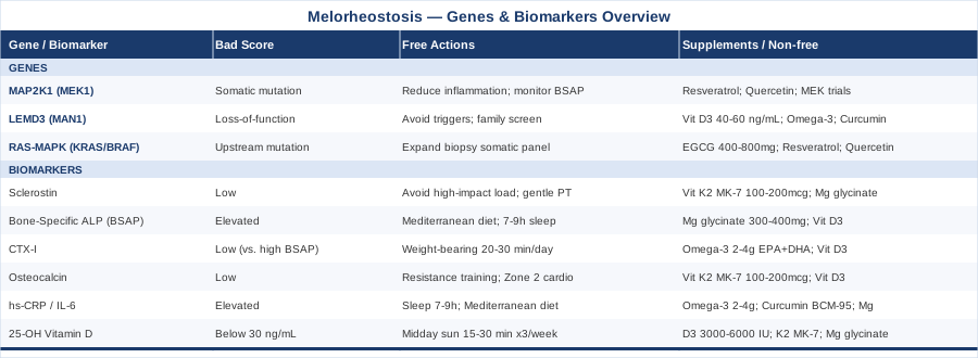
6 Biomarkers to Track in Melorheostosis
Biomarkers do not diagnose melorheostosis — imaging and biopsy do that. What biomarkers can do is give you a running picture of how active the underlying biology is, which pathways are most engaged, and whether your interventions are moving things in the right direction. The six markers below were chosen because they directly reflect the signaling pathways implicated in melorheostosis, are clinically accessible, and carry actionable interpretation. Peter Attia has consistently advocated for tracking bone turnover markers alongside standard panels in any longevity protocol; in a condition where bone formation is the central dysfunction, this is even more relevant.
Sclerostin: The Wnt Pathway Window
Sclerostin is a protein encoded by the SOST gene and secreted primarily by osteocytes — mature bone cells embedded in the bone matrix. Its primary function is to act as a natural brake on bone formation by blocking the Wnt/β-catenin signaling pathway. When sclerostin is low or absent, bone formation proceeds unchecked: this is the mechanism behind sclerosteosis and Van Buchem disease, both characterized by massive cortical bone overgrowth that shares phenotypic features with melorheostosis. While melorheostosis runs primarily through the MAPK pathway (MAP2K1), the Wnt and MAPK pathways interact downstream through shared transcription factors. Sclerostin levels therefore provide a useful surrogate for how actively the bone formation machinery is running in affected regions.
How to Measure It
Sclerostin is measured via ELISA in a fasting blood sample. It is not available as a routine standalone order at standard commercial labs, but specialty labs — including Immuno Diagnostic Laboratory and academic medical center reference labs — can process it. Cost ranges from $150 to $300 out of pocket. Reference ranges vary by lab, age, and sex; most healthy adults fall between 20 and 75 pmol/L.
If the Score Is Low: The Plan Without Supplements
Very low sclerostin in the context of melorheostosis suggests that the Wnt brake is not being applied effectively in lesion areas, and osteoblast activity is running high. The non-pharmacological response centers on mechanical load management: avoid repetitive high-impact stress on affected limbs, since mechanical strain is a known trigger for Wnt pathway activation. Gentle physiotherapy focused on range of motion and proprioception — rather than high-load strengthening of the affected segment — is the most evidence-consistent approach. Discuss the finding with a metabolic bone specialist; very low sclerostin in context might inform a pharmaceutical conversation.
If the Score Is Low: The Plan With Supplements or Equipment
No supplement directly and safely raises sclerostin. The pharmaceutical approach (romosozumab, an anti-sclerostin antibody) works in the opposite direction and is used for osteoporosis. However, vitamin K2 in the MK-7 form (100–200 mcg daily) supports balanced bone remodeling signaling and has shown modulation of osteoblast/osteoclast coupling in human studies. Take it daily with a fat-containing meal; no cycling is required. Side effects are minimal, but K2 interacts with warfarin — check with a physician if you are on anticoagulants. Magnesium glycinate (200–300 mg/day) also supports downstream Wnt signaling fidelity. Start low; loose stools are the main dose-limiting side effect.
Bone-Specific Alkaline Phosphatase: Measuring Formation Activity
Total alkaline phosphatase (ALP) appears on most standard blood panels, but it reflects isoforms from bone, liver, kidney, and intestine simultaneously, making it a noisy signal. Bone-specific alkaline phosphatase (BSAP) is produced exclusively by osteoblasts and directly quantifies how actively bone is being formed. In melorheostosis, where osteoblastic activity is constitutively elevated in affected regions due to MAPK pathway dysregulation, BSAP provides a dynamic measure of disease activity. Tracking it quarterly or biannually reveals whether the condition is stable, worsening, or — rarely — improving. Peter Attia includes bone formation markers in his extended metabolic panels as part of musculoskeletal longevity assessment.
How to Measure It
BSAP is available at Quest Diagnostics, LabCorp, and most commercial labs, either as a standalone test or as part of a bone remodeling panel. Cost: $30–$80 at commercial labs, often covered by insurance when ordered under a bone metabolism diagnosis code. Normal adult range is approximately 11–30 mcg/L, with variation by age and sex.
If the Score Is Elevated: The Plan Without Supplements
Elevated BSAP signals high osteoblast activity. The lifestyle interventions most supported by evidence for reducing systemic osteoblast-stimulating signals are: a Mediterranean-style dietary pattern (associated with lower bone remodeling markers in observational studies); sleep optimization (7–9 hours, consistent schedule), since sleep deprivation drives cortisol and inflammatory cytokines that can stimulate bone turnover; and careful aerobic exercise — moderate intensity, 3–5 days per week, avoiding high-impact loading of affected limbs. Bisphosphonates (pharmaceutical) are sometimes prescribed for symptomatic melorheostosis and have been reported to reduce BSAP; this is a specialist conversation.
If the Score Is Elevated: The Plan With Supplements or Equipment
Magnesium glycinate (300–400 mg/day) is commonly deficient in individuals with high bone turnover and supports enzymatic regulation of osteoblast activity. Vitamin D3 (2000–5000 IU/day, titrated to blood levels) regulates osteoblast-osteoclast balance; target serum levels of 40–60 ng/mL. Both are taken daily with meals; no cycling required. For vitamin D at higher doses, retest at 3 months. Magnesium at doses above 400 mg may cause loose stools — titrate gradually. Neither replaces specialist management of persistent elevation.
CTX-I: Tracking the Resorption Side of the Equation
C-terminal telopeptide of type I collagen (CTX-I) is the most widely used clinical marker of bone resorption — the breakdown side of the bone remodeling cycle. Healthy bone continuously couples formation and resorption; osteoblasts build, osteoclasts resorb, and the net result is bone that renews without accumulating abnormally. In melorheostosis, the formation signal is pathologically amplified, but resorption may not compensate, leading to net bone accumulation in lesion areas. When BSAP is high and CTX-I is low-normal, this uncoupled pattern is consistent with the melorheostosis phenotype. Tracking both together gives you a formation-to-resorption ratio that is more informative than either marker alone.
How to Measure It
CTX-I must be drawn fasting in the morning, before 10 a.m. — levels fluctuate significantly with food intake and exhibit a strong circadian rhythm. Available at most standard labs. Cost: $50–$100. Normal adult range: approximately 0.10–0.45 ng/mL; values are higher in women after menopause and in children, and lower with bisphosphonate use.
If the Score Is Low (Relative to Elevated BSAP): The Plan Without Supplements
The goal here is to restore some coupling between formation and resorption. Low-to-moderate weight-bearing activity — walking 20–30 minutes daily, or gentle resistance exercises to the unaffected limbs — provides mechanical cues that stimulate osteoclast recruitment. Mechanical loading is one of the few non-pharmacological signals that can up-regulate resorption activity. This should be adjusted based on pain levels and the distribution of affected bone — a physiotherapist familiar with skeletal dysplasias is the right person to tailor a program.
If the Score Is Low: The Plan With Supplements or Equipment
Omega-3 fatty acids (2–4 g EPA+DHA daily, from fish oil or algal oil) have shown effects on osteoclast activity and bone remodeling coupling in human observational and intervention studies. Take daily with a fat-containing meal; no cycling required. At doses above 3 g/day, be aware of mild blood-thinning effects — discuss with a physician if on anticoagulants or aspirin. Vitamin D3 (as noted above) also plays a regulatory role in osteoclast differentiation via RANKL signaling.
Osteocalcin: Bone Formation Marker and Metabolic Signal
Osteocalcin is produced exclusively by osteoblasts and is one of the most abundant non-collagen proteins in bone. What distinguishes it from other bone formation markers is its dual role: beyond its structural function in mineralizing bone, undercarboxylated osteocalcin released into circulation acts as a metabolic hormone, promoting insulin sensitivity, glucose metabolism, and even muscle contractility during exercise. This metabolic dimension matters in melorheostosis because insulin resistance and metabolic dysfunction can amplify inflammatory cytokines that feed back into bone signaling pathways. A low osteocalcin reading can indicate both impaired osteoblast function and metabolic dysfunction; an elevated osteocalcin alongside elevated BSAP suggests very active bone formation in line with lesion activity.
How to Measure It
Total osteocalcin is available at most commercial labs for $50–$100. Undercarboxylated osteocalcin is primarily a research assay and not routinely clinically ordered. Normal range for total osteocalcin: approximately 10–40 ng/mL in adults; higher in adolescents and postmenopausal women. Carboxylated fraction requires adequate vitamin K2.
If the Score Is Low: The Plan Without Supplements
Resistance exercise is the most consistently documented lifestyle driver of osteocalcin elevation in human trials. Two to three sessions per week of moderate resistance training — adapted with bands or bodyweight if affected limbs limit barbell work — is the starting protocol. Cardiovascular fitness improvements (Zone 2 cardio, 3–4 sessions/week) also support osteocalcin release by reducing insulin resistance, which in turn supports osteocalcin's metabolic function. Sleep quality improvement matters: cortisol from poor sleep suppresses osteoblast activity.
If the Score Is Low: The Plan With Supplements or Equipment
Vitamin K2 MK-7 (100–200 mcg daily) is the most direct lever: osteocalcin requires K2 for carboxylation and activation. Without adequate K2, osteocalcin is synthesized but remains biologically inert. Vitamin D3 (2000–5000 IU/day) supports osteocalcin gene expression. Both are taken daily with food; no cycling required. K2 interacts with vitamin K-antagonist anticoagulants — physician review needed. At supplemental doses of D3 above 4000 IU, retest at 3 months to avoid toxicity.
High-Sensitivity CRP and IL-6: The Inflammatory Load
Chronic low-grade inflammation plays an amplifying role in many bone conditions, and melorheostosis is no exception. Interleukin-6 (IL-6) is a cytokine with complex roles in bone biology: at physiological levels it supports normal remodeling, but in chronic elevation it promotes osteoclast activation and, critically, amplifies pain sensitization via central and peripheral mechanisms. High-sensitivity CRP (hs-CRP) is the most accessible clinical proxy for systemic inflammatory load. In melorheostosis, neither elevated CRP nor IL-6 directly drives the lesions — that is a MAP2K1/MAPK issue — but they can substantially worsen pain, fatigue, and functional capacity. Reducing systemic inflammation is one of the highest-yield modifiable targets in this condition.
How to Measure It
hs-CRP is routine, inexpensive ($20–$50), and available everywhere. IL-6 is a specialty order ($100–$200) available through LabCorp or hospital reference labs. Optimal hs-CRP target: below 1.0 mg/L (Peter Attia recommends below 0.5 mg/L as a longevity target). IL-6 reference range: typically below 7 pg/mL.
If the Score Is Elevated: The Plan Without Supplements
Three lifestyle interventions have the strongest human evidence for reducing hs-CRP: (1) sleep optimization — 7–9 hours consistently, since even one night of sleep deprivation acutely raises IL-6; (2) a Mediterranean-style dietary pattern rich in fatty fish, olive oil, leafy vegetables, and minimal ultra-processed food — associated with significantly lower hs-CRP in meta-analyses; (3) regular moderate aerobic exercise (3–5 sessions/week, 30 minutes each), which chronically lowers systemic inflammation despite transiently raising IL-6 during exercise itself.
If the Score Is Elevated: The Plan With Supplements or Equipment
Omega-3 fatty acids (2–4 g EPA+DHA/day) have among the strongest supplement evidence for reducing both hs-CRP and IL-6 — multiple RCTs confirm meaningful reductions at these doses. Take daily with a meal; no cycling needed. At higher doses monitor for easy bruising. Curcumin (500–1000 mg/day of a high-bioavailability form such as BCM-95 or Longvida) has shown IL-6 reduction in several human trials. Take with a fat-containing meal. A 2-week break every 3 months is a conservative precaution. Magnesium glycinate (300–400 mg/day) has also shown modest CRP-lowering effects in randomized trials in deficient individuals.
25-OH Vitamin D: The Foundational Bone Regulator
Vitamin D insufficiency is prevalent across the general population, and it has disproportionately large consequences for anyone whose bone metabolism is already under stress. In melorheostosis, adequate vitamin D is not a treatment — it does not inhibit MAP2K1 or correct abnormal bone formation — but its absence removes an important modulator of osteoblast-osteoclast balance, amplifies musculoskeletal pain directly through vitamin D receptor pathways in muscle and nerve tissue, and worsens the inflammatory background that the previous markers help characterize. This is the most affordable and highest-yield biomarker on this list to check and correct.
How to Measure It
25-hydroxyvitamin D (25-OH D) is a standard blood test available anywhere for $30–$80, often covered by insurance. Most conventional guidelines accept 20 ng/mL as sufficient; Peter Attia targets 40–60 ng/mL for metabolic and musculoskeletal optimization, a threshold also supported by many metabolic bone specialists. Below 30 ng/mL warrants active correction.
If the Score Is Low: The Plan Without Supplements
Midday sun exposure — 15–30 minutes on large body surface areas without sunscreen, 3–4 times per week — can raise levels, though this is highly dependent on latitude, season, and skin tone. Dietary sources (fatty fish, egg yolks, fortified foods) contribute but rarely suffice to correct a deficiency. Managing body weight is also relevant: vitamin D is fat-soluble and sequesters in adipose tissue, reducing circulating levels.
If the Score Is Low: The Plan With Supplements or Equipment
Vitamin D3 (3000–6000 IU/day to correct deficiency, then 1500–2000 IU/day for maintenance), always paired with Vitamin K2 MK-7 (100–200 mcg/day) to ensure calcium is directed to bone rather than soft tissue. Retest at 3 months; adjust dose to reach the 40–60 ng/mL target. Toxicity is a real but rare risk above 10,000 IU/day chronically. Note that magnesium is required enzymatically for vitamin D conversion to its active form — magnesium glycinate (200–400 mg/day) covers this gap if dietary intake is low. All three can be taken daily with dinner at modest additional cost.
Understanding the Genetics Behind Melorheostosis
The biomarkers above tell you what is happening in the body right now. Genetics tells you why. In melorheostosis, the molecular biology has come into focus rapidly since 2018, when researchers demonstrated that affected bone tissue — not peripheral blood — contains specific gain-of-function mutations in a kinase that controls a fundamental cell signaling cascade. This is a somatic (non-inherited) mutation story, which changes both the implications for family members and the strategy for intervention. The three genetic elements below cover the primary culprit, the secondary pathway, and the broader signaling architecture that contextualizes both.
MAP2K1 (MEK1): The Primary Somatic Driver
MAP2K1 encodes MEK1 (Mitogen-Activated Protein Kinase Kinase 1), a serine/threonine kinase that sits centrally within the RAS-RAF-MEK-ERK signaling cascade. Normally, MEK1 is activated transiently in response to growth factors and mechanical signals, then rapidly inactivated. Gain-of-function somatic mutations in MAP2K1 render MEK1 constitutively active — it keeps signaling continuously, driving osteoblasts toward unrelenting bone formation in the tissue where the mutation resides.
The critical point about MAP2K1 in melorheostosis is that this is a mosaic somatic mutation: it arose in a single cell during development and expanded into the affected bone tissue, but it is not present in blood or saliva. This means standard germline genetic testing will not detect it. Identifying a MAP2K1 mutation requires analysis of tissue from an affected bone lesion, typically via biopsy. The NIH's intramural research team has confirmed this mechanism in a landmark study, and it opens the door to a therapeutic question that researchers are actively exploring: MEK inhibitors, already approved in oncology for MAP2K1-mutated cancers (trametinib, cobimetinib), may have a role in suppressing lesion activity. This is not standard care today — it is early-stage rationale — but it represents a genuine mechanistic target.
If the Gene Is Affected: The Plan Without Supplements
Since MAP2K1 mutations in melorheostosis are somatic and tissue-specific, there is no systemic germline intervention. The practical steps are: (1) Request molecular testing on biopsy tissue if a surgical procedure is already planned — confirming the MAP2K1 mutation provides diagnostic clarity and opens clinical trial eligibility. (2) Track the biomarkers above (BSAP, sclerostin, osteocalcin) quarterly to monitor lesion activity non-invasively. (3) Discuss with a metabolic bone specialist or a rare disease center whether MEK inhibitor trials are accessible or applicable to your case. (4) Reduce systemic pro-inflammatory signals (sleep, diet, exercise) that can amplify MAPK pathway activity across tissues.
If the Gene Is Affected: The Plan With Supplements or Equipment
No supplement has demonstrated direct MAP2K1/MEK1 inhibition in human bone disease at this time. Some compounds with in vitro MAPK pathway modulation include resveratrol (250–500 mg/day of a bioavailable form) and quercetin (500 mg/day) — both have shown ERK1/2 modulation in cell studies, but human bone disease data are absent. If used, treat these as supportive anti-inflammatory agents rather than MAP2K1-specific therapies. Take resveratrol with a fat-containing meal; quercetin is better absorbed in phytosome form. No cycling required; both are generally well tolerated. Do not substitute these for specialist consultation or clinical trial evaluation.
LEMD3 (MAN1): The TGF-β/BMP Regulator
LEMD3 encodes MAN1, an inner nuclear membrane protein with a specific and important job: it binds R-SMAD proteins (the downstream effectors of TGF-β and BMP signaling) and prevents them from accumulating in the nucleus where they would activate bone formation genes. When LEMD3 function is lost, TGF-β and BMP signaling becomes over-active, driving aberrant bone formation. Germline loss-of-function mutations in LEMD3 cause Buschke-Ollendorff syndrome (BOS), which presents with osteopoikilosis (bone islands on X-ray) and connective tissue nevi — and a subset of BOS patients develop melorheostosis-like lesions. Unlike MAP2K1, LEMD3 mutations are germline and can be detected via standard blood-based genetic testing.
If you have a family history of bone islands (found incidentally on X-ray), skin connective tissue lesions, and melorheostosis, LEMD3 germline sequencing is worth discussing with a geneticist or rare disease specialist. The NIH Genetic and Rare Diseases Information Center (GARD) maintains a current profile on melorheostosis that can help navigate specialist referrals.
If the Gene Is Affected: The Plan Without Supplements
Germline LEMD3 variants mean that TGF-β and BMP pathways may be constitutively over-active in bone tissue. The practical response begins with genetic counseling for first-degree relatives. For the individual: (1) chronic environmental TGF-β activators should be minimized — these include cigarette smoke, chronic alcohol consumption, and high-load mechanical injury to affected areas; (2) family screening with skeletal radiographs is reasonable to detect BOS features early; (3) connecting with a rare skeletal disease center provides access to registry participation and emerging treatment options.
If the Gene Is Affected: The Plan With Supplements or Equipment
TGF-β pathway modulation with accessible supplements is limited. Vitamin D3 (titrated to 40–60 ng/mL) has demonstrated anti-TGF-β effects in multiple human tissues, including bone. Omega-3 fatty acids (2–4 g EPA+DHA/day) modulate SMAD3 phosphorylation in experimental models. Curcumin (BCM-95 form, 500–1000 mg/day) has shown TGF-β pathway inhibition in multiple human studies. None of these constitute treatment for a LEMD3 germline variant, but they represent a reasonable supportive anti-inflammatory and pathway-modulating approach. Frequency: daily, with meals; no cycling required; monitor for drug interactions as noted in the biomarker section.
The Broader RAS-MAPK Pathway: KRAS, BRAF, and ERK
MAP2K1 is the best-validated somatic driver of melorheostosis, but the RAS-RAF-MEK-ERK cascade has multiple components, and gain-of-function alterations elsewhere in this pathway can produce similar downstream effects. KRAS, BRAF, and RAF1 mutations have been associated with other RASopathies and overgrowth conditions; some melorheostosis patients without identifiable MAP2K1 mutations may harbor mutations in upstream pathway members. Additionally, the expression of ERK1/2 phosphorylation (the direct downstream output of MEK1 activity) in bone tissue is a research-level marker that could eventually become part of a clinical molecular profiling workflow for this condition.
If Pathway Mutations Are Suspected: The Plan Without Supplements
If a biopsy for melorheostosis is performed and MAP2K1 sequencing returns negative, requesting a broader RAS pathway panel (KRAS, BRAF, RAF1, NRAS, MAP2K2) through the same tissue sample is a reasonable next step. Most academic pathology departments can include this in a comprehensive somatic panel. This matters because different MAP kinase mutations may respond differently to available MEK or ERK inhibitors — precision matching of mutation to therapy is the evolving standard in oncology and is beginning to enter rare bone disease research.
If Pathway Mutations Are Suspected: The Plan With Supplements or Equipment
The same resveratrol and quercetin considerations noted for MAP2K1 apply here — both have shown multi-point MAPK pathway modulation, though with weak human bone-specific evidence. More relevantly, green tea extract (EGCG, 400–800 mg/day of a standardized extract) has shown ERK1/2 inhibitory effects in multiple human cell studies and modest anti-proliferative effects in MAPK-active tissues. Take with food; cycling (4 weeks on, 1 week off) is a common precaution given its hepatic processing at higher doses. Do not exceed recommended doses on labeling. Side effects at standard doses are minimal; high-dose EGCG extract has been associated with liver stress in rare cases.
What Peter Attia's Precision Medicine Framework Reveals About Rare Bone Conditions
Peter Attia's book Outlive: The Science and Art of Longevity (2023) is not written for melorheostosis patients specifically, but its framework — what Attia calls "Medicine 3.0" — is more directly applicable to a rare disease with a known molecular mechanism than to most common chronic conditions. The core argument is that modern medicine waits for disease to manifest, then manages it; a better approach tracks biology proactively, intervenes early at the level of mechanisms, and treats individuals rather than population averages. For melorheostosis, this is not philosophy — it is a practical guide.
1. Bone Density Is Not Enough — Track Turnover Markers
DEXA scans measure bone quantity. Bone turnover markers (BSAP, CTX-I, osteocalcin) measure bone activity. In a condition defined by abnormal formation, activity markers are more informative than density for monitoring disease trajectory. Attia advocates incorporating BSAP and CTX-I into any bone health protocol; for melorheostosis, this is not optional.
2. Inflammation Is the Amplifier — Always Measure hs-CRP
Attia consistently targets hs-CRP below 1.0 mg/L as a baseline longevity metric. In melorheostosis, elevated inflammation does not cause lesions but amplifies pain, fatigue, and the biological cost of the condition. Measuring and tracking hs-CRP every 6–12 months is low-cost, high-yield, and directly actionable.
3. Vitamin D Targets Should Be Personalized, Not Minimal
Attia argues that achieving the barely-sufficient threshold of 20 ng/mL recommended by some guidelines is not the same as optimization. He targets 40–60 ng/mL, a range associated with better musculoskeletal function, lower pain scores, and improved immune regulation. Correction to this range is inexpensive and should be verified by blood test, not estimated.
4. Zone 2 Cardio Is Anti-Inflammatory Medicine
Attia dedicates substantial attention to Zone 2 aerobic training (the intensity at which you can just barely hold a conversation) as the most durable anti-inflammatory exercise modality. Three to four sessions of 30–45 minutes per week of Zone 2 cardio produces chronic reductions in IL-6, TNF-alpha, and hs-CRP over 8–12 weeks. For melorheostosis patients, this can be adapted to cycling or swimming to minimize impact on affected limbs.
5. Resistance Training Preserves Musculoskeletal Function — With Adaptations
Attia considers resistance training the single most important exercise modality for healthspan, and its osteocalcin-raising, insulin-sensitizing effects are directly relevant. In melorheostosis, exercise programs must be adapted around affected limb distribution; band-based or water resistance training can preserve these benefits while minimizing stress on lesion areas.
6. Metabolic Health Shapes Bone Biology
Insulin resistance and metabolic syndrome elevate systemic cytokines that feed back into bone signaling pathways. Attia's work consistently links insulin resistance (tracked via fasting insulin, HOMA-IR, and continuous glucose monitoring) to elevated inflammatory markers that worsen bone and musculoskeletal health. Addressing metabolic health is part of a complete melorheostosis management strategy.
7. Sleep Is a Non-Negotiable Biological Regulator
Poor sleep acutely raises IL-6 and cortisol, both of which impair bone metabolism and amplify pain. Attia cites Matthew Walker's research extensively: seven to nine hours of quality sleep per night is not a luxury variable — it is a primary lever for systemic inflammation, metabolic function, and musculoskeletal health.
8. Every Rare Case Needs a Medical "Owner"
Attia argues that the most dangerous thing about complex or rare conditions is the absence of a single physician who owns the full case and coordinates across specialties. For melorheostosis, this means finding a metabolic bone specialist or rare skeletal disease center — not just managing it through a primary care physician who reviews imaging annually.
9. Precision Medicine Means Your Data, Not Population Averages
Lab ranges are derived from population distributions, not from what is optimal for you specifically. A sclerostin at the bottom of the reference range is not "normal" in the context of melorheostosis — it is a signal. Attia's framework trains patients to look for trends and patterns in their own longitudinal data, not just whether individual values cross a flagged threshold.
10. Biomarker Tracking Is a Feedback Loop, Not a One-Time Test
The value of the biomarkers in this article comes from tracking them over time, not from a single snapshot. Attia advocates for a structured quarterly review of key panels. For melorheostosis, a twice-yearly review of BSAP, CTX-I, hs-CRP, vitamin D, and osteocalcin gives you a longitudinal picture of disease activity and intervention response that no single test can provide.
Complementary Approaches Worth Exploring
The biomarker and genetics strategies above focus on measurable biology. The modalities below address what that biology produces most directly in daily life: pain, stiffness, and the functional limitations that erode quality of life. Each has meaningful human clinical evidence, adapted here to the specific context of melorheostosis.
Mindfulness-Based Stress Reduction (MBSR) for Chronic Bone Pain
MBSR is an 8-week structured program developed by Jon Kabat-Zinn at the University of Massachusetts that combines meditation, body scan practice, and mindful movement. Its relevance to melorheostosis lies in the neuroscience of chronic pain: persistent bone pain from dysplastic conditions is not purely nociceptive — it involves central sensitization, where the nervous system amplifies pain signals independent of peripheral tissue damage. MBSR addresses this central component by training attentional regulation and reducing pain catastrophizing, the cognitive pattern most strongly associated with disability in chronic pain conditions.
A landmark randomized controlled trial by Cherkin et al., published in JAMA (2016), compared MBSR, cognitive behavioral therapy, and usual care in chronic low-back pain — a pain type sharing central sensitization features with bone pain — and found that MBSR produced clinically meaningful and sustained reductions in pain interference and disability at 6 and 12 months. While no RCT has studied MBSR specifically in melorheostosis, the mechanism (reducing central sensitization) transfers directly to any chronic musculoskeletal pain condition.
Practically: participate in a structured 8-week MBSR course (widely available online through Palouse Mindfulness or hospital-affiliated programs, often free or low-cost) and follow with daily 15–20 minute body scan or breath awareness practice. Begin with the online MBSR curriculum if in-person access is limited. Expect 6–8 weeks before a consistent pain effect is noticed. No side effects; evidence for benefit accumulates over months with sustained practice.
Photobiomodulation (Low-Level Laser Therapy) for Localized Bone and Soft Tissue Pain
Photobiomodulation (PBM), also called low-level laser therapy (LLLT), uses specific wavelengths of red and near-infrared light (typically 630–1000 nm) to stimulate mitochondrial function, reduce local inflammation, and modulate pain signaling in treated tissue. Its relevance to melorheostosis is that it targets the periosteum and soft tissue immediately adjacent to affected bone — precisely where the pain and stiffness are most concentrated — without systemic effects or pharmaceutical burden.
A systematic review and meta-analysis published in Pain by Chow et al. (2009) examined 22 RCTs of LLLT for musculoskeletal pain and found significant short-term pain reduction across multiple pain types, with 830 nm near-infrared wavelength showing the most consistent results. Evidence is limited to musculoskeletal conditions generally — no melorheostosis-specific trials exist — but the anti-inflammatory and analgesic mechanisms are directly applicable to periosteal bone pain.
For practical application: seek a physiotherapy clinic or sports medicine practice that offers LLLT with a class IV therapeutic laser or a 830 nm diode device. A typical protocol is 5–10 minutes per treatment area, 3 sessions per week for 4–6 weeks, then maintenance as needed. At-home photobiomodulation panels (available at $300–$800) can extend access between clinic visits. There are no significant side effects at therapeutic doses; avoid treatment over areas with suspected tumors.
Biofeedback for Pain Regulation and Muscle Guarding
Biofeedback is a technique that uses real-time physiological monitoring (muscle tension via EMG, skin temperature, or heart rate variability) to teach individuals conscious regulation of normally involuntary functions. In melorheostosis, where bone abnormalities in a limb trigger protective muscle guarding — chronic overcontraction of muscles around affected areas — EMG biofeedback is directly relevant. Prolonged muscle guarding compounds pain and reduces range of motion beyond what the bone lesion itself would cause, creating a secondary functional limitation that biofeedback is specifically designed to address.
A Cochrane review on biofeedback for chronic pain conditions (Nestoriuc et al., 2008) found meaningful short-term pain reductions across multiple chronic pain presentations when biofeedback was delivered over 6–8 sessions by a trained therapist, with effects maintained at 6-month follow-up. Evidence for bone-specific conditions is limited; the effect mechanism (reducing secondary muscle hypertonicity) is most likely to generalize well to melorheostosis when the affected limb has visible guarding patterns.
Practically: seek a clinical psychologist or physiotherapist credentialed in biofeedback therapy (Association for Applied Psychophysiology and Biofeedback, AAPB, maintains a practitioner directory). A standard protocol is 8 sessions over 6–8 weeks, beginning with awareness training and progressing to active regulation exercises. Home EMG devices (Muse headband for HRV-based biofeedback, or Thought Technology surface EMG units at $200–$500 for clinical-grade) can extend practice between sessions. Effects are strongest when combined with the movement rehabilitation from a physiotherapist familiar with skeletal dysplasias.
Conclusion
Melorheostosis is a condition where the molecular story has become significantly clearer in recent years, even as treatment options remain limited. The MAP2K1 somatic mutation story, the interplay of LEMD3 and TGF-β signaling, and the trackable biomarkers that reflect bone formation activity and inflammatory load all represent real, actionable information — not speculative promises. Knowing what is happening at the pathway level, and tracking the markers that reflect it, puts you in a fundamentally different position than waiting passively for symptom progression.
The most practical next step is simple: start with the biomarkers. A basic panel covering BSAP, CTX-I, osteocalcin, hs-CRP, and 25-OH vitamin D can be ordered through any physician and interpreted in the context of this article for less than $200 total. If surgical intervention has been discussed, ask about somatic molecular profiling of biopsy tissue for MAP2K1. And if you have not yet connected with a metabolic bone specialist or rare skeletal disease center, that referral is worth pursuing — not because anything in this article replaces specialist care, but because the questions you bring to that conversation will be far sharper than before.
Musculoskeletal Endocrine & Metabolic
Musculoskeletal: Bone Conditions
Autoimmune: Inflammatory Conditions Connective Tissue Conditions