This article contains AI generated content.
· UpdatedMeniscal Calcification: 4 Genes And 6 Biomarkers To Track
Introduction
If you've been told that calcium deposits in your knee are simply a sign of aging, you have received an incomplete answer. Meniscal calcification — medically known as chondrocalcinosis or calcium pyrophosphate deposition disease (CPPD) — is driven by specific imbalances in how the body produces, transports, and clears a molecule called inorganic pyrophosphate. That process has identifiable metabolic and genetic drivers. Aging accelerates it, but aging alone does not cause it, and accepting that framing too quickly leads people away from interventions that could genuinely slow or interrupt the process.
What makes generic advice fall short here is that CPPD has multiple possible origins. Elevated parathyroid hormone, excess iron storage, low magnesium, and impaired alkaline phosphatase activity each push pyrophosphate metabolism in a different direction. Two people with identical X-ray findings may have completely different underlying problems. Treating them the same way — anti-inflammatory medication and activity modification — ignores the metabolic signal the calcification is sending.
This is not a fringe perspective. The secondary causes of chondrocalcinosis are documented in rheumatology guidelines worldwide. The problem is that metabolic workups are often skipped in clinical practice, either because time constraints limit investigation or because the symptom is assumed to be age-driven. The result is that reversible contributors go unidentified for years while the calcification slowly progresses.
This article gives you the information to ask better questions. The first and most detailed section covers six blood biomarkers that reveal which part of your mineral and inflammatory metabolism is off-balance, with specific action plans for each result. The second section covers four genes that regulate pyrophosphate metabolism and increase lifetime calcification risk when they carry common variants — including what you can realistically do about each one. Following that, a distillation of the most impactful scientific insights on metabolic inflammation and joint calcification offers a broader framework, and a final section covers four complementary modalities with relevant clinical evidence.
Summary
This article covers the six most actionable biomarkers for meniscal calcification — serum magnesium, ferritin, parathyroid hormone, hsCRP, alkaline phosphatase, and vitamin D — with testing costs, optimal targets, and specific plans depending on whether or not you use supplements. The genetics section maps four key genes (ANKH, ENPP1, ALPL, and VDR) that regulate pyrophosphate metabolism, with a concrete protocol for compensating each variant. Beyond that: ten science-based insights that challenge the standard view of joint calcification, and four complementary approaches — including photobiomodulation, tai chi, MBSR, and massage therapy — that have clinical evidence for joint pain and cartilage health. If you have been told calcification is just part of aging, this article will change how you see that.
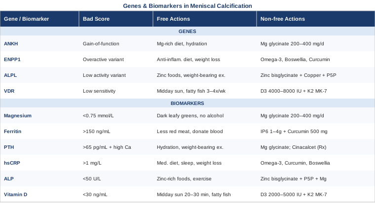
6 Biomarkers to Track If You Have Meniscal Calcification
The standard knee workup — X-ray, MRI, physical exam — tells you that calcification is present. These six blood tests ask a more useful question: which part of your pyrophosphate and mineral metabolism is out of range? Each biomarker is actionable, relatively affordable, and points toward a different intervention pathway.
1. Serum Magnesium and RBC Magnesium
Why it matters
Magnesium plays a direct and underappreciated role in suppressing calcium pyrophosphate crystal formation. At the cellular level, adequate magnesium reduces the concentration of free calcium and pyrophosphate available to form crystals, and magnesium ions compete with calcium during the crystal lattice formation step itself. Hypomagnesemia — chronically low magnesium — is one of the most consistently documented metabolic associations with chondrocalcinosis, and case series have shown that correcting deficiency reduces the frequency of acute CPPD attacks in some patients.
The limitation of standard serum magnesium testing is that it measures only the magnesium circulating in plasma, which represents less than 1% of total body magnesium. Serum levels can appear within the reference range while intracellular stores are significantly depleted. Red blood cell (RBC) magnesium is a more reliable indicator of tissue status and is worth requesting specifically.
How to measure it
A standard serum magnesium test costs $15–$40 at most labs and is included in many comprehensive metabolic panels. RBC magnesium costs approximately $50–$100 and requires a specific order. Optimal serum magnesium is generally above 0.85 mmol/L (2.1 mg/dL); anything below 0.75 mmol/L warrants attention. For RBC magnesium, functional practitioners often target values above 5.5 mg/dL.
If the score is low — the plan without supplements
Remove the biggest dietary drains on magnesium: excess alcohol, high caffeine intake, and ultra-processed foods. Increase consumption of foods genuinely dense in magnesium: cooked dark leafy greens (especially Swiss chard and spinach), pumpkin seeds, black beans, and 85%+ dark chocolate. Gut absorption matters as much as intake — chronic proton pump inhibitor use, gut dysbiosis, and inadequate stomach acid all reduce magnesium absorption even with an excellent diet.
If the score is low — the plan with supplements
Magnesium glycinate at 200–400 mg elemental magnesium per day is the most bioavailable oral form with the lowest risk of loose stools. Magnesium threonate crosses the blood-brain barrier more effectively and is preferable when cognitive symptoms or poor sleep accompany joint issues. Start at 200 mg at night and increase by 100 mg weekly until stools begin to soften, then reduce slightly to find tolerance. No strict cycling is required at these doses. Side effects include loose stools at higher doses and, rarely, mild hypotension. Retest RBC magnesium after 8–12 weeks of consistent use.
2. Serum Ferritin and Transferrin Saturation
Why it matters
Hemochromatosis — pathological accumulation of iron — is one of the best-documented secondary causes of CPPD. Iron deposits in synovial tissue, cartilage cells, and the joint capsule, where it alters chondrocyte function and stimulates pyrophosphate release. In population studies of chondrocalcinosis, patients with elevated iron stores have significantly higher rates of joint calcification than age-matched controls. The relationship extends beyond overt hereditary hemochromatosis: subclinical iron elevation, particularly ferritin consistently above 200–300 ng/mL in men or post-menopausal women, can accelerate cartilage damage over years.
Importantly, the joint calcification pattern in hemochromatosis tends to affect the knee and wrist early — sometimes before any other symptoms — making it a clinically actionable discovery even when the diagnosis is unexpected.
How to measure it
Order serum ferritin and transferrin saturation together. Ferritin reflects iron stores; transferrin saturation reflects how much circulating iron is bound to its carrier protein. Combined cost is typically $30–$80. Optimal ferritin for joint health purposes is generally 50–150 ng/mL. Transferrin saturation above 45% warrants further investigation for hereditary hemochromatosis, including HFE gene testing (approximately $100–$200).
If ferritin is elevated — the plan without supplements
Reduce dietary heme iron sources: red meat (especially organ meats), iron-fortified cereals, and avoid cooking in cast iron pans. Avoid supplemental iron entirely unless true deficiency is confirmed. Drinking coffee or tea with iron-rich meals significantly reduces iron absorption. Regular blood donation is the most effective and medically recognized way to reduce iron stores — a therapeutic phlebotomy program under physician supervision can normalize ferritin over 12–18 months in confirmed hemochromatosis. Avoid vitamin C supplements taken alongside iron-rich meals.
If ferritin is elevated — the plan with supplements
IP6 (inositol hexaphosphate) at 1–4 g/day has preliminary evidence as a mild iron chelator and antioxidant, though it does not replace phlebotomy in established hemochromatosis. Curcumin at 500–1,000 mg/day with piperine may reduce ferritin-associated oxidative stress in joint tissue and has modest iron chelation properties. If genetic hemochromatosis is confirmed, these supplements are supportive only — supplementation alone will not normalize iron stores. Cycling: IP6 can be used continuously at lower doses; take a four-week break every three months at higher doses. Side effects: IP6 at high doses may reduce zinc and calcium absorption — monitor mineral levels.
3. Parathyroid Hormone (PTH) and Serum Calcium
Why it matters
Primary hyperparathyroidism — overproduction of PTH by one or more parathyroid glands — is a major and reversible cause of chondrocalcinosis. PTH increases serum calcium, alters calcium flux into cartilage cells, and appears to stimulate pyrophosphate secretion by chondrocytes. In some clinical series, up to 10% of patients presenting with chondrocalcinosis have undiagnosed hyperparathyroidism as the underlying driver. Crucially, the longer PTH remains elevated, the more extensive the joint calcification becomes — making early detection genuinely impactful.
Always measure PTH alongside total calcium and ionized calcium. The combination tells you whether PTH is inappropriately elevated relative to the prevailing calcium level, which is the diagnostic key.
How to measure it
An intact PTH blood test combined with serum calcium costs approximately $50–$120. Ionized calcium adds $30–$60 and provides a more accurate read of biologically active calcium. Reference ranges: PTH should generally be 15–65 pg/mL; serum calcium 8.5–10.2 mg/dL. High PTH with high-normal or elevated calcium strongly suggests primary hyperparathyroidism. High PTH with low calcium suggests secondary hyperparathyroidism, often driven by vitamin D deficiency (see biomarker 6).
If PTH is elevated — the plan without supplements
The most important step is identifying the cause. A parathyroid imaging scan — sestamibi scintigraphy or 4D CT — can localize a parathyroid adenoma if primary hyperparathyroidism is confirmed. Surgical resection of an adenoma is often curative, and normalization of PTH post-operatively has been documented to halt progression of chondrocalcinosis in reported cases. If surgery is deferred, ensure optimal hydration, maintain moderate dietary calcium (restriction is not recommended and may worsen the imbalance), and prioritize weight-bearing exercise to support bone mineral regulation. Secondary hyperparathyroidism driven by vitamin D deficiency resolves with vitamin D optimization.
If PTH is elevated — the plan with supplements
Cinacalcet is a calcimimetic prescription drug that reduces PTH in primary hyperparathyroidism — not an over-the-counter option, but worth discussing with an endocrinologist. On the supplement side, ensuring optimal vitamin D (see biomarker 6) is critical for normalizing secondary hyperparathyroidism. Magnesium glycinate is also directly relevant: magnesium regulates PTH secretion, and hypomagnesemia can paradoxically raise PTH even without parathyroid pathology. No strict cycling is required for these supplements at physiological doses. Monitor serum calcium regularly if PTH is being actively managed.
4. High-Sensitivity C-Reactive Protein (hsCRP)
Why it matters
Acute CPPD attacks — clinically indistinguishable from gout flares — are fundamentally inflammatory events. Crystals shed from the meniscus into the joint space trigger a powerful neutrophil and macrophage response, flooding the joint with pro-inflammatory cytokines including IL-1β and TNF-α. But hsCRP is more than just a flare marker. Chronically elevated systemic inflammation — even mild elevations above 1 mg/L — accelerates cartilage degradation, worsens synovial tissue function, and creates a joint environment where crystal formation is more likely to persist and trigger repeated attacks.
Peter Attia and other longevity-oriented clinicians have argued that hsCRP below 0.5 mg/L should be the target for metabolically healthy individuals, not simply "within the normal reference range" (often set at <5 mg/L for standard clinical panels). At the levels relevant to meniscal calcification, hsCRP provides a real-time read on how actively the immune system is responding to existing crystal load.
How to measure it
hsCRP is widely available and costs $20–$60 at most labs; many preventive health panels include it. The target for joint and cardiovascular health purposes is below 1.0 mg/L, and ideally below 0.5 mg/L. Levels above 3 mg/L suggest moderate systemic inflammation; above 10 mg/L may indicate an active flare or concurrent infection and should be interpreted alongside clinical symptoms.
If hsCRP is elevated — the plan without supplements
A Mediterranean-pattern diet — rich in olive oil, fatty fish, legumes, and vegetables — has the strongest dietary evidence for reducing hsCRP. Addressing sleep quality is equally important: even one week of partial sleep restriction measurably increases circulating inflammatory markers in controlled human studies. Visceral fat is a major driver of chronic low-grade inflammation; even a 5–10% reduction in body weight produces meaningful hsCRP reductions. Eliminate alcohol in excess, trans fats, and highly processed seed oils.
If hsCRP is elevated — the plan with supplements
Omega-3 fatty acids (EPA + DHA) at 2–4 g/day are the most evidence-supported anti-inflammatory supplement, with replicated data on reducing hsCRP and joint inflammatory markers. Curcumin at 500–1,500 mg/day in a phospholipid or piperine formulation has multiple randomized trials supporting hsCRP reduction. Boswellia serrata (AKBA fraction) at 100–300 mg/day targets the 5-LOX inflammatory pathway relevant in crystal arthropathy. Cycling: omega-3s can be taken continuously; use caution with blood thinners. Curcumin is generally well-tolerated continuously. Boswellia: 8–12 weeks on, 4 weeks off is a practical approach. Side effects: omega-3s at high doses may prolong bleeding time; curcumin can reduce non-heme iron absorption if taken with iron-containing meals.
5. Alkaline Phosphatase (ALP) and Bone-Specific ALP
Why it matters
Alkaline phosphatase — encoded by the ALPL gene and also called tissue non-specific alkaline phosphatase (TNAP) — is the primary enzyme responsible for degrading extracellular inorganic pyrophosphate (PPi). When ALP activity is low, PPi accumulates in cartilage tissue and combines with calcium to form CPPD crystals. This is why hypophosphatasia — a rare genetic condition caused by severe ALPL loss-of-function mutations — is characterized by pathological calcification in both bones and joints.
Milder forms of ALP suppression, driven by zinc deficiency, liver dysfunction, hypothyroidism, or common ALPL gene variants, can contribute to the same pyrophosphate accumulation problem at a subclinical level. An ALP result at the low end of the reference range — often reported as "normal" at 40–44 U/L — may still reflect insufficient PPi clearance in cartilage tissue.
How to measure it
ALP is a standard component of the comprehensive metabolic panel (CMP), costing $15–$50. Bone-specific ALP, which separates liver from bone contributions to the total reading, costs approximately $80–$150. Standard reference range for adults is 44–147 U/L, but values below 50 U/L should prompt further evaluation in the context of joint calcification. Zinc levels and thyroid function should be tested alongside low ALP results, as both deficiencies significantly suppress ALP activity.
If ALP is low — the plan without supplements
Eliminate causes of ALP suppression first: have TSH tested for hypothyroidism, evaluate for zinc deficiency, and review medications that suppress ALP (corticosteroids, clofibrate, statins at high doses). Weight-bearing exercise increases ALP activity by stimulating osteoblast function. A nutrient-dense diet with adequate protein, zinc, and B vitamins supports enzyme synthesis. Reduce phytate-rich foods eaten without proper preparation — soaking or fermenting legumes and grains improves zinc bioavailability.
If ALP is low — the plan with supplements
Zinc bisglycinate at 15–30 mg/day can restore ALP activity in zinc-deficient individuals; always balance with copper at 1–2 mg/day (approximately a 10:1 zinc-to-copper ratio). Pyridoxal-5-phosphate (P5P, active B6) at 25–50 mg/day supports alkaline phosphatase synthesis — notably, severe B6 deficiency and hypophosphatasia share some clinical overlap. Magnesium glycinate at 200–400 mg/day supports the full cofactor profile for multiple PPi-metabolizing enzymes. Cycling: these are physiological-range nutrients; continuous use with quarterly monitoring of zinc, copper, and ALP levels is appropriate. Side effects: excess zinc (above 40 mg/day long-term) depletes copper — always co-supplement and check levels; B6 neuropathy risk emerges only at doses well above 200 mg/day.
6. 25-Hydroxyvitamin D
Why it matters
Vitamin D is the master regulator of calcium and phosphate homeostasis. It promotes calcium absorption from the gut, suppresses PTH secretion when levels are adequate, and modulates immune responses in joint tissue. Deficiency drives secondary hyperparathyroidism (see biomarker 3) and dysregulates the calcium gradient in cartilage. There is also evidence that vitamin D receptors in chondrocytes directly modulate how cartilage cells respond to mineral stress, making local vitamin D signaling relevant beyond its systemic calcium-regulating role.
While vitamin D deficiency does not directly cause CPPD, it creates the metabolic environment in which calcification is more likely to progress. Optimizing vitamin D is one of the lowest-cost, highest-leverage interventions in a metabolic approach to chondrocalcinosis.
How to measure it
The 25-hydroxyvitamin D blood test costs $30–$80. Peter Attia and most functional medicine practitioners consider optimal levels to be 40–60 ng/mL (100–150 nmol/L), significantly higher than the clinical deficiency threshold of 20 ng/mL. Levels below 30 ng/mL are common and warrant intervention. Test at baseline, then retest three months after starting supplementation.
If vitamin D is low — the plan without supplements
The most natural route is UVB exposure: 20–30 minutes of midday sun on arms and legs (without sunscreen on exposed skin) produces 10,000–20,000 IU of vitamin D in light-skinned individuals. Dietary sources include fatty fish (salmon, mackerel, sardines) and egg yolks, though dietary intake alone is rarely sufficient to normalize clinical deficiency. Outdoor exercise in daylight doubles the benefit.
If vitamin D is low — the plan with supplements
Vitamin D3 (cholecalciferol) at 2,000–5,000 IU/day is appropriate for most adults with deficiency below 30 ng/mL. Pair it with vitamin K2 (MK-7 form, 100–200 mcg/day) to direct the increased calcium absorption toward bones rather than soft tissues — this pairing is especially important in the context of meniscal calcification, where raising vitamin D without K2 could, in theory, increase circulating calcium without directing it appropriately. Retest after three months. Doses above 5,000 IU/day require physician supervision and periodic calcium monitoring. Side effects: vitamin D toxicity (hypercalcemia) is rare at doses below 10,000 IU/day but can occur in susceptible individuals; K2 at these doses has an excellent safety profile.
Understanding your biomarker profile answers the "what is happening right now" question. The next section goes one step further — asking why your metabolism may be wired toward this imbalance in the first place.
What Recent Genetics Research Reveals About Meniscal Calcification
Meniscal calcification runs in families — sometimes obviously, sometimes as a pattern that only becomes visible in retrospect when multiple relatives develop knee or wrist calcification in their 40s or 50s rather than their 70s or 80s. Four genes have been most consistently implicated in regulating pyrophosphate metabolism and calcification risk, and understanding your likely position on each one helps explain why the imbalance may exist — and what to prioritize.
The research is a mix of well-established mechanism (particularly from familial chondrocalcinosis studies) and emerging personalized genomics. Where evidence from human clinical data is strong, it is noted. Where it is preliminary, that is stated clearly.
ANKH — The Pyrophosphate Transporter Gene
What the gene does
The ANKH gene encodes a membrane protein that transports inorganic pyrophosphate (PPi) from inside the chondrocyte out to the extracellular matrix. In normal function, this controlled release regulates cartilage mineralization. Gain-of-function mutations in ANKH — where the transporter works too actively — cause excess PPi to accumulate in cartilage, where it crystallizes with calcium into CPPD. Several ANKH mutations have been identified in families with early-onset chondrocalcinosis (onset before age 50), and more common, milder variants likely contribute to elevated risk without causing florid familial disease.
If the gene variant is present — the plan without supplements
The ANKH-related calcification pathway is most active in the presence of excess calcium and phosphate in the joint environment. Maintaining serum magnesium in the upper normal range has strong mechanistic rationale — magnesium ions compete with calcium in crystal lattice formation, inhibiting CPPD crystal nucleation. Staying well-hydrated supports joint fluid dilution of mineral concentrations. Reducing refined carbohydrates and ultra-processed foods reduces the inflammatory burden on cartilage tissue.
If the gene variant is present — the plan with supplements
Magnesium glycinate at 200–400 mg/day is the primary supplement to consider: magnesium inhibits crystal formation at the physicochemical level and has the strongest mechanistic rationale in an ANKH-driven pyrophosphate excess scenario. Supporting TNAP enzyme function through zinc and B6 (see ALPL section) is complementary, as it increases PPi degradation to partially offset the transport excess. There are currently no supplements that directly inhibit ANKH protein activity in humans. Cycling: continuous magnesium at these doses is appropriate with periodic RBC magnesium monitoring. Retest the relevant biomarkers (magnesium, ALP) every six months.
ENPP1 — The Pyrophosphate Generator Gene
What the gene does
ENPP1 (ectonucleotide pyrophosphatase/phosphodiesterase 1) is the enzyme responsible for generating extracellular PPi in cartilage tissue. It converts ATP into AMP and PPi. Variants that increase ENPP1 activity push more PPi into the extracellular space, raising the substrate available for crystal formation. Conversely, loss-of-function ENPP1 variants reduce PPi, which paradoxically promotes a different form of calcification — hydroxyapatite deposition (calcific tendinopathy, arterial calcification) rather than CPPD.
Understanding which direction your ENPP1 variant pushes the system matters for choosing the right approach. Gain-of-function variants overlap mechanistically with ANKH-related pathology; loss-of-function variants present with a distinct pattern.
If ENPP1 is overactive — the plan without supplements
Chronic low-grade inflammation increases ENPP1 expression in chondrocytes, amplifying the pyrophosphate-generating pathway. An anti-inflammatory diet and actively optimizing hsCRP (see biomarker 4) are therefore directly relevant. Weight management reduces mechanical stress on the knee joint, which is itself a stimulus for ENPP1 upregulation in loaded cartilage tissue.
If ENPP1 is overactive — the plan with supplements
There are currently no approved supplements that specifically downregulate ENPP1 in humans. The indirect approach works through inflammation: omega-3 fatty acids (EPA + DHA, 2–4 g/day) and boswellia (AKBA fraction, 100–300 mg/day) reduce the inflammatory signaling that amplifies ENPP1 activity. Curcumin at 500–1,000 mg/day has been shown to reduce NF-κB-mediated ENPP1 upregulation in preclinical models; human data are limited but mechanistically plausible. Cycling: as described in the hsCRP section. Monitor joint symptoms and hsCRP every three to six months to assess response.
ALPL (TNAP) — The Pyrophosphate Degrader Gene
What the gene does
ALPL encodes tissue non-specific alkaline phosphatase (TNAP), the enzyme that breaks down extracellular PPi. Loss-of-function variants reduce PPi clearance, allowing it to accumulate and crystallize. Severe ALPL mutations cause hypophosphatasia — a rare disorder featuring pathological joint and bone calcification, premature tooth loss, and skeletal fragility. But milder ALPL variants, far more common in the general population, produce a reduced-activity enzyme that quietly allows PPi to build up at a slower rate over decades.
This gene is particularly actionable because TNAP requires specific cofactors to function at full capacity: zinc, magnesium, and pyridoxal-5-phosphate (the active form of B6) are all required. A person with a mild ALPL variant who is also zinc-deficient may experience an accelerated calcification trajectory that could be meaningfully slowed by correcting the cofactor deficiency — without targeting the gene directly.
If the ALPL variant is present — the plan without supplements
Prioritize dietary zinc through oysters, red meat in moderation, pumpkin seeds, and legumes. Reduce phytate-rich foods eaten without proper preparation — soaking legumes and fermenting grain-based foods meaningfully improves zinc bioavailability. Maintain adequate protein intake, as TNAP enzyme synthesis requires amino acid building blocks. Avoid chronic high-dose zinc supplementation paradoxically — extreme zinc excess depletes copper, which also impairs enzyme function.
If the ALPL variant is present — the plan with supplements
Zinc bisglycinate at 15–25 mg/day with copper at 1–2 mg/day directly supports TNAP function. Pyridoxal-5-phosphate (P5P) at 25–50 mg/day provides the active cofactor for alkaline phosphatase synthesis. Magnesium glycinate at 200–400 mg/day completes the cofactor profile. This combination addresses the enzymatic deficit at the functional level, though it cannot overcome severe loss-of-function mutations. Cycling: these are physiological-range nutrients; continuous use with quarterly monitoring of zinc, copper, and ALP levels is appropriate. Side effects: zinc at high doses depletes copper — always co-supplement and test copper every three months.
VDR — The Vitamin D Receptor Gene
What the gene does
The VDR gene encodes the vitamin D receptor, through which active vitamin D exerts its effects on calcium absorption, immune modulation, and gene expression in multiple tissues, including chondrocytes. Common VDR polymorphisms — including BsmI, TaqI, and FokI — affect receptor sensitivity and downstream gene expression. Individuals with reduced-sensitivity VDR polymorphisms may require significantly higher circulating vitamin D levels to achieve the same biological effect on calcium regulation and PTH suppression.
The connection to meniscal calcification is indirect but clinically meaningful: reduced VDR sensitivity impairs calcium regulation (raising PTH risk), reduces immune modulation in joint tissue, and may lower the inflammatory threshold at which CPPD crystal-induced synovitis becomes chronic and self-reinforcing.
If VDR variants are present — the plan without supplements
Maximize natural vitamin D sources: regular midday sun exposure on the largest possible skin area, fatty fish three to four times per week, and UV-exposed mushrooms (stem down, in direct sun). Outdoor exercise doubles the benefit — sun exposure plus mechanical joint loading. Reduce factors that impair VDR signaling further: obesity (adipose tissue sequesters circulating vitamin D), smoking, and excess alcohol all impair both vitamin D metabolism and receptor function.
If VDR variants are present — the plan with supplements
People with reduced-activity VDR polymorphisms often require vitamin D3 doses at the higher end of the supplementation range — 4,000–8,000 IU/day — to achieve 40–60 ng/mL serum levels. This must be confirmed with testing every three months when doses are being adjusted. Always pair with vitamin K2 MK-7 (150–200 mcg/day). Magnesium is essential for both vitamin D hydroxylation and VDR signaling — without adequate magnesium, supplemental vitamin D may not fully activate downstream targets. Cycling: no strict cycling needed; simply monitor levels quarterly. Side effects: doses above 8,000 IU/day require physician oversight and periodic calcium and parathyroid monitoring.
These genetic insights reframe the condition from a statistical coincidence into a predictable, metabolically mapped pattern. The following section distills the most impactful research findings across metabolic medicine and inflammation science into a practical knowledge framework.
10 Insights From Metabolic Research That Change How You See Joint Calcification
The science of pyrophosphate metabolism, calcium regulation, and joint inflammation has been examined in growing depth by researchers who sit outside the standard rheumatology model. Andrew Huberman's podcast has addressed inflammation, calcium metabolism, and sleep-driven repair in episodes that, taken together, offer a framework distinctly more actionable than what most people receive at a standard orthopedic consultation. Below are ten of the most impactful insights — each one grounded in published science and directly applicable to meniscal calcification.
1. Inflammation Is Not the Enemy — Unresolved Inflammation Is
Acute inflammation in response to crystal shedding into the knee joint is a normal immune function. The problem is when it fails to fully resolve. Specialized pro-resolving mediators (SPMs) — derived from EPA and DHA — actively terminate inflammation rather than simply suppressing it. A diet deficient in omega-3 fatty acids means the resolution phase is inadequate, and the joint remains in a low-grade inflammatory state that worsens cartilage integrity over time. Resolution is an active biological process, not just the absence of inflammation.
2. Calcium Follows Signaling, Not Just Diet
The assumption that dietary calcium causes joint calcification is incorrect. Calcium is directed to bones or soft tissues based on the presence of specific co-factors: vitamin K2 activates osteocalcin and matrix Gla protein (MGP), which physically blocks arterial and cartilage calcification. Without adequate K2 alongside vitamin D, circulating calcium defaults toward soft tissue deposition over time. Restricting dietary calcium without addressing the K2 and D3 deficit makes the problem worse, not better.
3. Pyrophosphate Balance Is the Core Issue — Not Calcium Alone
CPPD is fundamentally a pyrophosphate problem, not purely a calcium problem. Understanding that the body generates, transports, and degrades pyrophosphate through three distinct enzyme systems (ENPP1, ANKH, TNAP) shifts the entire conversation. Treating crystal-driven inflammation without investigating the pyrophosphate imbalance is the equivalent of mopping the floor without fixing the pipe.
4. Iron Overload Is the Most Underdiagnosed Driver
Hemochromatosis affects approximately 1 in 200 people of Northern European descent, and the majority remain undiagnosed. Joint calcification — often in the second and third metacarpophalangeal joints and the knees — is frequently the first clinical sign. A ferritin panel and HFE gene test cost less than $200 combined and can identify this reversible cause before more damage accumulates. Many patients are told their joint calcification is idiopathic when a simple iron panel would have revealed the underlying driver.
5. Sleep Deprivation Drives Joint Inflammation Directly
Six hours of sleep per night for two consecutive weeks produces inflammatory profiles in humans that resemble acute illness. Joint tissue — including cartilage — relies on the anti-inflammatory and repair cascade that occurs during slow-wave and REM sleep. Chronic sleep restriction increases TNF-α and IL-6, two cytokines directly involved in crystal-induced synovitis. This is not a soft association — it is a measurable physiological mechanism with direct implications for CPPD progression.
6. The Gut-Joint Axis Is a Real Pathway
Gut dysbiosis — imbalanced intestinal microbiota — produces systemic low-grade inflammation through impaired tight junction integrity and elevated lipopolysaccharide (LPS) in circulation. While CPPD specifically has less direct gut microbiome research than rheumatoid arthritis or gout, the pathway from gut inflammation to elevated hsCRP to lowered joint inflammatory threshold is mechanistically sound and increasingly supported across crystal arthropathy research.
7. Magnesium Deficiency Is a Silent Accelerant
An estimated 50–80% of the population is below optimal magnesium status, yet it rarely appears on standard blood panels because serum magnesium remains normal until deficiency is severe. Magnesium's role in over 300 enzymatic reactions includes PPi crystal inhibition, PTH regulation, vitamin D activation, and anti-inflammatory cytokine reduction. Correcting subclinical magnesium deficiency costs less than $30 per month and has no serious safety concerns at physiological doses.
8. Weight-Bearing Exercise Changes the Joint Chemical Environment
Cartilage has no blood supply — it receives nutrients through compression and decompression during movement. Immobility starves cartilage of the nutrient delivery required for repair and maintenance. Regular low-impact weight-bearing exercise (walking, cycling, swimming) promotes synovial fluid circulation, reduces intra-articular inflammatory mediators, and improves chondrocyte metabolic activity. Absence of this mechanical input accelerates cartilage degeneration independent of crystal load.
9. The Vitamin D Threshold Most Doctors Use Is Too Low
A 25-OH vitamin D level of 20 ng/mL — the standard clinical threshold — was set to prevent rickets and osteomalacia, not to optimize PTH regulation, immune modulation, or cartilage health. Emerging consensus among longevity clinicians places the functional optimal range at 40–60 ng/mL, a level many people with "normal" test results do not reach. Getting to this range typically requires consistent supplementation with D3 and K2 together, not sun exposure alone.
10. Early Detection Fundamentally Changes the Trajectory
Meniscal calcification identified at an early stage — when deposits are small, symptoms mild, and no secondary osteoarthritis changes have occurred — is a fundamentally different clinical situation than late-stage disease with bone erosion and joint space narrowing. The six biomarkers in this article provide a framework for catching and correcting metabolic drivers before structural joint damage becomes irreversible. A single targeted panel costs under $300 combined and can reveal the key drivers in most cases.
Complementary Approaches With Clinical Evidence for Joint Health
The following modalities have the most relevant clinical evidence for joint pain and crystal arthropathy management. None of them replaces metabolic optimization, but each offers a practical addition that may reduce symptoms and support joint tissue health.
Low-Level Laser Therapy (Photobiomodulation)
Low-level laser therapy (LLLT), also called photobiomodulation, delivers specific wavelengths of red or near-infrared light to tissue at intensities that stimulate cellular energy production without generating heat. In joint tissue, it reduces inflammatory mediators, increases ATP production in chondrocytes, and promotes tissue repair. For meniscal calcification, where ongoing crystal-induced synovitis accelerates cartilage breakdown, the anti-inflammatory and chondroprotective mechanisms of LLLT are directly relevant to slowing that cycle.
A randomized controlled trial published in Lasers in Medical Science evaluated 904 nm laser therapy applied to the knee in patients with knee osteoarthritis over 12 sessions and found significant reductions in pain scores and improved functional outcomes compared to sham treatment. While this study focused on osteoarthritis rather than specifically CPPD, the overlapping pathology — crystal-driven cartilage degradation and synovial inflammation — makes the evidence reasonably applicable. Protocol: 6–12 sessions of 5–10 minutes per application area, three times per week, at wavelengths of 810–904 nm.
A course of 8–12 clinical LLLT sessions from a physiotherapy or sports medicine clinic costs approximately $400–$900. Consumer-grade near-infrared devices (630–850 nm, $200–$600) offer a lower-intensity home option. Apply to the medial and lateral knee joint line for 10 minutes per area. Results accumulate gradually — expect 4–8 weeks of consistent use before assessing response. LLLT is safe for most individuals; avoid use over active implants or near suspected malignant tissue.
Tai Chi
Tai chi is a slow, controlled movement practice now widely recognized for its benefits in musculoskeletal conditions. For knee joint pathology, including chondrocalcinosis and associated osteoarthritis, tai chi provides low-impact joint loading, proprioceptive training, and anti-inflammatory benefits mediated partly through parasympathetic nervous system activation. Its slow, deliberate weight shifts through partial knee flexion make it particularly well-suited to joints with existing crystal pathology where high-impact movement triggers inflammation.
A landmark randomized controlled trial published in Annals of Internal Medicine (Wang et al., 2016) compared tai chi to physical therapy in patients with knee osteoarthritis. Participants practicing tai chi twice weekly for 12 weeks showed equivalent pain reduction to physical therapy, with additional improvements in depression scores and quality of life. The primary outcome — reduction of joint pain and stiffness in the context of degenerative knee cartilage — is directly applicable to the secondary OA changes that develop alongside long-standing meniscal calcification. Protocol: Yang-style tai chi twice weekly for 12 weeks, with daily home practice of key knee-friendly forms.
Most community centers and recreation facilities offer tai chi classes for $10–$20 per session. Free beginners' programs are widely available online. The key is consistency — twice weekly over at least 12 weeks before assessing benefit. Progress gradually: even 10 minutes of daily practice builds meaningful joint proprioception in the early weeks. People experiencing an acute CPPD flare should wait until pain is below a 5/10 before beginning or resuming practice.
Mindfulness-Based Stress Reduction (MBSR)
MBSR is an 8-week structured program that combines mindfulness meditation, body scan practices, and gentle movement to change the relationship between the nervous system and pain perception. In the context of meniscal calcification, chronic pain modulates the central nervous system in ways that amplify symptom intensity independent of the underlying pathology — a phenomenon called central sensitization. MBSR works upstream of symptom management by reducing the central nervous system amplification of peripheral joint pain signals.
A meta-analysis published in JAMA Internal Medicine (Goyal et al., 2014) reviewed 47 randomized controlled trials and found that mindfulness meditation programs produced moderate reductions in pain, anxiety, and depression in chronic pain populations. While CPPD is not isolated in this literature, chronic musculoskeletal pain driven by crystal arthropathy responds to the same central mechanisms. Protocol: the standard 8-week MBSR curriculum involves 2.5 hours of weekly group instruction plus 45 minutes of daily home practice; body scan and mindful movement components are particularly relevant for joint pain management.
MBSR programs are offered through hospitals, wellness centers, and major online platforms, costing $300–$600 for a full 8-week program. Apps and free platforms provide guided body scans and pain-specific meditations at minimal cost. Even 15–20 minutes of daily body scan practice has been shown to reduce pain catastrophizing, which independently predicts functional outcomes in chronic joint conditions. Use MBSR as a complement to — not a substitute for — the metabolic investigation this article describes.
Massage Therapy
Therapeutic massage applied to the muscles and connective tissue surrounding the knee — quadriceps, hamstrings, IT band, calf, and medial soft tissue — reduces muscle tension that mechanically overloads an already-compromised joint. In chondrocalcinosis, secondary muscle compensation patterns develop quickly around a painful knee: tight quadriceps increase patellofemoral load, tight hamstrings raise posterior capsular pressure, and altered gait patterns strain the meniscus unevenly. Massage therapy directly addresses these secondary muscular contributors that standard rheumatology management often overlooks.
A randomized controlled trial by Perlman et al. (2015, Annals of Family Medicine) found that multiple sessions of Swedish massage — one 60-minute session per week for eight weeks — significantly reduced pain and improved physical function in knee osteoarthritis patients compared to a light-touch control group. Effects were maintained at a 16-week follow-up with twice-monthly maintenance sessions. Protocol: Swedish or myofascial release techniques applied to quadriceps, IT band, and posterior knee structures; 45–60 minutes per session, weekly for eight weeks, then twice monthly for maintenance.
A qualified massage therapist familiar with musculoskeletal conditions can tailor techniques to the specific compensation patterns around a calcified knee. Cost: $60–$150 per session. Foam roller and tennis ball self-massage can address quadriceps and IT band tension at minimal cost between sessions. Avoid deep pressure directly over the medial or lateral joint line during an active CPPD flare. Daily five-minute quadriceps self-massage above the knee significantly extends the benefit of professional treatment.
Conclusion
Meniscal calcification rarely has a single cause, and it rarely responds to a single intervention. What the evidence makes clear is that pyrophosphate metabolism — the core biochemical process driving crystal formation — is shaped by identifiable, measurable, and often correctable factors: magnesium levels, iron stores, parathyroid hormone, inflammatory status, alkaline phosphatase activity, and vitamin D. Genetic variants in ANKH, ENPP1, ALPL, and VDR can accelerate this process, but they are not fixed destiny — each one has practical compensating strategies that work with existing biology.
The next smart step is a targeted panel of the six blood tests described here, followed by a conversation with a rheumatologist, endocrinologist, or metabolically oriented physician who can interpret these results in the context of your specific presentation. The information exists, the tests are affordable, and the interventions are evidence-grounded. Better information makes better decisions possible — and in the case of meniscal calcification, earlier decisions preserve more joint function over time.
Musculoskeletal: Bone Conditions Joint Conditions
Autoimmune: Inflammatory Conditions