This article contains AI generated content.
· UpdatedMeniscal Ossicle — 6 Biomarkers And 5 Genes To Track
Introduction
A meniscal ossicle is not something most people have heard of until they find themselves in an orthopedic surgeon's office, staring at an MRI and trying to make sense of a small, bony fragment sitting inside the knee meniscus — a structure made of fibrocartilage that has no business forming bone. This rare condition can range from a silent incidental finding to a genuinely disabling source of knee pain, locking, and swelling. And because it is uncommon, the clinical conversation often stops at diagnosis and surgical options, leaving most people without a biological roadmap for understanding why it happened and what is sustaining it.
Generic knee advice — lose weight, do physical therapy, take an anti-inflammatory — is not wrong, but it also falls short when the underlying problem is ectopic ossification in a specific tissue. The mechanisms behind a meniscal ossicle differ between individuals. For some, it follows trauma and a fibrocartilaginous repair response that went too far. For others, genetic predispositions to aberrant bone formation, disturbed calcium-phosphate signaling, or overactive inflammatory pathways are the primary drivers. Treating everyone identically ignores this heterogeneity.
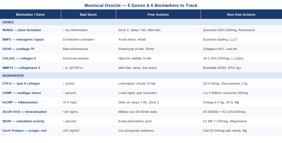
This article approaches the condition from two complementary angles: the biomarkers you can actually track — blood and urine markers that reveal what is happening at the tissue level right now — and the key genes associated with ectopic ossification, cartilage maintenance, and fibrocartilage integrity. Together, these frameworks give you and your clinical team a more precise picture of the underlying biology than symptoms alone ever could.
The goal is not to replace medical judgment but to raise the quality of the conversation you have with specialists. Better information leads to more targeted questions, sharper diagnostic workups, and ultimately smarter decisions. The biomarker section that follows will help you understand which measurable signals matter most and what to do when they are off. The genetics section will explain which inherited and epigenetically modified risk factors may have set the stage. Further down, you will find integrative tools with genuine clinical evidence and a synthesis of longevity medicine's most actionable principles for joint health.
6 Key Biomarkers to Track in Meniscal Ossicle
Understanding a meniscal ossicle through biomarkers means examining the biology from two overlapping angles: what is happening to the fibrocartilaginous tissue itself, and what is driving the mineralization and bone formation. Six markers cover this ground comprehensively, ranging from widely accessible tests to more specialized panels.
1. CTX-II — Cartilage Degradation in Real Time
What it is and why it matters
CTX-II (C-terminal crosslinking telopeptide of type II collagen) is a breakdown product of type II collagen, the structural protein that constitutes the bulk of cartilage — including the fibrocartilage of the meniscus. When this tissue is degrading, CTX-II concentrations rise in the urine. In the context of a meniscal ossicle, chronically elevated CTX-II suggests that the surrounding fibrocartilaginous tissue is under ongoing mechanical or inflammatory stress, which may be perpetuating the ossification cycle or causing secondary damage to adjacent articular cartilage.
Garnero and colleagues have consistently linked elevated urinary CTX-II to faster cartilage loss and worse structural outcomes in knee joint disease, work that has been replicated across multiple cohort studies. While direct meniscal ossicle-specific studies remain limited, CTX-II is one of the most clinically validated joint degradation markers available for capturing what is happening to type II collagen-rich tissues.
How to measure it
Urine sample, morning void preferred, analyzed by ELISA. Cost ranges from approximately $50–$120 depending on the laboratory. Some functional medicine panels (such as those offered by ZRT Laboratory) include it in joint-focused combinations. It is not uniformly covered by insurance; confirm with your provider before ordering.
If the score is high — the plan without supplements
Reducing mechanical load is the immediate priority. Shift to low-impact joint-loading activities: water therapy, stationary cycling, and elliptical training. Work with a physiotherapist on gait retraining if any varus or valgus knee alignment is present. Prioritize sleep, because collagen synthesis is predominantly nocturnal and sleep debt measurably impairs tissue repair. Formal physiotherapy 3 times per week for a minimum of 12 weeks. Recheck CTX-II at 3 months.
If the score is high — the plan with supplements or equipment
Undenatured type II collagen (UC-II form, 40 mg/day) works through oral tolerance mechanisms distinct from standard hydrolyzed collagen and has joint-specific evidence. Glucosamine sulfate at 1,500 mg/day has shown cartilage-protective properties in long-term randomized trials. Boswellia serrata standardized to AKBA content (100–200 mg/day) reduces MMP-driven fibrocartilage breakdown. Cycle all three for 3 months, then take a 4-week break before re-evaluating. Side effects: mild GI discomfort with glucosamine at the higher end; Boswellia may interact with warfarin — disclose to your physician.
2. COMP — A Stress Gauge for Fibrocartilage
What it is and why it matters
Cartilage Oligomeric Matrix Protein (COMP) is a pentameric glycoprotein released from chondrocytes under mechanical stress or damage. Serum COMP rises acutely with loading and remains chronically elevated when cartilage tissue is in a state of ongoing injury. For someone with a meniscal ossicle, COMP serves as an early-warning signal for chondral stress beyond the ossicle itself — the bony nodule creates abnormal biomechanical forces that can damage the adjacent articular cartilage over time, and COMP tracks this process before structural change is visible on imaging.
How to measure it
Fasting serum sample, analyzed by ELISA. Less commonly ordered than inflammatory panels; may require a specialty or academic rheumatology laboratory. Cost: approximately $80–$150. Reference ranges vary by lab and by age; fasting status matters for interpretation.
If the score is high — the plan without supplements
Immediate load management. This includes correcting asymmetric loading patterns identified during gait analysis, using an unloading knee brace if the ossicle creates focal compartment overload, and avoiding high-impact activities (running on hard surfaces, cutting sports, plyometrics). Add targeted joint-stability work: single-leg balance progressions, vastus medialis oblique strengthening, and hip abductor training to reduce medial compartment stress. Frequency: 4–5 sessions per week, 20–30 minutes each.
If the score is high — the plan with supplements or equipment
Home LLLT devices designed for joint applications (808–940 nm wavelength range, adequate power density) have shown reductions in cartilage stress markers in preliminary work. Curcumin at 500–1,000 mg/day with piperine for absorption enhances bioavailability and reduces chondrocyte apoptosis signaling. Cycle curcumin for 12 weeks on, 4 weeks off. Monitor COMP at 3-month intervals. Caution: curcumin at high doses may potentiate the effects of anticoagulant medications.
3. High-Sensitivity CRP — The Inflammation Signal
What it is and why it matters
High-sensitivity C-reactive protein (hsCRP) is a liver-produced acute-phase protein that rises in response to systemic inflammatory activity. Persistently elevated hsCRP in someone with a meniscal ossicle suggests that inflammatory pathways — driven by IL-6, TNF-alpha, and prostaglandin E2 — may be actively promoting periosteal and fibrocartilaginous ossification. Inflammation is not merely a downstream consequence of the ossicle; in a meaningful subset of patients, it may be both a cause and an amplifier of the mineralization process through epigenetic activation of osteogenic gene programs.
Peter Attia has consistently highlighted hsCRP as one of the most underutilized systemic health markers in standard care, noting that standard CRP assays frequently miss low-grade chronic inflammation that high-sensitivity testing would detect.
How to measure it
Standard fasting blood draw. Cost: $10–$30, widely covered. Optimal target: below 0.5 mg/L. Levels between 0.5 and 1.0 mg/L indicate low-grade inflammation; above 3.0 mg/L indicates significant systemic inflammatory burden. Do not test within 48 hours of strenuous exercise or during acute illness, as both temporarily elevate the reading.
If the score is high — the plan without supplements
The anti-inflammatory lifestyle foundation should be consolidated before adding supplements: - Replace refined seed oils with olive oil and avocado oil as primary cooking fats - Achieve 7–9 hours of quality sleep nightly (CRP rises measurably with sleep restriction) - Accumulate 150+ minutes per week of moderate-intensity aerobic activity (Zone 2 pace) - Practice a 12–16 hour overnight fast (time-restricted eating reduces inflammatory cytokines)
Re-check hsCRP at 6–8 weeks before adding any supplementation.
If the score is high — the plan with supplements or equipment
Omega-3 fatty acids (EPA + DHA combined at 2–4 g/day) have robust randomized trial evidence for lowering hsCRP. Correcting vitamin D deficiency to 50–70 ng/mL independently lowers inflammatory markers. Magnesium glycinate at 300–400 mg taken in the evening supports multiple anti-inflammatory enzymatic pathways. No cycling needed for omega-3 or vitamin D — maintain continuously and recheck CRP at 3 months. Side effect note: fishy aftertaste with standard omega-3 capsules can be avoided with enteric-coated formulations; rare loose stools at high omega-3 doses.
4. 25-OH Vitamin D — The Mineralization Regulator
What it is and why it matters
25-hydroxyvitamin D is the circulating storage form of vitamin D and the most clinically informative indicator of status. Vitamin D is not simply a bone-density nutrient — it is a master regulator of calcium-phosphate homeostasis, immune modulation, and chondrocyte function. Both deficiency and uncorrected excess dysregulate bone formation and mineral deposition. In the context of a meniscal ossicle, vitamin D deficiency elevates parathyroid hormone (PTH), which disturbs the calcium-phosphate balance that governs ectopic mineralization. Simultaneously, low vitamin D impairs immune tolerance and ramps up the inflammatory environment around the knee joint.
How to measure it
Standard serum blood test. Cost: $30–$60, frequently covered with a clinical indication. Optimal functional range per most longevity-focused clinicians: 40–60 ng/mL (100–150 nmol/L). Supplementing above 80 ng/mL without concurrent monitoring of serum calcium and PTH is inadvisable.
If the score is low — the plan without supplements
20–30 minutes of midday sun exposure on large skin surface areas (arms, legs, back) generates approximately 10,000–20,000 IU of vitamin D3 in fair-skinned individuals under optimal conditions, less with darker skin tones or higher latitudes. Dietary sources are limited but real: fatty fish (mackerel, wild salmon), egg yolks, and liver. Prioritize daily outdoor exposure during summer months, where geographically possible.
If the score is low — the plan with supplements or equipment
Vitamin D3 at 5,000 IU/day is a common therapeutic starting dose for documented deficiency. Co-supplement with vitamin K2 MK-7 at 100–200 mcg/day — K2 activates matrix Gla protein and osteocalcin, directing calcium toward bone matrix rather than soft tissue (directly relevant to ectopic ossification risk). Recheck 25-OH vitamin D and serum calcium at 3 months and adjust the dose to maintain the optimal range. Side effects: calcium toxicity is possible at very high unmonitored doses; K2 co-supplementation substantially reduces this risk.
5. Bone-Specific Alkaline Phosphatase — Osteoblast Activity
What it is and why it matters
Alkaline phosphatase has multiple tissue sources — liver, bone, intestine — but bone-specific alkaline phosphatase (BSAP) is an isoenzyme produced specifically by osteoblasts, the cells responsible for building new bone. Elevated BSAP indicates that the body is in an active state of bone formation. In a meniscal ossicle, BSAP elevation suggests that osteoblast-like activity may be ongoing within or around the ossicle, potentially driving further mineralization of periossicle tissue.
This marker is especially informative when interpreted alongside a bone resorption marker such as CTX (serum C-telopeptide of type I collagen). The ratio of formation to resorption signals tells you whether the ossicle is likely growing, stable, or in a state of slow remodeling — a clinically relevant distinction when deciding between watchful waiting and intervention.
Thomas Dayspring, a leader in metabolic biomarker analysis, has highlighted P1NP (Procollagen Type I N-Terminal Propeptide) as the gold-standard bone formation marker. Ordering BSAP and P1NP together provides the most complete bone turnover picture.
How to measure it
Fasting serum test. Bone-specific ALP, unlike total ALP, requires a dedicated panel request. Cost: $50–$100. Some endocrinology and sports medicine labs include it in bone turnover panels alongside P1NP. Reference ranges vary meaningfully by sex and age — interpret with a physician familiar with bone metabolism.
If the score is high — the plan without supplements
Immediately reduce activities that create excessive periarticular bone stimulation: plyometrics, running, and heavy axial loading of the affected knee. Favor pool-based exercise and controlled cycling, which maintain muscle function without high periarticular impact. If BSAP is substantially elevated, a whole-body DEXA scan is warranted to rule out systemic bone disease contributing to the finding. Reassess loading patterns immediately and retest BSAP at 6 months.
If the score is high — the plan with supplements or equipment
Vitamin K2 MK-7 at 200 mcg/day is the most directly relevant intervention: it activates osteocalcin and redirects calcium into appropriate bone matrix rather than ectopic sites. Magnesium glycinate or malate at 350–400 mg/day acts as a cofactor for alkaline phosphatase function and regulates bone enzyme activity. Strontium ranelate modulates bone turnover ratios but is prescription-only and not available in all countries — discuss with an endocrinologist if BSAP remains persistently high. Monitor BSAP every 6 months alongside calcium levels.
6. Serum Phosphate and Calcium-Phosphate Product — Ectopic Ossification Risk
What it is and why it matters
At its most fundamental level, ectopic ossification is a calcium-phosphate precipitation event. When the serum calcium × phosphate product (Ca × P, expressed in mg²/dL²) exceeds approximately 55, spontaneous mineralization in soft tissues becomes chemically favored. Most people with a meniscal ossicle will not have overtly abnormal Ca × P products, but subtle shifts — particularly elevated inorganic phosphate from heavy consumption of phosphate-additive-containing ultra-processed foods, or reduced fetuin-A (a natural mineralization inhibitor) — can tip the balance toward ectopic bone formation in vulnerable tissue.
Evaluating phosphate metabolism fully means looking at fasting serum phosphate, serum calcium, intact PTH (which regulates both), and when available, FGF-23, which controls renal phosphate excretion. This is a more advanced panel than most primary care physicians routinely order, but it is highly relevant when the etiology of an ossicle is unclear or when the ossicle appears to be growing.
How to measure it
A standard basic metabolic panel (BMP) covers both calcium and phosphate (cost: typically $20–$50, widely covered). Intact PTH: $30–$80. FGF-23: $100–$200 at specialty labs. Optimal fasting phosphate: 2.5–4.0 mg/dL. Elevated fasting phosphate above 4.0 mg/dL in the absence of kidney disease warrants dietary and metabolic investigation.
If the score is imbalanced — the plan without supplements
The single highest-impact dietary change for phosphate load is eliminating ultra-processed foods containing inorganic phosphate additives. Inorganic phosphate from food additives is absorbed at approximately 90–100%, compared to 40–60% for the organic phosphate in whole foods — a critical difference. Eliminate phosphoric acid-containing beverages (most cola-type drinks). Shift protein sources to whole, minimally processed options. Increase daily hydration, which supports renal phosphate clearance. Recheck fasting phosphate at 4–6 weeks to assess dietary impact.
If the score is imbalanced — the plan with supplements or equipment
Calcium carbonate taken with meals (500 mg per meal, not as a standalone supplement taken separately) can bind dietary phosphate in the GI tract before absorption — the same pharmacological principle used in chronic kidney disease management. Magnesium supplementation improves calcium-phosphate regulation through multiple mechanisms. These approaches should be discussed with a nephrologist or endocrinologist before initiating, particularly if any kidney function abnormalities are present. Side effects: calcium carbonate taken without food causes constipation; overuse risks hypercalcemia.
These six biomarkers together provide a practical, actionable biological map of the forces driving or maintaining a meniscal ossicle. The genetic layer below explains why certain individuals are more susceptible to this process in the first place.
Genetics and Epigenetics: 5 Genes That May Shape Your Risk
Dedicated genome-wide association studies specific to meniscal ossicle do not yet exist — this condition is simply too rare for large-scale genetic epidemiology studies. However, it sits at the intersection of two well-mapped biological domains: ectopic ossification and fibrocartilage integrity. The genetic architecture of both has been studied in meaningful depth, and five genes emerge as the most directly relevant based on their established roles in these pathways.
RUNX2 — The Master Switch for Bone Formation
What this gene does
RUNX2 (Runt-related transcription factor 2) is the master transcription factor governing osteoblast differentiation. Without functional RUNX2, osteoblasts cannot mature and bone cannot form. Conversely, overactive or constitutively upregulated RUNX2 — driven by inflammatory signals such as TNF-alpha and IL-1beta, by mechanical overload, or by epigenetic changes — can drive inappropriate bone formation in tissues that should not be mineralizing, including fibrocartilage.
Epigenetic research has shown that RUNX2 promoter methylation is reduced by chronic inflammatory environments. This means that persistent knee joint inflammation can effectively unmute RUNX2 expression in fibrocartilage cells that would normally never activate osteogenic programs. The implication: the inflammatory biomarkers above (hsCRP, IL-6 pathway) are not separate from genetic risk — they are the environmental inputs that determine whether a genetic susceptibility becomes a clinical problem.
If the gene pattern is concerning — the plan without supplements
The epigenetic layer that activates RUNX2 responds to the inflammatory environment. Anti-inflammatory aerobic exercise (150+ minutes per week at Zone 2 intensity), sleep optimization (inflammatory cytokines that epigenetically unmute RUNX2 peak with sleep restriction), and a Mediterranean-pattern diet create the systemic conditions in which RUNX2 is less likely to be inappropriately activated. These are not supplementary measures — they are the primary mechanism for managing epigenetic RUNX2 risk. Treat them as ongoing baseline habits, not temporary interventions.
If the gene pattern is concerning — the plan with supplements or equipment
Quercetin at 500–1,000 mg/day has demonstrated inhibition of RUNX2 expression in vascular smooth muscle cells in cellular studies — a mechanistically relevant finding for ectopic ossification contexts. Resveratrol at 100–500 mg/day modulates the epigenetic machinery (SIRT1 pathway) that influences RUNX2 transcriptional activity. Cycle both for 8 weeks on, 4 weeks off to avoid potential accumulation effects; high-dose resveratrol has mild estrogenic activity in some studies. Side effects: quercetin may interact with cyclosporine and some antibiotics — disclose to your prescribing physician.
BMP2 — The Bone Morphogenetic Signal
What this gene does
Bone Morphogenetic Protein 2 (BMP2) is among the most potent osteogenic signaling molecules identified. It is secreted by osteoblasts, chondrocytes, and periarticular stromal cells, and acts through SMAD intracellular pathways to promote mineral deposition and new bone formation. Variants in or near the BMP2 gene have been associated with differences in bone mineral density and with susceptibility to heterotopic ossification after trauma. In meniscal tissue, BMP2 is expressed in response to injury and may be the proximate molecular signal that initiates the ossification cascade following meniscal trauma or repetitive microinjury.
Understanding whether your BMP2 signaling pathway is constitutively more active helps explain why some individuals form ossicles after relatively minor injury while others with equivalent trauma do not.
If the gene pattern is concerning — the plan without supplements
Limit re-injury events that trigger BMP2 release from stressed fibrocartilage cells. This means modifying activities with high meniscal shear risk (pivot-dominant sports, kneeling on hard surfaces, deep squatting under load) and working with a sports medicine physician on a structured rehabilitation program designed to avoid secondary microtrauma to the meniscus. This is a permanent modification until structural stability is confirmed by interval MRI, not a temporary restriction.
If the gene pattern is concerning — the plan with supplements or equipment
Noggin is a natural extracellular BMP2 antagonist produced endogenously. Eccentric loading protocols — exercise where a muscle lengthens under tension — have been shown to modulate local BMP signaling in periarticular tissue more favorably than concentric-only training. Low-level laser therapy (LLLT) modulates BMP2-related downstream pathways in bone and connective tissue in several preclinical studies; while human evidence specific to ectopic ossification is early, LLLT's safety profile makes it a reasonable adjunct. No supplement directly and safely inhibits BMP2 in humans at present — the evidence remains preclinical for specific BMP2 antagonism.
SOX9 — The Cartilage Maintenance Gene
What this gene does
SOX9 is the master transcription factor for chondrocyte differentiation and maintenance. It drives expression of type II collagen, aggrecan, and other cartilage matrix proteins that define functional fibrocartilage. When SOX9 is silenced — by inflammatory cytokines, mechanical failure, or epigenetic changes — chondrocytes begin to dedifferentiate: losing their fibrocartilage-maintaining identity and progressively expressing osteoblast-like genes including RUNX2 and alkaline phosphatase. This process of chondrocyte-to-osteoblast identity drift through endochondral ossification mechanisms is now considered a plausible cellular mechanism underlying many meniscal ossicles. In short, the cells that should be making fibrocartilage start making bone instead.
If the gene pattern is concerning — the plan without supplements
Mechanical loading regulates SOX9 expression in chondrocytes, but the relationship is nonlinear: both complete immobilization and overloading suppress SOX9 activity. The therapeutic window is moderate, rhythmic, controlled loading. Swimming, stationary cycling, and aquatic physiotherapy provide compressive and shear forces within the range that maintains chondrocyte identity. Frequency: minimum 4 sessions per week, 30 minutes each, sustained for at least 12 weeks before expecting measurable benefit in connective tissue markers.
If the gene pattern is concerning — the plan with supplements or equipment
Hydrolyzed collagen peptides at 10–15 g/day taken with vitamin C (which is the rate-limiting cofactor for collagen cross-linking) support SOX9-driven extracellular matrix production by providing the amino acid substrate for the collagen genes SOX9 regulates. Oral hyaluronic acid at 80–200 mg/day has shown evidence of supporting chondrocyte identity and synovial joint homeostasis in some human trials. Cycle collagen peptides for 3 months on with a reassessment period; oral HA can be maintained continuously. Side effects: minimal with both; occasional GI discomfort with high-dose collagen peptides.
COL2A1 — Structural Integrity of Fibrocartilage
What this gene does
COL2A1 encodes the alpha-1 chain of type II collagen, the primary structural protein of both hyaline cartilage and fibrocartilage, including the meniscus. Variants in COL2A1 have been associated with early-onset joint disease and with impaired cartilage repair responses. When COL2A1-encoded collagen is structurally compromised — either through genetic variation or through post-translational modifications driven by oxidative stress, glycation from poor glucose control, or vitamin C deficiency — the fibrocartilaginous matrix of the meniscus is less able to withstand mechanical forces. This creates a tissue environment more prone to micro-tearing and aberrant healing, one established pathway through which ossicles form.
If the gene pattern is concerning — the plan without supplements
Reducing meniscal shear forces through hip and core stability training is the most evidence-based mechanical intervention. Weakness in the hip external rotator group is consistently associated with increased medial meniscal stress and poor lower-limb alignment during functional movements. Targeted resistance training of the hip abductors, external rotators, and gluteus medius at 3 sessions per week with a 6-week progressive loading protocol directly reduces meniscal compressive and shear forces.
If the gene pattern is concerning — the plan with supplements or equipment
Vitamin C at 500–1,000 mg/day is the rate-limiting cofactor for prolyl and lysyl hydroxylase enzymes that cross-link and stabilize collagen — essential for converting COL2A1 gene product into structurally functional collagen. L-Lysine at 1,000 mg/day provides a key structural amino acid for collagen synthesis that is often at the lower end of dietary intake. Undenatured type II collagen (UC-II, 40 mg/day) works through oral immunological tolerance to reduce autoimmune attack on existing type II collagen — relevant when immune-mediated mechanisms are contributing. Side effects: vitamin C above 2,000 mg/day may cause loose stools; lysine is very well-tolerated.
MMP13 — The Fibrocartilage-Degrading Enzyme
What this gene does
MMP13 (Matrix Metalloproteinase 13, collagenase 3) is the most chondrocyte-specific of the matrix metalloproteinases and the primary enzyme responsible for degrading type II collagen and aggrecan in cartilage breakdown. Overexpression of MMP13 — triggered by inflammatory cytokines (IL-1beta, TNF-alpha) or mechanical injury — creates the degenerative tissue environment in which fibrocartilage loses structural integrity and aberrant healing becomes more likely. Variants affecting MMP13 promoter activity and expression levels have been studied in osteoarthritis cohorts and likely influence susceptibility to fibrocartilage breakdown more broadly.
Ali Torkamani and colleagues at the Scripps Research Institute have contributed to understanding how variants in MMP-pathway genes interact with environmental inputs — particularly inflammation and mechanical stress — to create individual differences in joint degradation risk. The key insight from this work is that MMP13 genetic risk is highly modifiable through the same inflammatory inputs that are measurable via hsCRP.
If the gene pattern is concerning — the plan without supplements
IL-1beta and TNF-alpha are the primary transcriptional inducers of MMP13. Reducing these cytokines is the upstream lever. Without supplements, the most evidence-supported approach is: Mediterranean-pattern diet, 7–9 hours of sleep (cortisol from sleep restriction amplifies IL-1beta production), chronic stress reduction (psychological stress chronically elevates TNF-alpha), and elimination of ultra-processed foods and high-fructose corn syrup (both promote cytokine production independent of caloric effects). These are ongoing lifestyle practices, not short-term interventions.
If the gene pattern is concerning — the plan with supplements or equipment
Boswellia serrata standardized to AKBA content (100–200 mg/day) directly inhibits MMP13 at the cellular level through a mechanism distinct from its 5-LOX anti-inflammatory action. EPA from omega-3 fatty acids at 2+ g/day reduces IL-1beta transcription, cutting MMP13 at its upstream inducer. Sub-antimicrobial dose doxycycline has demonstrated direct MMP inhibition in osteoarthritis trials — this requires physician oversight and is not appropriate for long-term unsupervised use. Cycle Boswellia for 3 months on, 1 month off. Side effects: Boswellia occasionally causes GI upset and may interact with warfarin; high-dose EPA may modestly increase bleeding time.
Genetic information of this kind is most valuable when acted upon early and continuously. These five genes interact with each other and with the biomarker profile above — a person with epigenetically activated RUNX2 and persistently elevated hsCRP faces a qualitatively different challenge than someone with a COL2A1 structural variant and normal inflammatory markers. Personalization is the whole point.
Ten Ideas From Peter Attia's Outlive That Apply Directly Here
Peter Attia's Outlive: The Science and Art of Longevity (2023) devotes substantial attention to musculoskeletal health as a pillar of what he calls "healthspan" — not length of life but quality of physical function across the decades. While the book does not address meniscal ossicle directly, its biological framework for joint protection, metabolic inflammation, and preventive medicine maps onto this condition with unusual precision. These are the ten most actionable takeaways.
Zone 2 Cardio Directly Reduces Systemic Inflammatory Burden
Attia advocates for 3–4 weekly sessions of Zone 2 cardio — aerobic exercise at a pace where you can still hold a conversation — as the most evidence-backed method for reducing basal inflammatory cytokine levels. This is not incidental to meniscal ossicle management: hsCRP reduction, RUNX2 epigenetic modulation, and IL-1beta suppression all operate through the same metabolic channels that Zone 2 training improves.
Muscle Mass Around the Joint Is Load Protection
Atrophy of the quadriceps and hamstrings significantly increases compressive and shear forces transmitted to the meniscus with every step. Attia is emphatic that resistance training — specifically progressive, loaded eccentric work — is the most effective intervention for joint protection. Rest does not protect joints; appropriately loaded muscle does.
VO2 Max Correlates With Inflammatory Control
A higher VO2 max is associated with lower baseline inflammatory cytokine concentrations across multiple cohort studies. Building aerobic capacity through structured cardiorespiratory training is simultaneously an anti-inflammatory intervention for the joint environment — two goals served by one practice.
Sleep Is the Most Underrated Tissue Repair Tool
In Attia's framework, sleep is when collagen synthesis, immune regulation, and anti-inflammatory recovery are concentrated. Sleep debt acutely elevates IL-6 and TNF-alpha — both major drivers of MMP13 expression and RUNX2 epigenetic unsilencing. Seven to nine hours of quality sleep per night is a non-negotiable input, not a lifestyle luxury.
Glucose Dysregulation Stiffens Cartilage Through AGE Formation
Attia connects hyperglycemia to advanced glycation end-products (AGEs) that cross-link collagen abnormally, reducing tissue elasticity and impairing cartilage matrix integrity. Continuous glucose monitoring (CGM), which Attia recommends for many patients, identifies post-meal glucose spikes that contribute to AGE accumulation in fibrocartilaginous tissue over time.
Protein Intake Is Chronically Underestimated in Most Adults
Most adults consume substantially less protein than what is needed for musculoskeletal tissue maintenance and repair, particularly as they age. Attia recommends 1.6–2.2 g per kg of body weight daily — the amino acid substrate needed for SOX9-mediated collagen gene expression, chondrocyte function, and muscle maintenance around the joint.
The Omega-3 Index Is More Informative Than Dietary Estimates
Attia highlights the omega-3 index — EPA + DHA as a percentage of red blood cell membrane fatty acids — as more predictive of anti-inflammatory status than self-reported dietary intake. An omega-3 index above 8% correlates with systemic anti-inflammatory physiology, directly relevant to MMP13 and BMP2 pathway modulation in the knee joint.
Stability Training Matters More Than Flexibility for Joint Protection
Attia is clear that proprioception, neuromuscular control, and joint stability training protect joints far more effectively than passive flexibility work. For meniscal ossicle patients, reducing the abnormal shear forces that trigger both MMP13 release and BMP2 upregulation requires active neuromuscular stability — not passive stretching.
Bone Is a Metabolically Active Tissue That Responds to Input
Attia frames bone as continuously responsive to mechanical and hormonal signals rather than static. He recommends DEXA scanning to track bone density and bone turnover markers to guide resistance training prescription — a framework directly applicable to interpreting bone-specific alkaline phosphatase and BSAP trends in ossicle management.
The Informed Patient Changes the Clinical Relationship
Attia's central thesis is that patients willing to engage deeply with their own biology — tracking biomarkers, understanding genetic predispositions, arriving at appointments with data — receive meaningfully better care. For a condition as rare as meniscal ossicle, where most clinicians will have limited direct experience, this is not optional. It is the most impactful thing you can do at the next appointment.
Complementary Approaches With Meaningful Clinical Evidence
Low-Level Laser Therapy and Photobiomodulation
Low-level laser therapy (LLLT) — also called photobiomodulation — involves applying specific wavelengths of red or near-infrared light (typically 650–1,000 nm) to tissue. At the cellular level, it activates cytochrome c oxidase in mitochondria, increasing cellular energy production (ATP), reducing pro-inflammatory cytokine release, and modulating local bone remodeling signals. For meniscal ossicle, LLLT is relevant through three overlapping mechanisms: reduction of periarticular inflammation, modulation of osteogenic signaling in local cells, and analgesic effects that may reduce reliance on systemic anti-inflammatory medications.
A systematic review by Ezzati and colleagues examining LLLT for knee joint conditions found statistically significant reductions in pain and functional improvements compared to sham treatment. The World Association for Laser Therapy (WALT) has published dosing guidelines recommending 4–8 J/cm² per treatment point for musculoskeletal applications, with a treatment course of 2–3 sessions per week for 4–6 weeks as a standard starting protocol.
In practice, LLLT can be delivered through clinic-based devices (more powerful, therapist-guided) or FDA-cleared home devices in the 808–940 nm range (more accessible, lower cost). The key quality variable is power density — many consumer devices are significantly underpowered relative to study protocols. Contraindications are limited: active malignancy over the treatment area, and use directly over the abdomen during pregnancy. Side effects are minimal. Evidence for meniscal ossicle specifically is extrapolated from adjacent conditions; LLLT works best as part of a multimodal approach rather than as a standalone intervention.
Tai Chi for Knee Stability, Proprioception, and Inflammation
Tai chi is a slow, controlled movement practice that combines single-leg balance training, progressive lower-limb strengthening, and proprioceptive challenge in a format accessible to people with joint pain. For meniscal ossicle management, its relevance is primarily biomechanical: by improving neuromuscular control of the knee and hip, tai chi reduces the abnormal shear and compressive forces that amplify mechanical stress on periossicle fibrocartilage. Secondarily, its meditative and rhythmic elements activate parasympathetic tone and reduce cortisol, with downstream effects on inflammatory cytokine levels.
Wang and colleagues published a landmark randomized controlled trial demonstrating that 12 weeks of structured tai chi practice produced significant reductions in pain, improved functional capacity, and reduced inflammatory markers compared to a wellness education control group in patients with knee joint disease. This study, published in Arthritis & Rheumatism, remains among the strongest controlled trials of a movement-based intervention for knee pathology and has been replicated in subsequent cohort work.
For someone with a meniscal ossicle, the practical starting point is a beginner yang-style tai chi class — modified to minimize deep knee flexion — practiced 2–3 times weekly, 30–45 minutes per session. Begin under instructor supervision (in-person or high-quality structured video series) to ensure alignment is correct. Avoid styles with rapid weight shifts or low stances during initial rehabilitation. Progress the depth of knee flexion gradually over 8–12 weeks. This is a low-risk, low-cost intervention with documented benefit for the type of knee pain and instability this condition produces.
Mindfulness-Based Stress Reduction for Pain Modulation
Mindfulness-Based Stress Reduction (MBSR) is an 8-week structured program training present-moment awareness, body scanning, and non-reactive attention to physical and emotional experience. Its relevance for meniscal ossicle is multidimensional. First, it reduces pain catastrophizing — the cognitive amplification of pain signals that significantly worsens functional outcomes in musculoskeletal conditions independent of structural severity. Second, it modulates cortisol and downstream inflammatory cytokines, with modest but consistent reductions in IL-6 and CRP documented across several clinical trials. Third, it targets central sensitization, the neural process by which the knee becomes more painful than the structural pathology alone would predict — a common occurrence in chronic joint conditions.
Cherkin and colleagues published a well-powered randomized trial in JAMA Internal Medicine (2016) demonstrating that MBSR significantly outperformed usual care for functional outcomes in chronic pain conditions, with effects that persisted at 6- and 12-month follow-up. Inflammatory biomarker sub-studies from MBSR trials generally confirm modest, consistent reductions in systemic inflammatory markers relevant to the ossification-promoting pathways discussed above.
The standard MBSR curriculum involves weekly 2.5-hour group sessions plus daily home practice of 30–45 minutes for 8 weeks. It is widely available online; the Palouse Mindfulness program offers a free, standardized 8-week curriculum accessible to anyone. Realistic expectations are important: MBSR reduces pain severity and pain-related disability but does not structurally resolve the ossicle. It is most effective as part of a comprehensive approach alongside physical rehabilitation, biomarker tracking, and targeted supplementation — not as a replacement for mechanical or metabolic interventions.
Conclusion
Meniscal ossicle is rare, but the biology driving it is not unknowable. The six biomarkers covered here — CTX-II, COMP, hsCRP, 25-OH vitamin D, bone-specific alkaline phosphatase, and the calcium-phosphate product — give you a genuinely actionable biological picture of what is sustaining tissue degradation, driving mineralization, and amplifying inflammation in your specific case. The five genes — RUNX2, BMP2, SOX9, COL2A1, and MMP13 — add the upstream context, explaining why susceptibility differs between individuals and where epigenetic levers exist to modify genetic predispositions. Attia's framework for longevity medicine ties it together: sleep, protein, Zone 2 cardio, stability training, and glucose control are not vague lifestyle advice — they are the primary inputs that regulate every one of these biological pathways.
The clearest next step is concrete: arrange the biomarker panel with a sports medicine physician, rheumatologist, or functional medicine doctor willing to interpret results in context. Begin the lifestyle and nutritional foundations that support every strategy here — they have no downside and meaningful upside regardless of where your specific results land. Add LLLT, tai chi, or MBSR as circumstances allow. Come to your next clinical appointment with data in hand. None of this replaces the judgment of a qualified orthopedic surgeon, particularly for symptomatic ossicles that may require arthroscopic management — but an informed patient who understands their own biology is in a far stronger position to make those decisions wisely.
Musculoskeletal: Bone Conditions Joint Conditions Sports Injuries
Autoimmune: Inflammatory Conditions Connective Tissue Conditions