This article contains AI generated content.
· UpdatedMeniscal Root Tear: 5 Genes and 6 Biomarkers to Track
Introduction
A meniscal root tear is a specific and consequential injury — the meniscus detaches from its bony insertion, and with it, the mechanical function of the entire compartment changes. If you have been through this, you likely already know what the MRI report says. What it almost certainly does not say is why your tissue was vulnerable, why inflammation may be prolonged, or what your internal biology looks like right now in ways that would actually change what you do next.
Generic rehabilitation protocols are built for statistical averages. They do not account for whether your cartilage degradation is actively accelerating, whether your vitamin D is too low to support tissue repair, or whether a genetic variant has been quietly limiting the quality of your collagen since birth. Following a standard protocol without that information is not necessarily wrong — it is just incomplete.
This article does not rehash surgical options or basic anatomy. Instead, it focuses on two complementary lenses: what your blood and urine can reveal right now about your current state, and what your genetic profile might suggest about your longer-term connective tissue biology. Both approaches are grounded in published research. Neither replaces clinical care. Together, they shift the question from "what happened to my meniscus" to "what does my biology need to heal well."
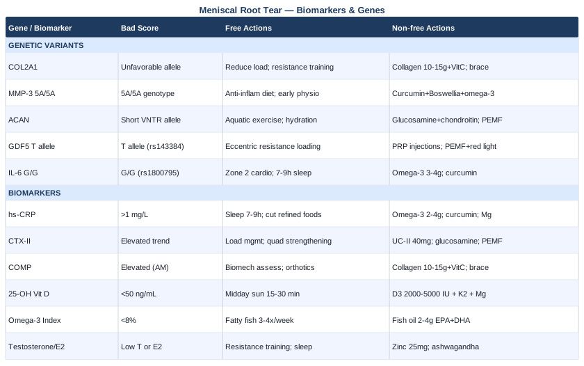
The first section covers six measurable biomarkers — from systemic inflammation to cartilage turnover to hormonal support — each with a practical protocol for intervention. The following section explores five genetic variants linked to connective tissue vulnerability and inflammatory tone, with specific plans whether or not you choose to use supplements. A summary of Peter Attia's Outlive and a set of evidence-supported complementary approaches round out the picture. The goal is better information leading to better decisions — not a shortcut, but a smarter path forward.
6 Biomarkers That Reveal What Is Happening in Your Knee
Standard orthopedic follow-up rarely includes targeted blood work. Yet several validated biomarkers can tell you whether your inflammatory state is working against healing, whether cartilage is actively degrading, and whether key nutrients and hormones are present in the amounts tissue repair actually requires. These six markers span different biological systems — none are exotic, most are affordable, and together they give a genuinely useful picture of your physiological environment after a meniscal root tear.
1. High-Sensitivity C-Reactive Protein (hs-CRP)
Why it matters
hs-CRP is the most widely used marker of systemic low-grade inflammation, and its relevance after a meniscal root tear is direct. Chronic low-grade inflammation does not just cause pain — it accelerates cartilage breakdown in the joint adjacent to the tear, impairs the quality of tissue remodeling, and slows the resolution of healing phases. Studies tracking patients with knee pathology consistently show that those with elevated hs-CRP lose cartilage faster over time and have worse functional outcomes. The meniscal root tear creates a structural vulnerability; an inflamed internal environment turns that vulnerability into ongoing degradation.
How to measure it
Request hs-CRP specifically — not standard CRP, which lacks the sensitivity to detect low-grade inflammation. Available at most standard labs. Cost: $10–$30. Optimal for musculoskeletal recovery: below 0.5 mg/L. Values between 1 and 3 mg/L indicate moderate inflammatory tone; above 3 mg/L suggests active systemic inflammation that will meaningfully impair healing. Test at baseline and retest every 60–90 days when implementing interventions.
If the score is elevated — the plan without supplements
The most impactful lifestyle levers: consistent sleep (7–9 hours, dark environment, consistent schedule), elimination of refined seed oils and ultra-processed foods from the diet, significant reduction of simple sugars, and management of chronic psychological stress via whatever mechanism works for you (structured breathing, outdoor time, social engagement). During active recovery from a root tear, prioritize low-impact movement — swimming, cycling, walking — over high-intensity training, which paradoxically elevates CRP acutely. Sleep is the single most undervalued anti-inflammatory intervention available without a prescription.
If the score is elevated — the plan with supplements or equipment
Omega-3 fatty acids (EPA + DHA combined, 2–4g daily with the largest meal, ongoing) have the strongest and most consistent evidence base for reducing hs-CRP. Curcumin with piperine (500–1000mg of standardized curcumin extract with 5–10mg piperine, daily, ongoing) has multiple RCT support for CRP reduction — but bioavailability varies significantly between products, so phospholipid-complexed or nanoparticle formulations are preferred. Magnesium glycinate (300–400mg before bed, ongoing) reduces inflammatory signaling and improves sleep architecture simultaneously. For equipment: red light therapy panels targeting 630–850nm wavelength (10–20 minutes daily over the affected knee and lower body) have emerging evidence for local and systemic inflammation modulation via mitochondrial mechanisms. Retest hs-CRP at 90 days to assess response.
2. CTX-II (C-terminal Crosslinked Telopeptide of Type II Collagen)
Why it matters
CTX-II is the most condition-specific urinary biomarker of type II collagen degradation — the collagen that forms articular cartilage and the fibrocartilage of the meniscus itself. When a meniscal root tear alters joint mechanics, the adjacent articular cartilage begins bearing abnormal compressive and shear forces, and CTX-II rises in the urine before any structural change is visible on imaging. Multiple prospective studies in knee osteoarthritis research have confirmed that elevated urinary CTX-II predicts the rate of subsequent cartilage loss. In post-tear monitoring, a rising CTX-II trend over three to six months signals that the biological degradation is outpacing repair — an early warning worth acting on before structural progression occurs.
How to measure it
CTX-II is measured from second morning urine, adjusted for creatinine to normalize for hydration. Less commonly ordered than CRP, but available through specialty functional medicine labs and some university-affiliated testing services. Cost: approximately $80–$150. The key clinical signal is trend, not a single number — compare values at baseline, three months, and six months. Age- and sex-matched reference ranges from your lab are essential context, as values rise naturally with age.
If the score is elevated — the plan without supplements
Load management is the primary intervention and the most mechanistically direct. Reducing tibiofemoral compressive activities — deep squats, loaded stair descent, prolonged standing on hard surfaces, high-impact sport — lowers the degradative stimulus at the source. Aquatic therapy and cycling allow meaningful range of motion training at low compressive load and should replace high-impact activities during this period. Consistent strengthening of the quadriceps and hip abductors reduces medial compartment load — directly relevant to medial root tears, which are by far the most common — and this has documented cartilage-protective effects in multiple clinical studies. Gait retraining with a physiotherapist to address compensatory patterns also reduces abnormal joint stress.
If the score is elevated — the plan with supplements or equipment
Undenatured type II collagen (UC-II, 40mg/day, taken on an empty stomach, ongoing) has shown effects on joint symptoms and functional outcomes in multiple controlled trials, with a plausible mechanism involving oral tolerance to collagen antigens — a fundamentally different mechanism from hydrolyzed collagen peptides. Glucosamine sulfate (1500mg/day, ongoing) has the largest long-term evidence base among joint supplements; the sulfate form consistently outperforms hydrochloride in RCTs. Unloader knee braces (custom or semi-custom, worn during weight-bearing activities) mechanically redistribute load away from the affected compartment and are supported by clinical guidelines for medial compartment knee disease. Pulsed electromagnetic field (PEMF) devices (8–30 minutes daily over the affected knee) have RCT evidence for symptom reduction and have demonstrated chondrocyte-level effects in controlled research settings. Reassess CTX-II at 90-day intervals.
3. COMP (Cartilage Oligomeric Matrix Protein)
Why it matters
COMP is a non-collagenous matrix protein integral to cartilage structure, and serum COMP is acutely sensitive to joint mechanical loading. After a meniscal root tear, the altered biomechanics create abnormal shear and compressive patterns across the joint surface. Serum COMP reflects the degree to which cartilage is being stressed relative to its repair capacity. In resting-state morning measurements, elevated COMP indicates that degradation is likely exceeding remodeling. While not yet a mainstream clinical marker, COMP is well-established in research and increasingly available through functional medicine labs. It provides a useful complement to CTX-II because it reflects current mechanical stress rather than just cumulative degradation products.
How to measure it
Serum COMP via ELISA, available through specialty and functional medicine labs. Cost: $100–$200. Standardize collection: always morning, before any physical activity — COMP rises transiently after exercise, so post-activity values are not comparable to resting values. Elevated COMP in a resting, fasted morning state is more clinically meaningful than any single post-exertion reading.
If the score is elevated — the plan without supplements
Biomechanical assessment is the first priority. A physiotherapist trained in movement analysis can identify compensatory loading patterns — often adopted unconsciously after injury — that increase stress on the affected compartment. Lateral wedge insoles or custom orthotics may redistribute load in cases of medial compartment overload associated with medial root tears. Cryotherapy post-activity (15 minutes, 2–3 times daily during active rehabilitation) reduces acute local inflammatory burden without impairing longer-term healing biology. Attention to movement quality during all daily activities, not just formal rehabilitation exercises, matters significantly for cumulative load over time.
If the score is elevated — the plan with supplements or equipment
Hydrolyzed collagen peptides (10–15g daily with 200–500mg vitamin C, taken 30–60 minutes before a loading session) have demonstrated increases in collagen synthesis markers in periarticular tissues when combined with mechanical loading — this timing protocol is supported by research from Dr. Keith Baar's group at UC Davis on tendon and ligament collagen synthesis. Vitamin C as a cofactor for collagen hydroxylation is often overlooked but is biochemically essential. PEMF devices overlap here with CTX-II protocols and are supported by the same mechanisms. For severe loading-related COMP elevation, temporary use of an unloader brace during high-demand activities allows continued movement without amplifying the degradative signal.
4. 25-OH Vitamin D
Why it matters
Vitamin D receptors are expressed on chondrocytes, tenocytes, fibroblasts, and immune cells throughout joint tissue. Low 25-OH vitamin D is independently associated with accelerated cartilage loss, elevated inflammatory cytokines, impaired muscle strength, and reduced tissue healing capacity — all directly relevant in the context of a meniscal root tear. Research published in Arthritis & Rheumatology identified low serum vitamin D as a predictor of joint space narrowing in knee osteoarthritis. Separately, imaging studies have demonstrated that vitamin D deficiency correlates with increased meniscal tissue degeneration on MRI. For recovery, deficiency creates a biological headwind that rehabilitation alone cannot overcome.
How to measure it
Standard serum 25-OH vitamin D test, widely available. Cost: $30–$60. Optimal for musculoskeletal health and tissue repair: 50–80 ng/mL. The commonly cited clinical minimum of 30 ng/mL is a threshold for bone disease, not for musculoskeletal resilience — sports medicine and functional medicine literature consistently points to the 50–80 range as protective. Test twice yearly (late winter and late summer) to capture seasonal variation, as levels can drop substantially in winter months at northern latitudes.
If the score is low — the plan without supplements
Direct midday sun exposure (15–30 minutes on arms and legs, 3–5 times weekly during summer months in full UVB exposure conditions) can meaningfully raise levels for individuals with lighter skin tones. For those with darker skin, during winter, or in northern latitudes, dietary and sun sources alone are insufficient to reach the therapeutic range. Dietary sources — wild-caught fatty fish, egg yolks, liver, fortified dairy — contribute modestly and cannot substitute for direct sun or supplementation when deficiency is present. Reducing habitual full-body sunscreen use during brief midday exposures on larger skin surfaces is a simple behavioral adjustment.
If the score is low — the plan with supplements or equipment
Vitamin D3 with MK-7 form of vitamin K2 (2000–5000 IU D3 with 100–200mcg K2 daily, taken with the largest fat-containing meal, ongoing). K2 is included because it activates proteins that direct calcium into bone rather than soft tissue — critical when supplementing D3 at meaningful doses. Magnesium is required as a cofactor for vitamin D conversion and activation; without adequate magnesium status, D3 supplementation has limited effect. If you are supplementing D3 and not improving, magnesium status is the first variable to check. Retest 25-OH vitamin D after 90 days of supplementation to confirm response and titrate dose accordingly.
5. Omega-3 Index
Why it matters
The omega-3 index measures the percentage of EPA and DHA in red blood cell membranes — reflecting months of dietary intake rather than a single-point plasma measurement — and was developed and validated by Dr. William Harris. In the context of joint injury, EPA and DHA support the production of specialized pro-resolving mediators (SPMs) including resolvins, protectins, and maresins. These molecules actively terminate inflammatory cascades rather than merely suppressing them — a biologically distinct and essential process for tissue healing. Adequate omega-3 status means the body can resolve inflammation rather than sustaining it chronically, which directly influences how cleanly a meniscal injury heals.
How to measure it
Home fingerprick test kits are available from OmegaQuant, the most validated commercial source for this test, developed by Dr. Harris himself. Cost: $50–$100. Optimal target: 8% or above. The average American has an index of 4–5%, representing a meaningfully pro-inflammatory state. Values below 4% indicate significant omega-3 insufficiency. The index should be retested after 90 days of dietary or supplementation intervention to confirm tissue-level incorporation — plasma levels change quickly, but cell membrane incorporation takes months.
If the score is low — the plan without supplements
Increase fatty fish consumption to 3–4 servings weekly: wild-caught salmon, sardines, Atlantic mackerel, and herring are the most efficient dietary sources. Simultaneously reducing omega-6 intake from seed oils (canola, sunflower, corn, soybean oil) matters as much as increasing omega-3 intake, because the omega-6:omega-3 ratio determines the downstream balance of pro-resolving versus pro-inflammatory signaling. Walnuts, flaxseed, and chia seeds contribute ALA, which converts to EPA and DHA only at about 5–10% efficiency but still supports the ratio modestly.
If the score is low — the plan with supplements or equipment
High-quality fish oil or krill oil (2–4g total EPA + DHA daily, with meals, ongoing). Choose brands with published IFOS third-party certification for purity and oxidation levels — rancid fish oil generates inflammatory byproducts that counteract the purpose entirely. Triglyceride-form fish oil absorbs approximately 70% better than the ethyl ester form used in most budget products; this is worth checking on the label. For plant-based individuals, algae-derived EPA + DHA is the only functionally equivalent alternative — avoid relying on ALA conversion alone. Retest omega-3 index at 90 days.
6. Testosterone (Total and Free) and Estradiol
Why it matters
Sex hormone status has direct and underappreciated effects on connective tissue quality and repair capacity. Estrogen maintains collagen synthesis in ligamentous and fibrocartilaginous tissue — the rapid rise in knee ligament and meniscal injuries following menopause is not incidental, and it is now well-documented in sports medicine literature. In men, testosterone supports the lean muscle mass that mechanically protects the knee, directly modulates inflammatory tone at physiological levels, and influences satellite cell activity relevant to periarticular tissue repair. Low testosterone in men is consistently associated with elevated systemic inflammation and slower musculoskeletal recovery. For both sexes, suboptimal hormonal status creates a biological environment that makes meniscal recovery harder and joint degradation faster.
How to measure it
Standard serum panel: total testosterone, free testosterone (calculated or direct), and estradiol. Cost: $50–$120 as a bundle. For men, optimal for musculoskeletal health: total testosterone 600–900 ng/dL, free testosterone above 15 ng/dL. For premenopausal women, estradiol below 50 pg/mL in the follicular phase may indicate suboptimal support. For peri- and postmenopausal women, context and symptoms must be discussed with a menopause specialist. Always test testosterone in the morning (7–10 AM), as it peaks during this window.
If the score is suboptimal — the plan without supplements
For men: prioritize 7–9 hours of high-quality sleep (the majority of testosterone is produced during sleep), ensure caloric intake is not significantly below maintenance (chronic deficit suppresses testosterone), incorporate heavy compound resistance training (deadlifts, squats, rows) three times weekly, and reduce chronic psychological stressors which elevate cortisol and suppress gonadotropin signaling. For women approaching or in menopause: discuss current HRT evidence with a menopause-specialist clinician — the risk-benefit picture has shifted significantly since the original Women's Health Initiative misinterpretation, and physical activity, particularly resistance training, also supports estrogen-dependent tissue quality during the peri-transition period.
If the score is suboptimal — the plan with supplements or equipment
Zinc bisglycinate (25–30mg daily, 8 weeks on / 2 weeks off, paired with 1–2mg copper) supports testosterone synthesis as a necessary enzymatic cofactor and is commonly depleted in individuals with high training volume or poor dietary intake. KSM-66 or Sensoril ashwagandha extract (300–600mg daily, 8–12 week cycles) has multiple RCTs demonstrating modest but consistent testosterone support in men with stress-related suppression, alongside cortisol reduction effects. For women with confirmed low estradiol considering HRT: bioidentical transdermal estradiol with micronized progesterone has the most favorable safety profile in current evidence — but this requires physician prescription and monitoring, not self-management. Supplement strategies are adjunctive; significant hormonal deficiency requires clinical evaluation.
With the biomarker picture clearer, the next layer to understand is genetic — specifically, which inherited variants may have contributed to your tissue's baseline vulnerability and how much influence diet, lifestyle, and targeted supplementation can exert against them.
The Genetics Behind Meniscal Root Vulnerability: 5 Key Variants
Genetic testing for musculoskeletal health is not yet standard clinical practice, but the research base supporting several specific variants has grown substantially in the past decade. Companies like Genomic Life (founded by Dr. Ali Torkamani) and direct-to-consumer options make these insights increasingly accessible. The premise — as Gary Brecka and others in functional medicine have emphasized — is not that genes are destiny, but that knowing your genetic tendencies allows you to compensate upstream rather than managing consequences downstream. For connective tissue health, five variants are particularly relevant to meniscal integrity, cartilage quality, and inflammatory tone.
1. COL2A1 — The Type II Collagen Blueprint
What this gene affects
COL2A1 encodes the alpha-1 chain of type II collagen — the dominant structural collagen in articular cartilage and fibrocartilage, which includes the meniscus. Variants in COL2A1 are associated with abnormal collagen fibril structure, premature cartilage degeneration, and increased susceptibility to chondrodysplasias. In the context of meniscal root tears, a structurally inferior collagen matrix at the root insertion site may reduce the tissue's ability to withstand normal mechanical loads over time, contributing to degenerative root tears in middle age. The evidence here comes primarily from OA and intervertebral disc disease genetics, with logical extension to fibrocartilaginous tissue more broadly.
If the variant is unfavorable — the plan without supplements
Reducing compressive and shear stress on articular surfaces throughout life is the primary compensatory strategy. Maintaining lean body mass without excess weight directly reduces tibiofemoral forces with every step; every kilogram of excess body weight generates approximately 3–6 kilograms of additional knee joint force during walking. Lifelong resistance training that develops strong quadriceps, hip abductors, and posterior chain musculature reduces the mechanical load placed on cartilage and fibrocartilage. Avoiding sustained end-range knee loading (prolonged kneeling, deep squatting under load) minimizes stress concentration at the root insertion sites. These are not temporary measures — with a structural collagen variant, they are a permanent lifestyle scaffold.
If the variant is unfavorable — the plan with supplements or equipment
Hydrolyzed collagen peptides (10–15g daily with vitamin C, ongoing) provide the amino acid substrate — particularly glycine, proline, and hydroxyproline — that cells need to synthesize new collagen. This does not correct the genetic variant, but it ensures the synthetic machinery is not substrate-limited. Vitamin C (250–500mg alongside collagen intake) is the essential cofactor for lysyl hydroxylase and prolyl hydroxylase enzymes that create stable crosslinks in collagen fibrils. Lysine supplementation (1–2g/day) supports collagen crosslink formation. For equipment: compression braces during high-load activities reduce mechanical stress at vulnerable insertion points and can extend functional tissue life over years.
2. MMP-3 (5A/6A Promoter Polymorphism) — Matrix Degradation Intensity
What this gene affects
Matrix metalloproteinase-3 (stromelysin-1) is a key enzyme in extracellular matrix degradation, including the cartilage and fibrocartilage of the meniscus. The promoter region of MMP-3 has a common insertion/deletion polymorphism: the 5A allele is associated with significantly higher MMP-3 gene expression compared to the 6A allele. Individuals with the 5A/5A genotype tend to have more aggressive matrix breakdown following joint injury or inflammation. In the context of a meniscal root tear, a 5A/5A carrier may degrade adjacent cartilage faster during the post-injury inflammatory period, making anti-inflammatory interventions more urgent and the timeline for structural deterioration potentially shorter. Several studies have linked MMP-3 5A with worse outcomes in ligament injury and knee OA progression.
If the variant is unfavorable — the plan without supplements
Rapid and sustained reduction of joint inflammation is the most direct compensatory strategy — because MMP-3 expression is driven by inflammatory cytokines including IL-1β and TNF-α. This makes all of the hs-CRP interventions described above doubly important for MMP-3 5A carriers. Early physiotherapy to reduce effusion and restore joint movement normalizes the cytokine environment inside the joint. Avoiding prolonged immobilization post-injury is important — a static inflammatory joint environment with ongoing cytokine signaling will drive MMP-3 expression more than graduated, controlled loading. Diet quality — specifically reducing refined carbohydrates and seed oils — directly lowers the NF-κB signaling that drives MMP gene transcription.
If the variant is unfavorable — the plan with supplements or equipment
Curcumin (500–1000mg of a high-bioavailability formulation, daily, ongoing) directly inhibits NF-κB and has demonstrated downregulation of MMP-3 expression in multiple in vitro and clinical studies — making it more mechanistically specific for this variant than for most other conditions. Omega-3 fatty acids work here via a different mechanism: EPA and DHA produce SPMs that resolve the cytokine environment driving MMP activation. Boswellia serrata (AKBA-standardized extract, 100–200mg AKBA daily, 8–12 week cycles) inhibits 5-LOX and has human clinical evidence for joint benefit and anti-MMP activity, with a good tolerability profile. Combination of curcumin + boswellia + omega-3 creates complementary mechanistic coverage for MMP-3 5A carriers. Side effects: curcumin may interact with anticoagulants at high doses; consult a prescriber if relevant.
3. ACAN (Aggrecan Variable Number Tandem Repeat) — Cartilage Load Capacity
What this gene affects
The ACAN gene encodes aggrecan — the large proteoglycan that gives cartilage its compressive resistance by attracting and retaining water within the extracellular matrix. A variable number tandem repeat (VNTR) polymorphism in ACAN is associated with intervertebral disc degeneration, early-onset OA, and reduced cartilage mechanical resilience. Shorter VNTR alleles correlate with reduced aggrecan content and therefore reduced ability to distribute compressive loads — directly relevant to the fibrocartilage of the meniscus and the articular cartilage it protects. While the specific evidence for meniscal root tear risk linked to ACAN is extrapolated from disc and OA data, the biological mechanism is directly applicable.
If the variant is unfavorable — the plan without supplements
Body weight management is the highest-leverage lifestyle intervention for an ACAN short-allele carrier, because every compressive force experienced by the knee is amplified when the cartilage matrix has reduced proteoglycan content and water retention. Aquatic exercise and cycling, which deliver cardiovascular and musculoskeletal benefit without high compressive loading, are particularly valuable as long-term training modalities for individuals with compromised cartilage matrix genetics. Avoiding excessive sitting with the knee in a flexed loaded position reduces the compressive dehydration of already-compromised cartilage. Staying well hydrated (cartilage is imbibition-dependent — it draws fluid from synovial fluid during unloaded periods) is a simple and effective daily habit.
If the variant is unfavorable — the plan with supplements or equipment
Glucosamine sulfate (1500mg daily, ongoing) is the most extensively studied supplement specifically aimed at maintaining the proteoglycan matrix — the direct product of ACAN function. The sulfate form consistently outperforms hydrochloride in long-term RCTs. Chondroitin sulfate (800–1200mg daily, ongoing) provides additional glycosaminoglycan substrate and may slow proteoglycan loss. Hyaluronic acid supplementation (oral, 80–200mg/day of high-molecular-weight HA) has growing evidence for joint lubrication support and may be of particular value when matrix proteoglycan content is genetically limited. PEMF devices have shown ability to stimulate chondrocyte proteoglycan synthesis in controlled studies — an equipment-based approach with direct mechanistic relevance to ACAN variants.
4. GDF5 (Growth Differentiation Factor 5) — Joint Maintenance and Repair
What this gene affects
GDF5 encodes a signaling protein belonging to the TGF-β superfamily that plays a critical role in embryonic joint formation, cartilage homeostasis, and repair signaling in adult tissue. The rs143384 T allele of GDF5 is one of the most replicated genetic risk factors for knee osteoarthritis across multiple large-scale genome-wide association studies (GWAS), including studies in European, Asian, and Ashkenazi Jewish populations. The T allele reduces GDF5 expression in joint tissue, impairing the signals that maintain cartilage quality and support repair after injury. For individuals who carry the T allele and sustain a meniscal root tear, the post-injury repair signaling environment may be inherently compromised — meaning that the same injury, in a GDF5 T allele carrier, may progress faster than in a non-carrier.
If the variant is unfavorable — the plan without supplements
Graduated mechanical loading is specifically relevant here: GDF5 expression in joint tissue is stimulated by appropriate mechanical signals. Resistance training — specifically the compressive and tensile forces of squats, lunges, and loaded carries, performed within a pain-free range — may upregulate endogenous GDF5 signaling through mechanoregulation, partially compensating for the genetic reduction in baseline expression. This is the biological rationale for why controlled loading (not rest) promotes better long-term outcomes in cartilage-related conditions. Eccentric loading protocols, used carefully under physiotherapy guidance, are particularly potent mechanical stimuli for connective tissue maintenance.
If the variant is unfavorable — the plan with supplements or equipment
Platelet-rich plasma (PRP) injections — administered by an orthopedic or sports medicine physician — deliver high concentrations of growth factors including TGF-β family members that partially overlap with GDF5 signaling pathways. Multiple RCTs support PRP for knee OA, and its use post-meniscal injury is an active area of clinical research. Cost: $500–$2000 per injection, typically not covered by insurance. Collagen peptides (10–15g with vitamin C) support the repair matrix that GDF5 signaling normally helps maintain. For equipment: PEMF and red light therapy both have evidence for stimulating chondrocyte synthetic activity and may partially compensate for reduced endogenous repair signaling. These equipment interventions are low-risk and can be used long-term.
5. IL-6 (-174G/C, rs1800795) — Inflammatory Response Intensity
What this gene affects
The IL-6 gene promoter polymorphism at position -174 strongly influences how much interleukin-6 a person produces in response to an inflammatory trigger. The G allele is associated with higher IL-6 expression; individuals with the G/G genotype mount more intense and more prolonged inflammatory responses. IL-6 is a pleiotropic cytokine — it has both pro-inflammatory roles during acute injury and regulatory roles in repair — but chronically elevated IL-6 promotes joint synovitis, cartilage degradation, and pain sensitization. After a meniscal root tear, a G/G carrier may experience more intense swelling, slower resolution of the inflammatory phase, and greater risk of ongoing synovial inflammation driving secondary cartilage damage.
If the variant is unfavorable — the plan without supplements
The lifestyle factors that most directly attenuate IL-6 overexpression are sleep quality, dietary patterns, and aerobic fitness. Regular moderate aerobic exercise (Zone 2 cardio — 180 minus age as an approximate heart rate target, 3–5 sessions weekly) paradoxically reduces resting IL-6 levels over time despite acutely raising them. The chronic adaptation is a trained anti-inflammatory phenotype with reduced baseline cytokine tone. Sleep deprivation is one of the most potent known stimulants of IL-6 production — a G/G carrier who consistently under-sleeps is adding a genetic amplifier to a behavioral trigger. Eliminating processed food, refined sugar, and smoking also reduces NF-κB-driven IL-6 transcription.
If the variant is unfavorable — the plan with supplements or equipment
High-dose omega-3s (3–4g EPA + DHA daily, ongoing) are specifically relevant here: EPA and DHA directly suppress IL-6 production through inhibition of arachidonic acid conversion pathways. Curcumin (as above) inhibits NF-κB and AP-1, two transcription factors that drive IL-6 gene expression in response to inflammatory stimuli — mechanistically very well-matched to this variant. Tart cherry extract (480mg anthocyanins daily, 8–12 week cycles, break of 4 weeks) has emerging evidence for reducing inflammatory cytokines including IL-6 and may be a lower-cost adjunct. For equipment: cold water immersion (10–15 minutes in water at 10–15°C, 3–4 times weekly) acutely reduces inflammatory cytokine levels and has growing evidence for anti-inflammatory adaptation with regular use — caution in individuals with cardiovascular conditions.
Biomarkers and genetics together form a powerful individual picture. The next layer comes from synthesizing what is now known about long-term musculoskeletal health into an actionable framework — something Peter Attia's Outlive addresses with unusual depth and clarity.
What "Outlive" by Peter Attia Reveals About Joints That Most Doctors Don't Discuss
Peter Attia's 2023 book Outlive: The Science and Art of Longevity draws on decades of research across metabolic medicine, exercise physiology, and longevity science. While not a book about meniscal injuries specifically, its framework for musculoskeletal health as a primary longevity pillar — and its challenge to reactive, symptom-driven medicine — is directly applicable to anyone navigating joint injury. Ten insights from the book are particularly worth carrying into a meniscal root tear recovery and long-term joint protection context.
1. Muscle Mass Is a Metabolic and Mechanical Protector
Attia argues forcefully that muscle mass is among the most important predictors of long-term health and survival — and for joints, the mechanism is both metabolic (muscle is the major glucose sink that controls systemic inflammation) and mechanical (strong periarticular muscles reduce compressive load on cartilage). Building and maintaining muscle around the knee is not rehabilitation; it is long-term joint insurance.
2. VO2 Max and Zone 2 Training Affect Joint Tissue Directly
Aerobic capacity influences tissue perfusion, mitochondrial density in fibroblasts and chondrocytes, and the body's ability to resolve inflammatory states. Attia recommends consistent Zone 2 training (low-intensity, sustainable aerobic work) not as weight loss strategy but as a mitochondrial health intervention. Better-functioning cellular energy machinery in joint tissue means more capacity for repair and less susceptibility to degradative stress.
3. Inflammation Is Not Binary — You Need Numbers
One of Outlive's most practical messages is that inflammation exists on a spectrum, and clinical medicine too often waits for disease-level inflammation before intervening. Attia explicitly recommends hs-CRP as a routine tracking marker — the same biomarker listed first in this article. Below 0.5 mg/L is where tissue health is well-supported; most of his patients with chronic injury histories were hovering between 1 and 3 mg/L without knowing it.
4. The Supplement Gaps Are Real — But Omega-3 and Vitamin D Are Under-Dosed
Attia is skeptical of most supplements but specifically advocates for omega-3s at doses that achieve a meaningful omega-3 index (above 8%), not the low 1g doses commonly sold. Similarly, he targets 25-OH vitamin D at 60 ng/mL rather than the bare clinical minimum. Both overlap precisely with the biomarkers described above, and both are typically under-dosed in conventional practice.
5. Protein Intake for Connective Tissue Repair Is Higher Than Most Eat
Attia recommends 1.6–2.2g of protein per kilogram of body weight for individuals seeking to maintain or build muscle — considerably higher than conventional recommendations. For connective tissue repair specifically, adequate glycine and proline intake (found in collagen-containing foods and collagen peptide supplements) is an additional consideration that standard protein targets do not address.
6. Sleep Is the Most Underestimated Recovery Tool
Attia dedicates an entire chapter to sleep, citing evidence that consistent, high-quality sleep of 7–9 hours drives growth hormone release, reduces cortisol, reduces IL-6, repairs tissue, and consolidates neuromotor learning — all relevant to joint recovery. He argues that optimizing sleep often delivers more recovery benefit than any supplement protocol.
7. Stability and Balance Are As Important As Strength
Beyond muscle mass, Attia emphasizes neuromuscular stability — the ability to control load effectively through joints at end ranges. For knee health specifically, proprioceptive training, single-leg stability work, and controlled eccentric loading are as important as raw strength for joint protection. These are not rehabilitation exercises; they are permanent training priorities.
8. Senescent Cells Drive Joint Degradation — and Are Modifiable
Cellular senescence — cells that stop dividing but release inflammatory signals (the senescence-associated secretory phenotype, or SASP) — is an accelerating contributor to joint degeneration as we age. Attia covers emerging research on senolytics (compounds that clear senescent cells) including quercetin and dasatinib, noting that while the evidence is early in humans, the mechanism is biologically compelling for conditions involving chronic joint inflammation.
9. Reactive Medicine Misses the Decade Before the Diagnosis
Perhaps Outlive's most important framing is the concept of early intervention — in what Attia calls "Medicine 3.0." Meniscal root tears often occur in a tissue that has been degenerating quietly for years. By the time the root tears, the cartilage adjacent to it may already show early OA changes. The biomarkers and genetic insights described in this article are precisely the kind of tools Attia argues should be in every health-conscious person's baseline — not ordered after the injury, but tracked continuously.
10. Individualized Medicine Over Population Averages
Population-level recommendations — what the average person needs — routinely miss the specific biology of the person in front of you. Attia's framework is that the most valuable medical information is personalized: your hs-CRP, your omega-3 index, your hormonal panel, your genetic variants. This is not about becoming obsessed with biomarkers — it is about having enough data to make genuinely informed decisions rather than following generic guidelines that were not built for your specific biology.
Complementary Approaches With Clinical Support
Beyond biomarkers and genetics, several non-pharmaceutical modalities have accumulated meaningful human clinical evidence for conditions involving knee pain, cartilage health, and joint recovery. The three below were selected specifically because their evidence is condition-relevant, not just theoretically plausible.
Low-Level Laser Therapy and Photobiomodulation
Low-level laser therapy (LLLT) — also called photobiomodulation — uses specific wavelengths of red and near-infrared light (typically 630–1000nm) to stimulate mitochondrial function, reduce inflammatory cytokine production, and promote tissue repair at the cellular level. In the context of a meniscal root tear, the mechanism is particularly relevant: photobiomodulation increases ATP production in chondrocytes and fibroblasts, stimulates collagen synthesis, and reduces local prostaglandin E2 levels — all of which directly support the repair environment in a damaged joint.
A 2012 meta-analysis by Bjordal JM et al., published in BMC Musculoskeletal Disorders, reviewed multiple RCTs of LLLT for knee OA and found significant improvements in pain and function with treatment at appropriate wavelengths and doses. A separate RCT by Hegedus B et al. demonstrated that knee OA patients receiving active LLLT showed measurable improvements in pain scores and walking time compared to sham treatment, with effects persisting beyond the treatment period.
For practical application: clinical-grade laser therapy devices operated by a physiotherapist or sports medicine specialist offer the most reliable dosing. Home-use red light therapy panels (660nm and 850nm combination, delivering 20–40mW/cm² at skin surface) offer a more affordable option for regular adjunctive use — 10–20 minutes daily or every other day directly over the affected knee. The evidence is for genuine effect at appropriate doses, not all consumer products deliver therapeutic irradiance; check the device specifications before purchasing. Start conservatively and monitor for any local skin sensitivity.
Tai Chi
Tai chi is a low-impact movement practice combining slow, controlled postures, continuous weight-shifting, and conscious breathing. For knee joint health specifically, it works through multiple complementary mechanisms: building quadriceps and hip stabilizer strength, improving neuromuscular proprioception around the knee, reducing the psychological stress burden that elevates systemic inflammatory markers, and providing graduated articular loading that stimulates cartilage health without the compressive peaks of higher-impact activities.
A landmark randomized controlled trial published in the New England Journal of Medicine (2010) by Wang C et al. found that patients with knee OA who practiced tai chi twice weekly for 12 weeks experienced significantly greater improvements in pain, function, and quality of life compared to physical therapy. A subsequent meta-analysis confirmed these findings across multiple trials, with tai chi showing consistent superiority over control conditions for knee pain and physical function in OA populations. For meniscal root tear recovery, the evidence is extrapolated from these OA studies — direct RCT data for root tears specifically does not yet exist, but the mechanical and neuromuscular mechanisms are directly applicable.
Practically: most beginner tai chi programs involve 2–3 sessions weekly (30–60 minutes each) for 8–12 weeks before benefits consolidate. Sun-style or Yang-style short forms are most commonly used in clinical research and are accessible for deconditioned individuals. Practicing in a group class under qualified instruction reduces injury risk and improves adherence. Avoid sessions where pain above a 4/10 is generated — the practice should feel effortful but not painful at the knee joint.
Massage Therapy
Manual soft tissue therapy — specifically therapeutic massage targeting the musculature around the knee (quadriceps, hamstrings, calf, hip flexors) — addresses an underappreciated contributor to poor outcomes after meniscal injury: the reflex muscle guarding, trigger points, and myofascial tension that develop as secondary compensations around a painful joint. These soft tissue adaptations alter gait mechanics, increase joint compressive forces, and perpetuate abnormal loading patterns even after the primary injury is stabilized.
A systematic review published in Manual Therapy found that massage therapy targeting the periarticular musculature of the knee produced significant improvements in pain and function in individuals with knee OA, with effects persisting at follow-up. While direct RCT evidence for meniscal root tear is not available, the mechanism — reducing the soft tissue contributions to abnormal joint loading — is condition-relevant and supported by the broader knee pain evidence base.
For application: targeted deep tissue or myofascial release to the quadriceps (particularly the vastus medialis), hamstrings, IT band, and hip external rotators, performed by a licensed massage therapist or physiotherapist with manual therapy training, 1–2 times weekly during the acute and subacute phases. Self-massage with a foam roller or percussion device (targeting the same muscle groups, 5–10 minutes daily) can maintain gains between professional sessions. The goal is not to treat the torn root directly — massage cannot address the structural injury — but to optimize the mechanical environment around it.
Conclusion
A meniscal root tear is a structural problem, but how it heals — and whether the surrounding joint deteriorates or remains stable over the following years — is substantially determined by biology that is measurable and, in many cases, modifiable. The six biomarkers covered here give you a map of your current internal state: inflammation, cartilage turnover, nutritional sufficiency, hormonal support, and omega-3 status. The five genetic variants give you a deeper picture of your baseline vulnerabilities. Neither set of information replaces the guidance of a good orthopedic surgeon or physiotherapist — but together they give you something more valuable than a generic rehabilitation plan: a picture of what your specific biology actually needs.
The most productive next step is not overhauling everything at once. Choose one biomarker to measure first — hs-CRP and 25-OH vitamin D are the most accessible and impactful starting points. Bring the results to your treating clinician or a sports medicine physician who is comfortable interpreting them. Build from there. Better information leads to better decisions — and the decisions you make in the months following this injury will likely determine whether the knee you have in ten years is a functional one.
Musculoskeletal: Joint Conditions Tendon & Ligament Conditions Sports Injuries
Autoimmune: Inflammatory Conditions Connective Tissue Conditions