This article contains AI generated content.
· UpdatedMeniscofemoral Ligament Tear — 5 Genes And 6 Biomarkers To Track
Introduction
A meniscofemoral ligament tear is not the kind of injury that announces itself with a clean diagnosis and a clear roadmap. These small but structurally important ligaments — the ligaments of Humphrey and Wrisberg — connect the posterior horn of the lateral meniscus to the medial femoral condyle, and their damage is frequently underdiagnosed or bundled into broader ACL or meniscal injury narratives. If you are dealing with persistent knee instability, recurring inflammation, or a recovery that keeps stalling, it is worth asking whether the biology underneath the injury is being taken seriously enough.
The frustrating reality is that most rehabilitation protocols treat ligament tears as if everyone heals at the same rate. Rest, physiotherapy, and time are the default answers. But two people with anatomically similar injuries can have dramatically different healing trajectories — and increasingly, the research points to measurable biological factors that explain why. Genetic predisposition, systemic inflammation levels, cartilage turnover rates, and connective tissue quality all influence how a torn ligament responds to treatment.
This article takes a different approach. Instead of offering another generic guide to RICE protocol and quad sets, it focuses on the underlying biology you can actually measure and, in many cases, improve. That includes specific blood biomarkers that reflect how well your body is managing the injury, and specific genetic variants that may be quietly working against you — without you or your clinician knowing it.
The goal is not to replace medical care. It is to give you the kind of information that makes your medical conversations smarter and more targeted. When you understand what is driving your recovery (or stalling it), you can make better decisions — about nutrition, supplementation, movement, and timing. That is a meaningful shift from passively hoping the injury heals to actively supporting the biology that makes healing possible.
Summary
This article covers 6 key biomarkers and 5 genetic factors relevant to meniscofemoral ligament tear recovery. For each biomarker, you will find how to measure it, what a bad result means in practical terms, and what you can do about it — with and without supplements. The genetics section explains which gene variants are most associated with connective tissue vulnerability, slow healing, and elevated injury risk, along with specific action plans for each. Beyond the science, the article includes a deep dive into the Andrew Huberman podcast content most relevant to connective tissue recovery — covering collagen synthesis, loading protocols, and tissue remodeling insights you are unlikely to hear from your orthopedic surgeon. It closes with complementary approaches that have the strongest evidence base for knee ligament recovery, from photobiomodulation to targeted manual therapy. If your recovery has been slower or more complicated than expected, the answers may be measurable and addressable.
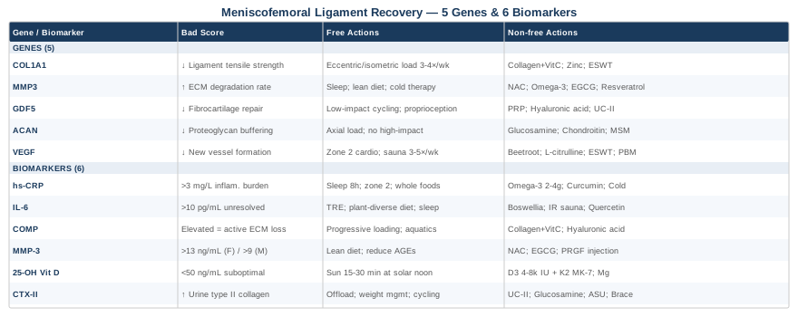
6 Biomarkers to Track for Meniscofemoral Ligament Tear Recovery
Biomarkers give you a live read of what is happening inside the knee and across the whole body. For connective tissue injuries, certain markers are particularly informative: they can tell you whether inflammation is being resolved, whether collagen is breaking down faster than it is being built, and whether your nutritional foundation supports tissue healing at all. The following six biomarkers offer the clearest picture of where you stand — and where to intervene.
1. hs-CRP: The Inflammation Control Tower
High-sensitivity C-reactive protein (hs-CRP) is the most widely used marker of systemic low-grade inflammation. After a meniscofemoral ligament tear, the body mounts an acute inflammatory response that is initially necessary for healing. The problem arises when this inflammation does not resolve — a state sometimes called chronic low-grade inflammation, which has been directly associated with impaired ligament and tendon healing, delayed collagen maturation, and increased risk of further joint degeneration. Elevated hs-CRP is also independently associated with worse outcomes following knee surgery and injury in multiple prospective cohort studies.
How to measure it: A standard blood draw. hs-CRP is differentiated from regular CRP by the assay's sensitivity to lower concentrations. Most labs include it in a cardiovascular panel. Cost typically ranges from $10–$40 depending on provider. Ideal range: below 0.5 mg/L. Values above 3 mg/L indicate a high-risk inflammatory burden. Peter Attia considers anything above 1 mg/L worth investigating and treating aggressively in the context of long-term tissue and cardiovascular health.
If the score is bad, the plan without supplements: The most powerful non-supplemental levers for hs-CRP reduction are sleep quality and duration (targeting 7.5–9 hours), elimination of ultra-processed foods and refined seed oils, regular moderate-intensity aerobic activity (zone 2 cardio, 3–4 sessions per week of 30–45 minutes), and stress reduction. Sleep deprivation alone can raise hs-CRP significantly within days. For knee injury recovery specifically, maintaining gentle movement — even seated leg exercises or non-weight-bearing cycling — helps prevent the inflammatory burden of complete immobilization.
If the score is bad, the plan with supplements or equipment: Omega-3 fatty acids are among the best-studied anti-inflammatory supplements. A dose of 2–4 g EPA/DHA per day with food has repeatedly demonstrated hs-CRP reduction in clinical trials. Cycle: 12 weeks on, then reassess. Curcumin (as a high-bioavailability formulation such as Meriva or with piperine) at 500–1000 mg/day has shown hs-CRP reductions in several randomized controlled trials. Magnesium glycinate at 300–400 mg/night supports both sleep quality and inflammatory resolution. Cold water immersion (10–15 minutes at 10–15°C, 3–4 times per week) has mechanistic evidence for reducing systemic inflammation, though the timing relative to training matters — avoid immediately after connective tissue loading sessions.
2. IL-6: The Acute Phase Regulator
Interleukin-6 (IL-6) is a cytokine that orchestrates the acute inflammatory cascade following tissue injury. In the first 24–72 hours post-injury, elevated IL-6 is appropriate and pro-healing: it recruits immune cells, initiates fibroblast activity, and drives the early phases of collagen deposition. However, chronically elevated IL-6 — which can persist for weeks or months after injury in some individuals — shifts the balance toward tissue degradation, activates matrix metalloproteinases (which break down the extracellular matrix), and inhibits satellite cell function relevant to supporting structures like the quadriceps.
How to measure it: A blood draw for serum IL-6. Less commonly ordered than hs-CRP and typically requires a specific request. Cost: $50–$120 at most labs. Optimal: below 3 pg/mL. Levels above 10–15 pg/mL in a non-acute phase suggest unresolved inflammation. Note that IL-6 is highly variable and should ideally be assessed at rest and more than 48 hours after any significant physical activity.
If the score is bad, the plan without supplements: Reducing visceral adipose tissue is one of the most powerful IL-6 interventions, since adipose tissue is a primary source of chronic IL-6 secretion. Time-restricted eating (8–10 hour eating window) combined with moderate caloric deficit has been shown to reduce IL-6 meaningfully over 8–12 weeks. Sleep optimization remains critical: IL-6 is regulated by circadian rhythms, and disrupted sleep architecture directly increases secretion. Gut microbiome health also matters: a high-fiber, plant-diverse diet reduces IL-6 through butyrate-producing bacterial communities.
If the score is bad, the plan with supplements or equipment: Boswellic acids (Boswellia serrata extract, 300–500 mg standardized to 65% boswellic acids) have shown IL-6 reduction in inflammatory joint conditions in randomized trials. Frequency: daily, in 8–12 week cycles with a 2–4 week break. Side effects are mild at recommended doses but may include gastrointestinal discomfort. Infrared sauna (20–30 minutes, 3 times per week) has mechanistic evidence for improving cytokine profiles and is increasingly used in athletic recovery contexts. Quercetin at 500–1000 mg/day has shown IL-6 modulation in human studies, particularly in combination with vitamin C.
3. COMP: The Connective Tissue Integrity Signal
Cartilage Oligomeric Matrix Protein (COMP) is one of the most informative biomarkers in musculoskeletal medicine. It is a structural protein found in cartilage, tendons, ligaments, and meniscal tissue. When these tissues are under mechanical stress or undergoing degradation, COMP is released into synovial fluid and eventually into the bloodstream. Serum COMP levels therefore reflect how aggressively connective tissue — including the meniscofemoral ligament complex — is being broken down. Research published in the European Journal of Sport Science and several orthopedic journals has validated COMP as a practical marker for monitoring knee tissue health over time. It is particularly useful for tracking whether a rehabilitation program is supporting tissue remodeling or inadvertently accelerating degeneration.
How to measure it: Serum COMP via a specialized lab request. Not available at every standard lab — often requires ordering through sports medicine, rheumatology, or a functional medicine provider. Cost ranges from $80–$200. Optimal values depend on the reference range of the assay used, but in the context of recovery monitoring, the direction of change over weeks is often more informative than a single number. COMP rises acutely after exercise — always measure at least 24 hours post-activity.
If the score is bad, the plan without supplements: Load management is the most direct non-supplemental intervention. Connective tissue responds poorly to both complete immobilization (which reduces collagen synthesis) and excessive loading (which accelerates COMP release). The sweet spot is progressive mechanical loading — isometric exercises, then isotonic, then dynamic — following a structured periodization plan. Eliminating impact loading and rotational stress on the knee during active degradation phases is essential. Aquatic therapy provides mechanical stimulus without high joint compression forces.
If the score is bad, the plan with supplements or equipment: Collagen peptides (10–15 g/day, ideally type I and type II combined) taken 30–60 minutes before loading exercise have demonstrated improved COMP profiles and connective tissue markers in multiple studies, including those from the Keith Baar lab at UC Davis. Vitamin C (500–1000 mg co-administered with collagen peptides) is required for hydroxylation of proline residues in collagen synthesis — the combination is more effective than either alone. Cycle: continuous during active rehabilitation. Side effects at these doses are minimal. Hyaluronic acid supplementation (oral, 80–200 mg/day) has also shown connective tissue support in preliminary human trials.
4. MMP-3: The Matrix Degradation Accelerator
Matrix metalloproteinase-3 (MMP-3, also called stromelysin-1) is an enzyme that degrades multiple components of the extracellular matrix, including fibronectin, proteoglycans, and non-fibrillar collagens. In the context of ligament injury, elevated MMP-3 activity is a sign that tissue breakdown is outpacing repair. MMP-3 is regulated both systemically (by inflammatory cytokines like IL-1β and TNF-α) and genetically — the MMP3 gene has well-documented variants that affect baseline MMP-3 activity, which is covered in the genetics section of this article. Serum MMP-3 is used clinically in rheumatoid arthritis management, but its relevance to ligament and meniscal injury makes it worth considering in any persistent knee injury that is not resolving as expected.
How to measure it: Serum MMP-3 via blood draw, typically ordered through rheumatology or functional/integrative medicine panels. Cost: $80–$150. Reference range: typically 3.3–13.3 ng/mL in women, 2.5–9.3 ng/mL in men, though lab ranges vary. Elevated values in the context of ligament injury suggest ongoing ECM catabolism.
If the score is bad, the plan without supplements: Controlling upstream inflammatory drivers (IL-1β, TNF-α) is the foundational approach. This ties back to the diet, sleep, and movement interventions described for hs-CRP and IL-6. Reducing dietary advanced glycation end products (AGEs) — found primarily in high-heat-cooked animal proteins and ultraprocessed foods — has specific mechanistic relevance to MMP-3 modulation. Maintaining a lean body composition reduces the inflammatory signaling from adipose tissue that drives MMP-3 upregulation.
If the score is bad, the plan with supplements or equipment: N-acetylcysteine (NAC) at 600 mg twice daily has shown MMP-3 modulation in oxidative stress and inflammatory contexts. Cycling: 8 weeks on, 2–4 weeks off; consult a physician if using alongside other medications. Green tea extract (EGCG, 400–800 mg standardized) has demonstrated MMP inhibitory properties in human and cell studies. Doxycycline at sub-antimicrobial doses (prescription only) is used in research for MMP suppression, though this requires medical supervision. Plasma-rich in growth factors (PRGF) injections administered by an orthopedic or sports medicine specialist can modulate local MMP activity at the injury site — an increasingly studied regenerative approach.
5. 25-OH Vitamin D: The Musculoskeletal Foundation
Vitamin D is not a trend supplement — it is a steroid hormone with receptors in virtually every tissue, including ligaments, tendons, and the synovial lining of joints. Deficiency is extraordinarily common (estimates suggest 40–70% of people in northern latitudes are deficient), and it directly impairs collagen cross-linking, fibroblast function, immune regulation, and muscle force production. For someone recovering from a meniscofemoral ligament tear, suboptimal vitamin D is one of the most common and most fixable barriers to healing. Several studies have associated vitamin D insufficiency with higher rates of ligament laxity, delayed soft tissue healing, and increased knee injury susceptibility in athletes.
How to measure it: A 25-OH vitamin D blood test, available at any standard lab. Cost: $30–$60, often covered by insurance. Optimal range (per Attia and functional medicine practitioners): 50–70 ng/mL (125–175 nmol/L). Many conventional references accept 30 ng/mL as adequate, but research on tissue healing and immune function suggests higher targets are more appropriate.
If the score is bad, the plan without supplements: Direct sun exposure to large body surface areas (arms, torso, legs) for 15–30 minutes around solar noon produces meaningful vitamin D synthesis, though this varies enormously by skin tone, latitude, season, and age. This approach alone is rarely sufficient to correct deficiency in a clinically meaningful timeframe and may not be practical during injury recovery.
If the score is bad, the plan with supplements or equipment: Vitamin D3 supplementation, co-administered with vitamin K2 (MK-7 form, 90–180 mcg/day) to direct calcium appropriately and avoid arterial calcification. Typical correction doses: 4,000–8,000 IU/day for 12–16 weeks, then retest and reduce to a maintenance dose of 2,000–4,000 IU/day. Always take with the fattiest meal of the day for optimal absorption. Retest every 3–4 months until stable. Side effects at these doses are rare but excessive supplementation above 10,000 IU/day long-term warrants physician oversight. Magnesium is required for vitamin D activation — supplement magnesium if not already doing so.
6. CTX-II: The Cartilage and Fibrocartilage Degradation Marker
C-terminal telopeptide of type II collagen (CTX-II) is released into the urine as type II collagen — the primary structural collagen in cartilage and fibrocartilaginous structures, including the meniscus — is degraded. In the context of a meniscofemoral ligament tear, the meniscus and articular cartilage are almost always co-stressed, and early cartilage degeneration is a known downstream consequence of ligamentous instability. Monitoring urinary CTX-II gives you a read on whether this silent degradation process is occurring faster than repair. Research published in Osteoarthritis and Cartilage has validated CTX-II as a predictive marker for knee OA progression, making it directly relevant to anyone managing long-term outcomes after a knee ligament injury.
How to measure it: Second morning urine sample, corrected for creatinine. Can be ordered through functional medicine labs or research-focused panels. Cost: $100–$200. Lower is generally better; elevated urinary CTX-II in a non-acute phase suggests ongoing fibrocartilage catabolism.
If the score is bad, the plan without supplements: Joint offloading strategies — correcting gait mechanics, optimizing footwear (reducing heel strike forces), and integrating non-impact exercise modes like cycling and swimming — reduce compressive type II collagen degradation. Body weight management is critically important: each kilogram of excess body weight adds approximately 4 kg of compressive force per step on the medial knee compartment. A modest 5–10% weight reduction can produce measurable CTX-II improvement. Proprioceptive training to restore knee stability also reduces abnormal joint loading patterns that accelerate cartilage catabolism.
If the score is bad, the plan with supplements or equipment: Undenatured type II collagen (UC-II, 40 mg/day) has been specifically studied for its ability to modulate immune-mediated type II collagen degradation through oral tolerance mechanisms. This is mechanistically distinct from collagen peptides and the two approaches can be stacked. Glucosamine sulfate (1500 mg/day) — specifically the sulfate form, not hydrochloride — has the most evidence for reducing cartilage breakdown markers including CTX-II. Cycle: continuous during active recovery phase; reassess at 6 months. Avocado/soybean unsaponifiables (ASU, 300 mg/day) have demonstrated cartilage-protective effects in randomized trials, particularly when combined with glucosamine. A knee offloading brace (valgus or varus depending on compartment) prescribed and fitted by an orthopedic specialist can mechanically reduce loading on the affected compartment and slow CTX-II elevation.
The Genetic Picture: What Your DNA May Reveal About Ligament Vulnerability
Understanding the biomarker picture is powerful, but genetics adds another layer: why some people are constitutionally more prone to ligament injury, slower healing, and higher MMP activity. Genetic testing (through services like 23andMe, with interpretation via tools such as Genetic Genie, StrateGene, or clinicians trained in nutrigenomics) can identify specific single nucleotide polymorphisms (SNPs) that meaningfully shift your risk profile. The following five genes are among the most clinically relevant for connective tissue integrity and ligament health.
COL1A1: The Collagen Architecture Gene
COL1A1 encodes the alpha-1 chain of type I collagen, the most abundant structural protein in ligaments, tendons, and bone. The most studied variant is the Sp1 binding site polymorphism (rs1800012), where the T allele is associated with reduced type I collagen production, lower tensile strength of ligaments, and a significantly elevated risk of ACL and other soft tissue injuries. Meta-analyses including data from multiple athletic populations have consistently identified this variant as a meaningful risk factor. Carriers of the TT genotype have approximately 2–3 times the ligament injury risk of GG homozygotes.
If the gene is bad, the plan without supplements: Prioritize progressive mechanical loading of connective tissue structures throughout life, not just during rehab. Tendons and ligaments adapt to load over months, not weeks — the stimulus for collagen remodeling requires repeated sub-maximal tension. This means long-term resistance training with appropriate rest intervals, not crash programs. Avoid long periods of complete immobilization. The loading frequency matters: 3–4 times per week of connective-tissue-targeted loading (slow eccentric contractions, isometric holds) appears to be the sweet spot. Evidence from the Baar lab suggests that 3–4 sets of 30-second isometric holds at 70% maximum voluntary contraction are a strong stimulus for tendon/ligament collagen synthesis.
If the gene is bad, the plan with supplements or equipment: Collagen peptides (10–15 g/day, type I/III blend) + vitamin C (500–1000 mg) taken 45–60 minutes before connective tissue loading is the most evidence-supported protocol for upregulating collagen synthesis despite a COL1A1 disadvantage. Glycine supplementation (3–5 g/day) is rate-limiting for collagen production — most adults are mildly deficient. Zinc (15–30 mg elemental zinc/day) is a cofactor for collagen cross-linking enzymes. Cycle collagen peptides continuously through rehabilitation; zinc for 8–12 weeks then reassess. Extracorporeal shock wave therapy (ESWT), delivered by a sports medicine or physiotherapy clinic, has shown connective tissue remodeling effects independent of genetic background.
MMP3: The Tissue Degradation Risk Gene
The MMP3 gene contains a functionally important promoter variant at rs679620 and a well-studied 5A/6A polymorphism at the promoter region. The 5A allele is associated with higher baseline MMP-3 transcription, meaning carriers produce more of the tissue-degrading enzyme at rest and particularly in response to inflammatory signals. This directly impacts ligament integrity: research in connective tissue biology consistently links elevated MMP-3 to accelerated degradation of the extracellular matrix components that give ligaments their structural strength. Carriers of the MMP3 5A allele appear to have higher rates of knee instability and slower soft tissue healing after injury.
If the gene is bad, the plan without supplements: The priority is controlling the upstream inflammatory signals (IL-1β, TNF-α) that trigger MMP-3 transcription. This directly overlaps with the IL-6 and hs-CRP lifestyle interventions: sleep quality, body composition management, dietary quality, and stress reduction all reduce the inflammatory milieu that activates MMP-3 expression. Cold water immersion and contrast therapy have mechanical evidence for reducing local MMP activity through vasoconstriction-mediated mechanisms.
If the gene is bad, the plan with supplements or equipment: NAC at 600 mg twice daily, omega-3 fatty acids at 3–4 g EPA/DHA per day, and green tea extract (EGCG) at 400–600 mg standardized — the combination targets MMP-3 upregulation through complementary anti-inflammatory pathways. Resveratrol at 250–500 mg/day has shown MMP-3 inhibitory properties in human tissue studies. Cycling: 12 weeks on, 4 weeks off. Side effects are generally minimal; resveratrol may interact with blood thinners. For those with persistently elevated serum MMP-3 despite lifestyle and supplement interventions, a regenerative medicine consultation for PRP or PRGF therapy targeting the injured ligament site is worth exploring.
GDF5: The Joint Development and Repair Gene
Growth Differentiation Factor 5 (GDF5), encoded by the GDF5 gene, is a member of the TGF-β superfamily and plays a fundamental role in joint morphogenesis, cartilage maintenance, and soft tissue repair. The most studied variant, rs143384 (C to T substitution in the promoter region), reduces GDF5 expression and has been consistently associated with knee osteoarthritis risk across multiple genome-wide association studies (GWAS) and confirmed in meta-analyses of European and Asian populations. Lower GDF5 expression impairs the body's ability to synthesize and maintain the fibrocartilaginous and synovial components of the knee, including the meniscal tissue closely associated with the meniscofemoral ligament complex.
If the gene is bad, the plan without supplements: Prioritizing movement that stimulates synovial fluid production and joint tissue nutrition — low-impact, cyclical movement such as walking, cycling, and swimming — is critical for GDF5-deficient individuals. Joint fluid circulation is the primary delivery mechanism for nutrients to avascular structures like the meniscus and ligaments. Reduced GDF5 signaling makes proactive joint loading even more important, not less. Proprioceptive and balance training (single-leg stance, wobble board protocols) reduces abnormal joint mechanics that accelerate degradation in GDF5-compromised knees.
If the gene is bad, the plan with supplements or equipment: Platelet-rich plasma (PRP) injections at the injury site contain growth factors that partially compensate for reduced endogenous GDF5 signaling. This is increasingly used in orthopedics for meniscal and ligamentous injuries, with growing evidence in randomized trials for functional improvement. Hyaluronic acid (oral 80–200 mg/day or intra-articular injection) supports synovial environment quality. Collagen type II (undenatured, UC-II, 40 mg/day) supports joint tissue homeostasis. For equipment, a continuous passive motion (CPM) device used during early recovery periods promotes joint fluid circulation and tissue repair — often prescribed post-surgically but relevant in conservative management too.
ACAN: The Aggrecan and Proteoglycan Gene
ACAN encodes aggrecan, the primary proteoglycan of cartilage and fibrocartilaginous tissues. Aggrecan provides the compressive resistance of joint tissues by attracting water through its glycosaminoglycan side chains. Variants in ACAN have been associated with intervertebral disc degeneration, early-onset joint degeneration, and increased soft tissue laxity. For the knee specifically, compromised aggrecan function reduces the viscoelastic buffering of the meniscal tissue that surrounds the meniscofemoral ligament, predisposing both to injury and to slower post-injury recovery. Some ACAN variants are also associated with short stature — a clinical clue that occasionally appears in the context of early joint degeneration.
If the gene is bad, the plan without supplements: Glycosaminoglycan synthesis is stimulated by appropriate mechanical loading of joint tissue. The same progressive loading protocols described above apply here — with emphasis on compressive loading (axial loading exercises, closed kinetic chain movements) which specifically stimulates proteoglycan synthesis in meniscal and cartilaginous tissue. Eliminating high-impact repetitive loading (long-distance running on hard surfaces, heavy plyometrics) during active recovery protects against disproportionate aggrecan loss.
If the gene is bad, the plan with supplements or equipment: Glucosamine sulfate (1500 mg/day) directly provides a substrate for glycosaminoglycan synthesis and has been specifically studied for its effects on aggrecan content in joint tissues. Chondroitin sulfate (800–1200 mg/day) complements glucosamine by contributing sulfated glycosaminoglycans. The GAIT trial and subsequent meta-analyses provide the most robust human data for this combination, though effect sizes are modest. MSM (methylsulfonylmethane, 1500–3000 mg/day) provides bioavailable sulfur for proteoglycan side chain synthesis. These three are commonly combined in joint support formulas. Continuous use for 6+ months is typically required before meaningful benefit is observed; side effects are minimal.
VEGF: The Vascularization and Healing Gene
Vascular Endothelial Growth Factor (VEGF) is the master regulator of angiogenesis — the formation of new blood vessels. This matters directly for ligament healing because the meniscofemoral ligament, like most ligaments, is a relatively avascular structure. The small capillary supply it does receive is critical for oxygen and nutrient delivery during repair. VEGF variants — particularly at rs2010963 and rs3025039 — are associated with altered baseline angiogenic capacity. Lower VEGF expression impairs the vascularization response following injury, which slows fibroblast recruitment, collagen deposition, and ultimately the maturation of repair tissue. This is one reason why some ligament injuries in VEGF-variant carriers take far longer to consolidate than imaging findings might suggest.
If the gene is bad, the plan without supplements: Exercise itself is the most powerful VEGF upregulator: aerobic exercise consistently increases VEGF gene expression and circulating VEGF protein. Zone 2 aerobic exercise (30–45 minutes, 3–5 times per week) is the most potent systemic angiogenic stimulus. Heat exposure (sauna, warm water immersion) also upregulates VEGF through heat shock protein pathways. Hypoxic breathing techniques (altitude training, reduced-oxygen breath holds) are a more advanced method — used in athletic contexts to stimulate VEGF, though care is needed with any cardiovascular condition.
If the gene is bad, the plan with supplements or equipment: Beetroot extract or dietary nitrates (150–300 mg nitrate/day through beetroot juice or extract) enhance nitric oxide bioavailability and support microvascular perfusion independent of VEGF. L-arginine (3–6 g/day) and L-citrulline (3–5 g/day) are NO precursors with similar rationale. Extracorporeal shock wave therapy (ESWT) directly stimulates VEGF upregulation in the treated tissue through mechanical signaling — one of the key mechanisms behind its tissue healing effects. Photobiomodulation (low-level laser therapy) also upregulates VEGF expression locally, which is discussed in the complementary approaches section. Cycle L-citrulline continuously; monitor blood pressure if combining with other vasodilatory approaches.
What the Huberman Lab Podcast Reveals About Connective Tissue Recovery
Andrew Huberman's podcast has, across multiple episodes, built one of the most referenced public frameworks for science-based tissue repair. While no single episode is dedicated exclusively to meniscofemoral ligament tears, the protocols he outlines — drawing from researchers including Keith Baar, Andy Galpin, and Stu McGill — directly apply to connective tissue recovery. The following are the ten most impactful insights from his body of work on this topic.
1. Tendons and Ligaments Respond to a Different Loading Frequency Than Muscle
Huberman frequently cites Keith Baar's research showing that connective tissues have a much slower turnover rate than muscle. While muscle protein synthesis peaks around 24 hours post-training, collagen synthesis in tendons and ligaments peaks at around 6 hours post-loading and requires 36–48 hours to return to baseline. This means training connective tissue more than every other day does not allow adequate recovery and synthesis time — a key reason why daily rehabilitation attempts often stall.
2. The Collagen + Vitamin C Pre-Loading Protocol
Baar's work, repeatedly referenced in Huberman episodes, demonstrates that consuming [BOLD]10–15 g of collagen peptides with 500 mg of vitamin C approximately 45–60 minutes before exercise[/TITLE] increases collagen synthesis in connective tissues more effectively than post-exercise consumption. The mechanism involves elevated circulating glycine and proline at the time of mechanical stimulation, optimizing ribosomal collagen production in fibroblasts.
3. Isometric Exercise as the Most Effective Early-Phase Ligament Stimulus
Huberman discusses how isometric contractions — particularly at 70–80% of maximum voluntary contraction, held for 20–45 seconds — generate significant mechanical signaling in connective tissue without producing joint shear forces. For knee ligament injuries, this means exercises like wall sits, leg press holds, and terminal knee extension isometrics can drive ligament adaptation while protecting the healing tissue from excessive deformation forces.
4. Heat Exposure and Connective Tissue Repair
Huberman covers the evidence for sauna and heat exposure in multiple episodes. Locally applied heat increases blood flow to connective tissue — addressing one of the core challenges of ligament healing in avascular structures. Heat also upregulates heat shock proteins that assist in collagen folding and crosslinking. His practical recommendation: 20 minutes of sauna or local heat application post-rehabilitation session, not immediately before loading.
5. Sleep Is Non-Negotiable for Tissue Repair
Across many episodes, Huberman makes the case that growth hormone — released primarily in the first few hours of deep sleep — is the primary driver of connective tissue anabolism. Compromising sleep duration or quality halts repair regardless of any other intervention. He advocates for a consistent sleep schedule, dark sleeping environment, and avoiding screens within 60–90 minutes of sleep onset as foundational practices.
6. The Role of Inflammation Management in Healing
Huberman has discussed the nuanced distinction between productive acute inflammation (which should not be aggressively suppressed in the first 48–72 hours post-injury) and chronic unresolved inflammation (which significantly impairs healing). He specifically cautions against overuse of NSAIDs in the first phase of injury, citing evidence that they may blunt the inflammatory signals that initiate fibroblast recruitment — essentially slowing the repair process in exchange for short-term pain relief.
7. Omega-3 Fatty Acids as the Core Anti-Inflammatory Supplement
In episodes covering recovery science, Huberman consistently identifies EPA/DHA (omega-3 fatty acids) as having the most robust evidence base for supporting tissue recovery through anti-inflammatory mechanisms. He cites doses of 2–3 g EPA/DHA per day as meaningful for systemic inflammatory reduction without the tissue-level interference seen with NSAIDs.
8. Vitamin D and Testosterone/Estrogen in Tissue Healing
Huberman has discussed how sex hormones — particularly testosterone and estrogen — affect connective tissue mechanical properties and healing rates. Women are at higher ligament injury risk at certain phases of the menstrual cycle due to estrogen-driven changes in collagen relaxation. He also covers how optimizing vitamin D supports the hormonal environment for tissue repair, noting that VDR (vitamin D receptor) expression in connective tissue fibroblasts makes vitamin D a direct modulator of ligament biology.
9. Blood Flow Restriction Training for Connective Tissue Recovery
Blood flow restriction (BFR) training — applying a tourniquet-like cuff to reduce venous outflow from a limb while performing low-load resistance exercise — is increasingly discussed in Huberman episodes as a tool for maintaining muscle mass during rehabilitation when high-load exercise is contraindicated. Relevant here: BFR also triggers local growth factor release including IGF-1 and VEGF, which support connective tissue repair. BFR for the knee requires proper equipment and ideally clinical supervision initially.
10. Neurological Component of Ligament Recovery
One of Huberman's most frequently overlooked insights is that ligaments contain mechanoreceptors (Ruffini endings, Pacinian corpuscles, Golgi tendon organ analogs) that feed into proprioceptive loops. Ligament tears disrupt these feedback signals, creating proprioceptive deficits that persist even after mechanical healing. His recommendation aligns with sports medicine evidence: balance training, single-leg coordination exercises, and perturbation training must be integrated into rehabilitation protocols — not as an afterthought, but as a core neuroscience-based component.
Complementary Approaches Worth Considering
The following modalities have meaningful evidence in the context of knee ligament and soft tissue recovery. They are not alternatives to rehabilitation or medical care — they are evidence-supported additions that address biological targets the standard care model often misses.
Photobiomodulation (Low-Level Laser Therapy)
Photobiomodulation (PBM) involves the application of red and near-infrared light (typically 630–1000 nm) to biological tissue, where it is absorbed by mitochondrial chromophores and triggers a cascade of cellular effects: increased ATP production, reduced oxidative stress, modulation of inflammatory cytokines, and upregulation of VEGF and collagen synthesis genes. For connective tissue injuries specifically, PBM has the strongest evidence base of any physical modality for accelerating fibroblast activity and collagen remodeling — addressing the core biology of ligament repair.
A 2017 systematic review and meta-analysis in Photomedicine and Laser Surgery, summarizing 36 randomized controlled trials, found significant effects of PBM on collagen deposition and tensile strength in soft tissue repair models. For meniscal and ligamentous knee injuries specifically, smaller RCTs have shown reduced pain, improved function, and histological evidence of improved tissue organization compared to sham treatment. The effect sizes are meaningful but not dramatic — PBM works best as an adjunct to loading exercise, not as a standalone intervention.
Practical application for meniscofemoral ligament recovery: seek a physiotherapy or sports medicine clinic offering Class IV or Class IIIb laser therapy, targeting the posteromedial knee compartment. Treatment duration of 6–10 minutes per session, 2–3 times per week for 6–8 weeks is a common evidence-based protocol. Home devices exist (red light panels and handheld devices in the 630–850 nm range), though clinical devices typically offer more precise dosing. Avoid applying over areas of active acute hemorrhage. Begin after the acute injury phase (day 3–5 post-injury).
Massage Therapy
Manual soft tissue therapy — whether classified as sports massage, deep tissue massage, or instrument-assisted soft tissue mobilization (IASTM) — addresses several recovery-relevant targets: it promotes lymphatic drainage of post-injury edema, reduces protective muscle guarding around the knee (particularly the hamstrings, gastrocnemius, and IT band complex), improves local vascularization, and prevents the formation of adhesive scar tissue that can restrict joint mechanics over time. In the context of a meniscofemoral ligament tear, neighboring structures are almost always in spasm or compensation patterns — addressing them directly reduces secondary pain and restores more normal joint kinematics.
A 2014 Cochrane-affiliated review in the Journal of Orthopaedic and Sports Physical Therapy found that massage combined with exercise rehabilitation produced superior outcomes in soft tissue injuries compared to rehabilitation alone, with meaningful improvements in pain and functional range of motion. For knee injuries specifically, there is good evidence for massage reducing protective hamstring tension — which places abnormal posterior shear forces on the knee, stressing healing ligamentous structures.
Realistic application: two sessions per week during the first 4–6 weeks post-injury, focusing on the surrounding musculature (hamstrings, quadriceps, IT band, and calf) rather than directly over the injury site during acute phases. Transition to one session per week in the subacute phase as loading is increased. Communicate clearly with your massage therapist about the injury location — direct manipulation over an actively healing ligament is contraindicated. Self-massage with a foam roller can supplement formal sessions for the surrounding tissues.
Tai Chi
Tai chi is a low-impact movement practice combining slow, deliberate weight-shifting, joint-loading movement, and balance challenge. Its relevance to ligament recovery lies in two mechanisms: first, it provides the proprioceptive training discussed in the Huberman section above — repeatedly challenging the neurological feedback loops disrupted by ligament injury. Second, it does so in a form that is inherently controlled, low-shear, and self-paced, making it accessible even in the subacute and later recovery phases when more aggressive exercise is inappropriate.
A robust randomized controlled trial published in Annals of Internal Medicine (Wang et al., 2010, accessible at PubMed) demonstrated that tai chi produced significant improvements in knee pain, stiffness, and physical function in patients with knee osteoarthritis compared to a control condition, with effects maintained at 24 weeks. While this study targeted osteoarthritis rather than ligament tear specifically, the proprioceptive and joint stability mechanisms are directly transferable. Multiple subsequent trials have confirmed tai chi's unique benefit for knee neuromuscular control.
For meniscofemoral ligament recovery: a supervised tai chi class twice per week, progressing from seated or half-weight-bearing postures in early recovery to full standing forms as healing progresses. The slow single-leg weight shifts and knee-tracking movements directly address the proprioceptive deficit that follows ligament injury. At home, even 10–15 minutes daily of the opening movement sequences provides meaningful neurological and joint-loading stimulus. Avoid deep knee bends until cleared by a physiotherapist.
Mindfulness Meditation and MBSR
Mindfulness-Based Stress Reduction (MBSR) addresses an often-neglected dimension of ligament recovery: pain chronification. Persistent pain following knee injuries is not always proportional to tissue damage — central sensitization, catastrophizing, and fear-avoidance behaviors frequently develop during recovery and significantly worsen long-term outcomes. MBSR targets these central nervous system contributions by training sustained attention to present-moment sensory experience, reducing the cortical amplification of pain signals and the psychological distress that drives avoidance of therapeutic movement.
A 2013 meta-analysis in JAMA Internal Medicine found that mindfulness meditation programs produced moderate, consistent reductions in pain, psychological distress, and disability — effects that were maintained at follow-up and comparable to other active behavioral interventions. In musculoskeletal medicine, fear of re-injury is one of the most consistent predictors of poor return-to-activity after knee ligament injury, and MBSR directly addresses the cognitive-affective dimensions underlying it.
An 8-week MBSR program (the standard validated format: weekly 2.5-hour group sessions plus 30–45 minutes of daily home practice) is the most evidence-based delivery method and is now widely available in clinical, community, and online settings. For someone recovering from a ligament tear, the most practical integration is a 15–20 minute daily body scan practice — attending to physical sensations in the injured knee without judgment or reactivity — which supports pain regulation while simultaneously reinforcing engagement with the healing tissue rather than avoidance of it.
Conclusion
Meniscofemoral ligament tears sit in an uncomfortable space in the injury landscape — significant enough to disrupt function and require real recovery time, but often underappreciated in the standard clinical encounter. What this article has tried to show is that your recovery is not entirely opaque. You can measure the inflammatory burden driving your MMP-3 activity. You can assess whether your vitamin D is adequate for collagen synthesis. You can screen for the COL1A1 or GDF5 variants that may explain why your tissue heals more slowly than expected. And you can act on each of these findings with specific, evidence-grounded interventions — not just generic advice.
The path forward is not about finding a single silver bullet. It is about identifying the specific biological bottlenecks in your particular case and addressing them methodically. Start with the most accessible biomarkers: hs-CRP, vitamin D, and COMP are all available at standard labs, affordable, and immediately actionable. If recovery continues to stall, genetic testing adds another layer of insight. Complement all of this with the loading protocols, sleep optimization, and collagen synthesis strategies the evidence consistently supports.
The next smart step is a targeted conversation with a sports medicine physician or functional medicine practitioner who can order the relevant panels and integrate the results into your rehabilitation plan. Bring this framework with you. The more specific your questions, the better the care you will receive.
Musculoskeletal: Joint Conditions Tendon & Ligament Conditions Sports Injuries
Autoimmune: Inflammatory Conditions Connective Tissue Conditions