This article contains AI generated content.
· UpdatedMetaphyseal Chondrodysplasia — 5 Genes And 6 Biomarkers To Track
Introduction
Receiving a diagnosis of metaphyseal chondrodysplasia — or trying to make sense of one — puts you in a frustrating position. The condition is rare enough that most general practitioners have never managed a case, and even specialists often encounter only a handful of presentations across an entire career. Information tends to swing between two unhelpful extremes: too generic to be actionable, or so deep in molecular biology that it leaves parents and patients no closer to knowing what to actually do next.
What makes metaphyseal chondrodysplasia particularly difficult to navigate is its fundamental heterogeneity. This is not a single disease but a family of genetically distinct skeletal dysplasias that share a radiological hallmark — irregular, frayed metaphyses — while differing substantially in molecular cause, severity, and associated features. The Schmid type, the Jansen type, cartilage-hair hypoplasia, and Shwachman-Diamond syndrome all fall under the MCD umbrella yet require entirely different management approaches. Generic bone health advice designed for osteoporosis or nutritional rickets does not translate cleanly to a condition rooted in specific disruptions of growth plate biology.
That is the gap this article addresses. The first major section examines the five genes most commonly implicated in MCD subtypes, drawing on current genomic evidence — including the kind of translational genomics work championed by researchers like Ali Torkamani — to explain what each gene does, what goes wrong when it is disrupted, and what targeted strategies have meaningful support for each genetic context. The second section covers six biomarkers worth tracking regularly: not because they change the underlying genetic situation, but because they reveal how the body is responding at any given moment and where secondary problems may be quietly developing.
Understanding your specific gene mutation and monitoring the right metabolic signals will not promise a cure. But it gives you better questions to ask specialists, clearer benchmarks for what is working, and a more precise picture of what your body actually needs — a meaningful step forward from a one-size-fits-all protocol that was not designed with your diagnosis in mind.
Summary
What this article covers, and why it matters:
- 5 key genes in metaphyseal chondrodysplasia — COL10A1 (Schmid), PTH1R (Jansen), RMRP (cartilage-hair hypoplasia), SBDS (Shwachman-Diamond), and FGF23 — with a targeted intervention plan for each, including what to do with and without supplements, along with cycling guidance and side effect notes - 6 biomarkers to monitor: alkaline phosphatase, Vitamin D, PTH, IGF-1, calcium-to-phosphorus ratio, and CTX — with measurement costs, optimal ranges, and specific correction plans drawn from the clinical frameworks of Peter Attia and Thomas Dayspring - Huberman Lab protocols distilling ten findings on bone biology, growth hormone, and IGF-1 optimization that challenge how most people approach skeletal health - Three evidence-backed complementary modalities — including photobiomodulation for bone tissue repair, tai chi for balance and fall prevention, and microbiome-directed strategies for the immune complications of cartilage-hair hypoplasia
If the diagnosis has been confirmed but the next steps feel unclear, this article bridges the gap between knowing the gene and knowing what to actually do about it.
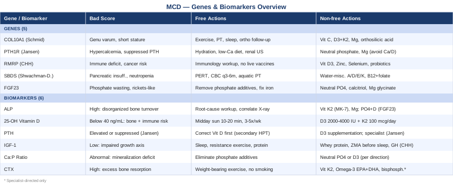
The 5 Genes That Define Metaphyseal Chondrodysplasia Subtypes
The genetic architecture of MCD is what separates informed management from guesswork. Each subtype has a distinct molecular cause, distinct metabolic vulnerabilities, and a distinct set of secondary complications to watch for. The five genes below account for the well-characterized MCD presentations most frequently encountered in genetic and metabolic bone clinics, covering a wide range of severity from manageable structural conditions to life-threatening immune deficiencies.
COL10A1 — The Schmid Type Blueprint
What this gene does: COL10A1 encodes type X collagen, a short-chain collagen expressed almost exclusively in hypertrophic chondrocytes at the growth plate. It forms a structural lattice in the zone where cartilage is being converted to bone and appears to coordinate the vascular invasion and mineral deposition that allow orderly new bone formation. It is expressed transiently during growth and is essentially absent from mature bone. MedlinePlus: COL10A1
What goes wrong: Heterozygous mutations in COL10A1 — either loss-of-function or dominant-negative variants — cause Schmid-type metaphyseal chondrodysplasia (MCDS), the most prevalent MCD subtype. The growth plate becomes disorganized; on X-ray, the metaphyses of long bones appear cupped, irregular, and sclerotic. Clinically, this produces short stature (typically 140–160 cm in adults), bowed legs (genu varum), coxa vara, and a characteristic waddling gait appearing in the second year of life. Intelligence and internal organs are unaffected, and the condition is generally non-progressive after skeletal maturity.
Mechanistic insight: Research in mouse models has established that misfolded COL10A1 protein accumulates in the endoplasmic reticulum of hypertrophic chondrocytes, triggering the unfolded protein response (UPR). This cellular stress cascade may amplify growth plate dysfunction beyond the simple structural absence of normal collagen — creating a low-grade inflammatory microenvironment within the growth plate itself that compounds the structural defect.
If the gene is disrupted — the plan without supplements
The underlying genetic deficit cannot be reversed through lifestyle alone, but the metabolic environment around bone formation can be meaningfully optimized:
- Low-impact, weight-bearing exercise, 20–30 minutes daily: Walking, swimming, or cycling activates osteoblasts through mechanotransduction, encouraging bone mineral density at metaphyseal sites without the high-impact stress that compromised growth plates cannot absorb. Avoid running on hard surfaces and jumping activities, particularly in childhood. - Physical therapy for gait correction, 2–3 sessions per week during childhood: Compensatory gait patterns in MCDS load hips and knees asymmetrically, accelerating secondary arthritis. Gait analysis followed by targeted hip abductor and quadriceps strengthening addresses this before deformity becomes fixed. Transitions to home maintenance in adulthood. - Annual orthopedic surveillance: Yearly AP pelvis X-ray in childhood to track coxa vara progression. Early identification allows guided growth procedures (hemiepiphysiodesis or corrective osteotomy) before irreversible deformity develops. - Sleep optimization: Growth hormone secretion is tightly coupled to slow-wave sleep. Consistent sleep schedules (8–10 hours in children, 7–9 in adults), dark rooms, and minimized screen light after sunset maximize endogenous GH pulsatility — directly supporting IGF-1-mediated growth plate activity even in a structurally compromised plate.
If the gene is disrupted — the plan with supplements
- Vitamin C (ascorbic acid), 250–500 mg daily: Vitamin C is essential for prolyl and lysyl hydroxylation, the post-translational modifications that give collagen its structural stability. Adequate Vitamin C supports functional COL10A1 output from the intact allele in this autosomal dominant condition. Side effect: doses above 1000 mg/day cause osmotic diarrhea; no cycling required at these lower doses. - Vitamin D3 + K2, 1000–2000 IU D3 with 100 mcg MK-7 daily: D3 drives calcium absorption; K2 (MK-7 form for its longer half-life) activates osteocalcin and ensures calcium deposits in bone rather than soft tissue. Monitor 25-OH Vitamin D; target 40–60 ng/mL. Recheck every 6 months. Side effects minimal at these doses. - Magnesium glycinate, 200–400 mg daily (evening): Cofactor for alkaline phosphatase, the primary enzyme driving bone mineralization, and required for Vitamin D activation in the liver and kidney. Glycinate form is better tolerated than oxide. Minimal side effects; no cycling required. - Bioavailable silicon (orthosilicic acid), 10–25 mg daily: Silicon stimulates collagen type I synthesis and bone mineralization in several human studies. Large RCTs are still lacking, but the mechanism is directly relevant for a COL10A1 context. Cycle 3 months on, 1 month off as a precautionary practice given limited long-term data.
PTH1R — The Jansen Type Paradox
What this gene does: PTH1R encodes the shared receptor for both parathyroid hormone (PTH) and PTH-related peptide (PTHrP). At the growth plate, PTHrP signaling through this receptor acts as a molecular brake on chondrocyte differentiation — preventing hypertrophic chondrocytes from maturing too quickly. This keeps the growth plate populated with proliferating chondrocytes capable of continued expansion. MedlinePlus: PTH1R
What goes wrong: Unlike most MCD genes, PTH1R mutations in Jansen-type MCD are activating (gain-of-function) — the receptor fires constitutively without needing its ligand. The result is severe metaphyseal dysplasia, profound hypercalcemia with paradoxically suppressed PTH, hypophosphatemia, and marked short stature. Jansen MCD is extraordinarily rare (fewer than 30 confirmed cases documented worldwide), but its biochemical signature is unmistakable when present and requires entirely different management logic from other MCD subtypes.
Clinical caution: The constitutive calcium-mobilizing effect of the hyperactive receptor causes nephrocalcinosis in most affected individuals — a serious complication requiring lifelong renal monitoring.
If the gene is disrupted — the plan without supplements
Management focuses entirely on controlling the downstream metabolic consequences of constitutive receptor activation:
- High fluid intake, 30–40 ml/kg/day: Sustained hydration promotes renal calcium clearance and reduces nephrocalcinosis and nephrolithiasis risk. - Dietary calcium calibration: Not elimination, but moderation. Avoid supplemental calcium and excessive dairy. Collaborate with a pediatric dietitian to keep dietary calcium within normal range without excess. - Annual renal ultrasound: Monitoring for nephrocalcinosis is non-negotiable throughout life given chronic hypercalciuria. - Bisphosphonate therapy (specialist-directed only): Intravenous pamidronate or zoledronic acid may be used in severe cases to reduce pathological bone resorption. This is a specialist-managed decision, not a self-directed intervention.
If the gene is disrupted — the plan with supplements
The supplementation logic here is about avoidance and cautious phosphate support, not augmentation:
- Avoid standard calcium supplements and high-dose Vitamin D: Both worsen hypercalcemia in Jansen MCD. Standard multivitamins containing calcium should be reviewed and often discontinued. Active Vitamin D analogs require specialist oversight in this context. - Neutral phosphate (specialist-guided), 500–1000 mg elemental phosphorus in divided doses: Hypophosphatemia is common and requires targeted phosphate support. Common side effect is osmotic diarrhea; titrate slowly over weeks. - Magnesium, 150–200 mg daily: Supports renal function and partially counterbalances calcium excess. Monitor levels alongside calcium.
RMRP — Cartilage-Hair Hypoplasia and the Immune Complexity
What this gene does: RMRP is unusual in that it encodes not a protein but a non-coding RNA — the RNA component of the mitochondrial RNA processing endoribonuclease (RNase MRP) complex. This ribonucleoprotein complex participates in ribosomal RNA processing, cell cycle regulation through cyclin B2 mRNA cleavage, and mitochondrial DNA replication initiation. Its role is therefore fundamental to cell division itself — which is why its disruption affects so many rapidly dividing cell populations simultaneously. MedlinePlus: Cartilage-Hair Hypoplasia
What goes wrong: Biallelic mutations in RMRP (autosomal recessive) cause cartilage-hair hypoplasia (CHH), also known as McKusick-type MCD. This is one of the most phenotypically complex MCD subtypes, combining metaphyseal dysplasia, fine sparse hair, combined immunodeficiency in approximately 80% of patients, macrocytic anemia, and a markedly elevated lifetime risk of non-Hodgkin lymphoma and skin cancers. The immune deficiency ranges from mild antibody insufficiency to severe combined immune deficiency (SCID) and is the primary driver of early mortality in CHH.
If the gene is disrupted — the plan without supplements
- Comprehensive immunological workup at diagnosis: Complete lymphocyte subsets (CD3, CD4, CD8, CD19, NK cells), immunoglobulin levels, and vaccine response titers. This defines the immune deficiency tier and is the foundation of all clinical management decisions. - Avoid live vaccines in CID-confirmed cases: BCG, live-attenuated influenza, MMR, and varicella can cause disseminated infection in immunodeficient CHH patients. Specialist review of every vaccine decision is mandatory. - Hematopoietic stem cell transplantation (HSCT) referral for severe CID: HSCT can restore immune function and dramatically improves survival for severe cases. Outcomes are best when performed before major infections occur — early specialist referral is strongly recommended. - Cancer surveillance, annual: CBC to screen for early lymphoma signs; annual dermatology review for basal cell carcinoma given elevated UV sensitivity in this population.
If the gene is disrupted — the plan with supplements
- Vitamin D3, 1000–2000 IU daily: Vitamin D has direct immunomodulatory effects — it shapes T-cell development and innate immune responses — beyond its skeletal role. Low Vitamin D measurably worsens immune dysfunction. Target 40–60 ng/mL; monitor every 6 months. Side effects minimal at these doses. - Zinc, 15–30 mg daily: Critical for T-cell development, thymic function, and NK cell activity — all impaired in CHH. Cycle 4–6 weeks on, 1 week off to prevent secondary copper depletion. Take with food to minimize nausea. - Selenium, 100–200 mcg daily: Supports NK cell function and provides antioxidant protection relevant to the elevated cancer risk in CHH. Do not exceed 400 mcg/day. Cycle 3 months on, 1 month off as standard precaution. - Multi-strain probiotics, 10–50 billion CFU daily: CHH immune deficiency affects gut-associated lymphoid tissue, which constitutes the majority of the body's adaptive immune activity. A well-formulated multi-strain probiotic (Lactobacillus, Bifidobacterium, Streptococcus thermophilus) supports mucosal immunity with a very low risk profile. Daily; no cycling required.
SBDS — Shwachman-Diamond and the Skeletal Overlap
What this gene does: SBDS encodes the Shwachman-Bodian-Diamond syndrome protein, which is required for ribosome biogenesis — specifically late-stage maturation and release of the 60S ribosomal subunit from the nucleolus. Cells that divide rapidly are disproportionately affected by ribosome assembly defects: bone marrow progenitors, pancreatic acinar cells, and growth plate chondrocytes are the primary casualties. MedlinePlus: SBDS
What goes wrong: Biallelic SBDS mutations cause Shwachman-Diamond syndrome (SDS), with a clinical triad of exocrine pancreatic insufficiency, neutropenia, and skeletal abnormalities. Metaphyseal changes — particularly hip metaphyseal irregularity — occur in approximately 50% of cases. Hematological complications dominate clinical urgency: neutropenia drives recurrent infections, and the risk of myelodysplastic syndrome or acute myeloid leukemia is substantially elevated across the lifespan.
If the gene is disrupted — the plan without supplements
- Pancreatic enzyme replacement therapy (PERT): Exocrine insufficiency causes fat malabsorption that depletes all fat-soluble vitamins — A, D, E, and K — essential for bone health. PERT is the non-optional foundation of nutritional management in SDS. Doses are titrated by gastroenterology. - Regular CBC with differential, every 3–6 months: Neutropenia monitoring; G-CSF (filgrastim) may be prescribed for severe neutropenia. - Annual bone marrow aspirate: Surveillance for MDS or AML is a lifelong clinical commitment given elevated leukemic transformation risk. - Aquatic physiotherapy for hip mechanics: Hip abductor strengthening in a low-impact medium minimizes load through abnormal metaphyseal architecture while building the functional muscle support that compensates for structural deficit.
If the gene is disrupted — the plan with supplements
- Fat-soluble vitamins in water-miscible forms (A, D, E, K): Standard fat-soluble vitamin preparations are poorly absorbed in exocrine insufficiency. Water-miscible formulations significantly improve bioavailability. Dose each based on measured serum levels; do not supplement blindly. - Vitamin D3, 1000–4000 IU daily (malabsorption-adjusted): Higher doses are often required because of malabsorption. Monitor 25-OH Vitamin D every 3–6 months to titrate; target 40–60 ng/mL. - Methylcobalamin (B12, 1000 mcg sublingual) and methylfolate (400–1000 mcg) daily: Malabsorption and neutropenia-driven immune stress both impair B vitamin status. Methylated forms bypass potential conversion inefficiencies. Daily; no cycling required.
FGF23 — The Phosphate Governor
What this gene does: FGF23 encodes fibroblast growth factor 23, a phosphaturic hormone secreted by osteocytes that reduces phosphate reabsorption in the kidney and simultaneously suppresses the 1-alpha-hydroxylase enzyme that activates Vitamin D. FGF23 is the primary negative feedback signal in the phosphate homeostasis axis — it rises in response to elevated phosphate and iron, reducing both phosphate absorption and active Vitamin D production. MedlinePlus: FGF23
What goes wrong: Excess FGF23 activity — from gain-of-function FGF23 mutations or from secondary elevating conditions — causes chronic phosphate wasting combined with impaired Vitamin D activation. The resulting hypophosphatemia and skeletal undermineralization generate metaphyseal changes on X-ray that closely resemble structural MCD, making FGF23-related conditions (X-linked hypophosphatemia, autosomal dominant hypophosphatemic rickets) critically important to distinguish from the structural subtypes. Notably, iron deficiency independently elevates FGF23 through impaired proteolytic cleavage — a connection that is frequently missed in standard workups.
If the FGF23 pathway is disrupted — the plan without supplements
- Eliminate phosphate additives from diet: Inorganic phosphate additives in processed foods (phosphoric acid, sodium phosphate, polyphosphates) are nearly 100% bioavailable and directly raise FGF23. Whole-food phosphate sources have lower net absorption and are far less disruptive. This single dietary change can meaningfully shift the phosphate load within weeks. - Correct iron deficiency if present: Iron deficiency elevates FGF23 through impaired FGF23 cleavage. Correcting iron status can lower FGF23 and partially restore phosphate reabsorption in iron-deficient individuals — this is frequently overlooked. - Burosumab (specialist-directed): The anti-FGF23 monoclonal antibody has become first-line therapy for confirmed FGF23-excess conditions. Subcutaneous injection every 2–4 weeks; initiated and monitored by metabolic bone or endocrinology specialists. It has fundamentally transformed outcomes in X-linked hypophosphatemia.
If the FGF23 pathway is disrupted — the plan with supplements
- Oral neutral phosphate (if burosumab is not indicated), 20–40 mg/kg/day in 4–5 divided doses: Correcting the phosphate deficit reduces the compensatory FGF23 elevation. Common side effect: osmotic diarrhea; titrate slowly over 2–3 weeks. Never co-administer with calcium supplements (calcium-phosphate precipitation reduces absorption of both). - Calcitriol (active Vitamin D) or alfacalcidol, 0.25–1 mcg daily (specialist-directed): Because FGF23 blocks 1-alpha-hydroxylase, standard Vitamin D3 supplementation is insufficient — the activated form must be used. Monitor serum calcium closely; hypercalcemia is the primary risk without concurrent phosphate replacement. - Magnesium glycinate, 200–400 mg daily: Required as a cofactor in phosphate metabolism pathways and frequently depleted in phosphate wasting conditions. Glycinate form preferred for tolerability. No cycling required.
Knowing which gene is implicated tells you why the bone-forming machinery is compromised. But genes are static — they do not tell you how well the body is compensating right now, whether an intervention is working, or where secondary complications are quietly developing. That is precisely what biomarker tracking addresses.
6 Biomarkers to Track Alongside Your Genetic Diagnosis
These six markers form a practical surveillance panel for MCD — covering bone turnover, mineral homeostasis, growth axis function, and long-term structural risk. Some are available on any basic metabolic panel; others require a specific order. All deliver meaningful clinical information when interpreted against the backdrop of the specific MCD subtype.
Biomarker 1: Alkaline Phosphatase (ALP)
Why it matters: ALP is the most universally available marker of bone metabolism. Osteoblasts release ALP as they work to mineralize new bone matrix; elevated levels reflect either active, healthy bone formation or — in the wrong clinical context — abnormal bone turnover. In MCD, ALP elevations may indicate accelerated but disorganized mineralization at metaphyseal sites. In Shwachman-Diamond syndrome, a liver-derived ALP contribution may confound interpretation, making bone-specific ALP the more informative marker in that subtype.
How to measure it
Part of any comprehensive metabolic panel. Cost: $15–50 within a standard panel. Bone-specific ALP (bALP) isolates the bone-derived fraction and is more informative when liver disease may confound total ALP — particularly relevant in SDS. bALP costs approximately $80–150 as a standalone test. Draw fasting, morning, for consistency across serial measurements.
If the score is bad — the plan without supplements
An unexpectedly elevated ALP demands root-cause thinking before intervention: has the MCD subtype been genetically confirmed? Is there an active fracture, accelerating bone turnover, or a secondary condition? Correlate with X-ray findings and the full mineral panel before modifying management. An elevated bALP in the context of low phosphate in an FGF23 case is an entirely different problem from an elevated total ALP with normal bALP in a Shwachman-Diamond patient.
If the score is bad — the plan with supplements or equipment
When elevated ALP reflects deficient mineralization despite active osteoblast activity (as in FGF23-related phosphate wasting), the goal is to restore the mineral substrates: phosphate replacement and active Vitamin D as outlined in the FGF23 section. When ALP is low alongside undermineralization, Vitamin K2 (MK-7, 100–200 mcg daily) enhances osteocalcin carboxylation and directly stimulates osteoblast matrix production. Magnesium (200–400 mg daily) is a cofactor for the ALP enzyme itself and should be assessed alongside bone ALP in any ALP workup.
Biomarker 2: 25-OH Vitamin D
Why it matters: Despite the vitamin label, 25-OH Vitamin D functions as a steroid hormone governing calcium absorption, modulating both innate and adaptive immunity, and directly regulating gene expression relevant to bone metabolism and cell cycle control — all implicated in multiple MCD subtypes. Peter Attia consistently highlights 25-OH Vitamin D as one of the most correctable and clinically impactful variables in metabolic bone disease; insufficiency is ubiquitous in modern populations and measurably worsens outcomes across skeletal and immune conditions simultaneously.
How to measure it
Serum 25-hydroxyvitamin D via standard blood draw. Cost: $30–80 standalone; frequently included in broader functional panels. Optimal target for metabolic bone and immune support: 40–60 ng/mL. Below 30 ng/mL is insufficient; below 20 ng/mL is deficient by any meaningful clinical standard. Test every 6 months when supplementing. Special note for Jansen MCD: supplementing Vitamin D with constitutively active PTH1R signaling requires specialist oversight — standard protocols do not apply and can worsen hypercalcemia.
If the score is bad — the plan without supplements
10–20 minutes of midday sun exposure on arms and legs, without sunscreen, three to five times per week during summer months at latitudes below 35°N. Above this latitude in winter, sun-based production approaches zero. For CHH patients with elevated skin cancer risk, shorter, more frequent exposures with post-exposure sun protection represent a reasonable balance between Vitamin D synthesis and photoprotection.
If the score is bad — the plan with supplements or equipment
Vitamin D3 (cholecalciferol) is the preferred supplemental form. Adults: 2000–4000 IU daily to correct deficiency; maintenance at 1000–2000 IU once in optimal range. Children: 1000–2000 IU daily, weight-adjusted. Always pair with Vitamin K2 (MK-7, 100 mcg) to reduce the theoretical risk of soft tissue calcium deposition as levels rise. Recheck at 8–12 weeks to confirm response. Side effects at these doses are minimal; toxicity above 150 ng/mL is rare below 10,000 IU daily.
Biomarker 3: Parathyroid Hormone (PTH)
Why it matters: PTH sits at the nexus of calcium, phosphate, and Vitamin D regulation. In MCD, monitoring PTH serves two distinct purposes: detecting secondary hyperparathyroidism (elevated PTH in response to low calcium or Vitamin D — common and highly correctable) and identifying the Jansen MCD pattern (paradoxically suppressed PTH with hypercalcemia — a diagnostic red flag). Thomas Dayspring emphasizes that PTH is always a contextual marker — interpreting it without simultaneous calcium and phosphate produces misleading conclusions.
How to measure it
Intact PTH (iPTH) via morning fasting blood draw. Cost: $50–100. Always order simultaneously with calcium and phosphate. Normal range approximately 10–65 pg/mL, though laboratory reference ranges vary. Elevated PTH with low calcium signals secondary hyperparathyroidism; elevated PTH with high calcium signals primary hyperparathyroidism; suppressed PTH with high calcium in MCD context is the Jansen alarm pattern.
If the score is bad — the plan without supplements
If PTH is elevated with low or normal calcium and low Vitamin D: correct Vitamin D status first — this resolves secondary hyperparathyroidism in the majority of cases within 8–12 weeks without any other intervention. If PTH is suppressed with hypercalcemia in a Jansen MCD context: specialist referral is immediate and mandatory — this is not self-manageable.
If the score is bad — the plan with supplements or equipment
For secondary hyperparathyroidism from Vitamin D insufficiency: D3 supplementation as described in the previous biomarker section, with a recheck of both 25-OH Vitamin D and PTH at 8–12 weeks. For PTH elevation in phosphate replacement therapy contexts (particularly FGF23-related disease): ensure concurrent calcium and Vitamin D status are adequate, as phosphate supplementation without Vitamin D optimization can worsen PTH-driven calcium mobilization from bone.
Biomarker 4: IGF-1 (Insulin-like Growth Factor 1)
Why it matters: IGF-1 is the primary effector of growth hormone action at the growth plate — it drives chondrocyte proliferation and differentiation, the exact process disrupted by MCD gene mutations. Beyond its growth-plate role, IGF-1 is a reliable proxy for overall anabolic status, nutritional adequacy, and sleep quality. A depressed IGF-1 in an MCD patient may indicate a compounding GH axis insufficiency layered on top of the structural genetic problem — a treatable secondary factor that clinicians may overlook when focused on the primary diagnosis. Peter Attia considers IGF-1 one of the most actionable yet underused panels in precision health monitoring.
How to measure it
Serum IGF-1 via standard blood draw. Cost: $50–120. Critical: always interpret against age- and sex-specific reference ranges — IGF-1 normally peaks in mid-puberty and declines progressively with age; raw numbers without this context are meaningless. Some laboratories report as a standard deviation score (SDS), which is more clinically useful for tracking individual progress in pediatric MCD patients over time.
If the score is bad — the plan without supplements
- Optimize sleep architecture: GH — and therefore IGF-1 — is driven primarily by slow-wave sleep. Consistent bedtimes, a cool dark room, no screens in the 60 minutes before bed, and stable wake times reliably improve GH pulse amplitude. This is one of the most impactful free interventions available for anyone with bone or growth concerns. - Progressive resistance exercise, 3 sessions per week minimum: Even low-impact resistance training generates local mechanical strain that drives IGF-1 expression directly in bone and muscle, independent of systemic GH levels. The exercise stimulus must be progressive over time to remain effective. - Protein adequacy, 1.2–1.6 g/kg/day: IGF-1 production is strongly protein-dependent — a relationship well established in nutrition research. Focus on complete amino acid profiles from animal sources or well-combined plant proteins.
If the score is bad — the plan with supplements or equipment
- Whey protein concentrate, 20–30 g post-exercise: The most direct dietary lever for IGF-1; whey has a particularly strong IGF-1-stimulating effect compared with other protein sources in head-to-head human studies. - Zinc and magnesium (ZMA formulation) before sleep, 25 mg zinc + 400 mg magnesium: Both minerals support GH secretion and pituitary function. Take on an empty stomach 30 minutes before bed. Cycle 8 weeks on, 2 weeks off to prevent copper competition with zinc. - Growth hormone therapy (endocrinology-directed): In cartilage-hair hypoplasia specifically, GH deficiency is documented in a meaningful subset of patients, and GH therapy has shown moderate benefit for height velocity in published pediatric studies. Requires formal GH stimulation testing and specialist management.
Biomarker 5: Calcium-to-Phosphorus Ratio
Why it matters: Bone mineral (hydroxyapatite) requires both calcium and phosphorus in approximately a 2:1 weight ratio for proper crystal structure. The serum calcium-to-phosphorus ratio (using mg/dL units) reflects the real-time balance of these minerals across intestinal absorption, renal handling, and bone deposition. An abnormal Ca:P ratio is one of the earliest detectable metabolic signals in both rickets and MCD-associated mineralization defects. Tracking this ratio over time is more sensitive than either mineral value alone. The ideal serum Ca:P ratio is approximately 2.2–2.7.
How to measure it
Both calcium and phosphate are included in any basic or comprehensive metabolic panel. Cost: $15–50 within a standard panel. Calculate the ratio from the reported values — most labs do not report it automatically. Always measure fasting; post-meal phosphate rises transiently and can falsely normalize a low ratio.
If the score is bad — the plan without supplements
If the ratio is low (excess phosphate relative to calcium): audit dietary phosphate carefully. Phosphate additives in packaged and processed foods (labeled as phosphoric acid, sodium phosphate, calcium phosphate, pyrophosphate, polyphosphate) are nearly fully absorbed and are invisible in standard nutritional databases. Eliminating processed foods and phosphate-containing sodas often shifts the ratio meaningfully within two to four weeks — without any supplementation.
If the score is bad — the plan with supplements or equipment
If the ratio is high (low phosphate relative to calcium — as in FGF23 excess): neutral phosphate supplementation as outlined in the FGF23 gene section. If the ratio is low with adequate phosphate but low calcium: prioritize Vitamin D optimization before adding calcium supplements. Vitamin D3 supplementation will improve calcium absorption from existing dietary sources and avoids the risks associated with supplemental calcium in the absence of adequate Vitamin D — a combination that can increase arterial calcification risk.
Biomarker 6: CTX (C-terminal Telopeptide of Type I Collagen)
Why it matters: CTX is a bone resorption marker measuring fragments of type I collagen released when osteoclasts degrade aged bone matrix. While MCD is primarily a disease of abnormal bone formation, secondary excess resorption compounds the structural problem over time, driving lower bone mineral density than the genetic deficit alone would predict. Thomas Dayspring recommends pairing CTX with bone-specific ALP as a "bone balance" assessment: high bALP with low CTX indicates net bone formation; high CTX with low bALP indicates net resorption — the more clinically concerning pattern in a pre-existing structural bone disease.
How to measure it
Serum CTX (also called beta-CrossLaps or sCTX). Cost: $60–150. This marker has strict sampling requirements: must be drawn fasting, before 10 AM. Eating reduces CTX by 20–25%; caffeine also suppresses it. Reference ranges are age- and sex-specific; growing children and postmenopausal women have physiologically higher values that must be interpreted accordingly.
If the score is bad — the plan without supplements
Elevated CTX in a non-growing adult context indicates excess bone resorption. The single most effective non-pharmacological intervention is weight-bearing exercise — 30 minutes minimum, five times per week — which reduces osteoclast activity through mechanical signaling pathways. Smoking cessation and alcohol limitation both reduce CTX measurably: smoking activates osteoclasts through inflammatory cytokines; alcohol suppresses osteoblast function and increases resorption. Addressing both is among the highest-leverage free interventions.
If the score is bad — the plan with supplements or equipment
- Vitamin K2 (MK-7), 100–200 mcg daily: K2 directly reduces osteoclast activity and activates osteocalcin carboxylation. Multiple clinical studies and meta-analyses confirm that K2 supplementation reduces bone resorption markers in adults; the mechanism is directly relevant regardless of underlying bone disease etiology. Daily; no cycling required. - Omega-3 fatty acids (EPA + DHA combined), 2–4 g daily: Omega-3s reduce IL-6 and other osteoclast-activating inflammatory cytokines. Human intervention studies show moderate reductions in bone resorption markers. Take with the largest meal of the day to maximize absorption and minimize GI discomfort. No cycling required. - Bisphosphonates (specialist-directed): In pediatric MCD with documented excess bone resorption — particularly when compounding structural metaphyseal disease — pamidronate infusions have been used cautiously in the literature. Specialist-only given risk of atypical fracture and jaw osteonecrosis with prolonged use.
Translating genetic knowledge and biomarker tracking into daily practice is where most clinical guidance falls short. The following section distills key insights from the Huberman Lab's work on bone biology, growth factors, and metabolic optimization — findings that add practical depth to the interventions already outlined and challenge several assumptions in standard care.
Bone Biology Insights That Change How You Think About This Condition
The Huberman Lab — led by neuroscientist Andrew Huberman at Stanford — has distilled an extensive body of peer-reviewed research on physical performance, bone health, growth factor biology, and circadian neuroscience into actionable protocols. Drawing from specialists including Dr. Peter Attia, Dr. Andy Galpin, and endocrinology researchers, the following ten findings are particularly relevant to managing MCD and represent some of the most under-applied insights in bone health practice.
1. Sleep Is the Most Potent Growth Hormone Stimulus Available at No Cost
Growth hormone is not secreted continuously — the largest pulse occurs approximately 90 minutes after sleep onset, during the first slow-wave sleep cycle. Missing or fragmenting this window through late bedtimes, alcohol, or light exposure directly reduces the GH amplitude that drives overnight IGF-1 production. For MCD patients trying to maximize every available anabolic signal for bone maintenance, optimizing this single behavioral variable has no ceiling for improvement and no cost.
2. Resistance Exercise Drives IGF-1 Locally in the Bone Being Loaded
Progressive resistance exercise generates mechanical strain that drives IGF-1 expression locally in the loaded bone and muscle tissue, independent of systemic GH levels. This local IGF-1 stimulates chondrocyte and osteoblast activity specifically in exercising limbs — meaning targeted exercise can create a bone-building signal precisely where it is most structurally needed in an asymmetric condition like MCD.
3. Morning Sunlight Sets the Circadian Clock That Times Hormone Pulsatility
Huberman's work on circadian biology establishes that morning sunlight exposure (within the first hour after waking, even on cloudy days) sets the master pacemaker that governs GH pulsatility timing, cortisol rhythms, and sleep architecture that night. Consistent morning light — even 10 minutes — has measurable downstream effects on the hormonal environment relevant to bone remodeling.
4. Cold Water Immersion After Exercise Blunts the Bone Adaptation Response
Immediate post-exercise cold water immersion — a popular recovery practice — suppresses the inflammatory signaling cascade that drives adaptation in bone and muscle. For MCD patients trying to maximize the osteogenic response to exercise, cold exposure is best delayed by at least four hours after a session or reserved entirely for non-training days. This finding challenges the reflexive cold-plunge-after-every-workout habit in athletic communities.
5. Repeated Sauna Use Transiently Elevates GH by 200–300%
Finnish research cited extensively by Huberman shows that sauna use (80–100°C, 20 minutes, 3–4 sessions per week) transiently elevates GH by 200–300%, mediated through thermal and dehydration-triggered signaling. Combined with adequate rehydration, this protocol represents an accessible GH-augmenting practice for adolescents and adults with MCD who cannot access pharmaceutical GH therapy.
6. Protein Distribution Across Meals Sustains IGF-1 Through the Day
Distributing protein across three to four meals rather than concentrating it in one or two maintains elevated circulating amino acids that sustain muscle protein synthesis and IGF-1 production over a larger fraction of the day. For bone-building applications, the sustained anabolic signal is particularly relevant for conditions where the bone-forming machinery is already compromised at the structural level.
7. Vitamin D Deficiency Undermines Every Bone-Building Intervention
Huberman frames Vitamin D not as a supplement but as a prerequisite. Virtually every bone-building signal — calcium absorption efficiency, PTH regulation, osteoblast differentiation, immune modulation, IGF-1 receptor sensitivity — operates measurably less efficiently when 25-OH Vitamin D is below 40 ng/mL. Correcting Vitamin D status before layering in other interventions is the first optimization step, not the last or optional one.
8. Zone 2 Aerobic Exercise Improves Mitochondrial Function in Cartilage
Low-intensity aerobic exercise maintained at the highest pace at which conversation is still possible (Zone 2) drives mitochondrial biogenesis in all tissues including articular cartilage. This is directly relevant for CHH patients, where RMRP mutations impair the mitochondrial RNA processing complex. Zone 2 cardio at 30–45 minutes, four times per week, improves the metabolic environment of cartilage at a level independent of the genetic defect.
9. Chronic Stress and Cortisol Elevation Directly Suppress Bone Formation
Chronically elevated cortisol — even at sub-clinical levels — is one of the strongest suppressors of osteoblast activity available outside pharmacology. Huberman cites multiple human studies showing that stress management and cortisol normalization improve bone formation markers. For families navigating a complex rare disease — with its diagnostic odysseys, surgical decisions, and daily management burden — this is a genuine bone-biology argument for stress reduction, not just quality-of-life advice.
10. Omega-3s Reduce the Inflammatory Brake on Bone-Forming Cells
EPA and DHA consistently rank among the most evidence-backed anti-inflammatory interventions in Huberman's coverage of the literature. For MCD specifically, reducing IL-6 and TNF-alpha — both elevated in the UPR-mediated growth plate inflammation of Schmid type and the immune activation of CHH — removes an active brake on osteoblast function. Omega-3s address an inflammation component that standard orthopedic management does not target.
Beyond what can be measured in blood and trained in a gym, there are several evidence-backed modalities that address the functional gaps most relevant to MCD — balance and gait stability, tissue energy metabolism, and immune regulation in CHH. The following three approaches have clinical evidence with human data supporting their use.
Three Evidence-Backed Complementary Approaches
Low-Level Laser Therapy for Bone and Cartilage Tissue
What it is and why it may be relevant: Low-level laser therapy (LLLT) — also known as photobiomodulation — uses near-infrared and red light wavelengths (typically 600–1000 nm) at non-thermal intensities to increase cellular ATP production via cytochrome c oxidase in the mitochondrial electron transport chain. This mitochondrial stimulation enhances osteoblast, chondrocyte, and fibroblast metabolic activity — the precise cell types whose function is disrupted in MCD. In bone and cartilage tissue, photobiomodulation has shown the ability to increase collagen synthesis, reduce pro-inflammatory cytokines, and accelerate mineralization in multiple in vitro studies and high-quality animal models, with emerging human evidence. The mechanism aligns particularly well with the UPR-driven chondrocyte stress in Schmid type and the mitochondrial processing dysfunction in CHH.
Supporting evidence: A 2017 systematic review and meta-analysis in Lasers in Medical Science (Ferrão Júnior and colleagues) examined photobiomodulation on bone defect healing and found significant improvements in ALP activity, osteocalcin expression, and collagen density across multiple human and high-quality animal studies. While MCD-specific RCTs do not yet exist, the mechanistic overlap is sufficiently strong to make this a plausible adjunct. Evidence for this specific condition is preliminary — it should be framed as supportive, not primary treatment.
How to apply it: For home use, devices in the 660–850 nm range at 10–50 mW/cm² irradiance applied directly over metaphyseal joint areas (hips, knees) for 10–20 minutes per session, three to five times per week. Professional treatment in a physiotherapy or sports medicine setting offers higher-power devices with more precise dosing. Begin with three sessions and assess local tissue response. An important caution: direct application over actively growing growth plates in young children should be discussed with a specialist before proceeding, given the theoretical concern about influencing growth plate biology.
Tai Chi for Balance and Fall Prevention in Skeletal Dysplasia
What it is and why it may be relevant: Tai chi is a Chinese movement practice combining slow, flowing weight shifts, balance challenges, and proprioceptive loading into a continuous sequence that can be scaled to virtually any mobility level. For individuals with MCD, where shortened limb proportions, altered hip geometry, and gait deviations shift the center of gravity and compromise dynamic balance, the proprioceptive and postural demands of tai chi have direct functional relevance. Falls in MCD carry higher mechanical consequences than average, given pre-existing bone quality issues at metaphyseal sites — and the risk increases with age as compensatory muscle strength declines.
Supporting evidence: A 2016 Cochrane systematic review of tai chi for fall prevention (Sherrington and colleagues) found significant reductions in fall rate and fall risk across multiple populations with balance impairments, supported by dozens of randomized controlled trials. A 2019 review in Osteoporosis International found tai chi specifically beneficial for bone mineral density maintenance in populations with metabolic bone disease, attributing this partly to sustained low-impact mechanical loading and partly to reduced fall-associated fracture incidence. The proprioceptive mechanisms are not population-specific and apply broadly across skeletal conditions.
How to apply it: Yang-style tai chi is the most widely studied and accessible form, with established adaptive versions for people with mobility limitations. Starting with a certified instructor for the first four to six weeks ensures correct body mechanics before independent practice. Target 20–30 minutes, three times per week — the minimum effective dose from RCT evidence. For MCD patients with significant short stature, posture modifications are usually straightforward; seated tai chi programs exist for more severely affected individuals and provide equivalent proprioceptive benefits without requiring full-standing balance.
Microbiome-Directed Therapy for CHH-Related Immune Dysfunction
What it is and why it may be relevant: Microbiome-directed therapies encompass dietary fiber optimization, targeted probiotic supplementation, and — in specific clinical contexts — fecal microbiota transplantation (FMT). In cartilage-hair hypoplasia, where approximately 80% of patients have some degree of combined immune deficiency driven by RMRP-related cell cycle disruption, the gut microbiome's role in training and sustaining immune function is directly relevant. Gut-associated lymphoid tissue (GALT) houses the largest concentration of immune cells in the body; the microbial composition of the gut directly shapes T-cell development, NK cell activity, and the balance between regulatory and effector immune responses — all impaired in CHH.
Supporting evidence: A 2022 meta-analysis in Frontiers in Immunology examining probiotic interventions in primary immunodeficiency conditions found consistent improvements in infection frequency, mucosal immunity markers, and inflammatory cytokine profiles with multi-strain probiotic supplementation. Direct CHH-specific RCTs are lacking, but the mechanistic pathway — microbiome → GALT activation → adaptive immune function — is well established in basic and clinical immunology. Dietary microbial diversity (targeting 30+ different plant species per week) is the most evidence-supported method for increasing microbiome diversity and GALT stimulation according to the American Gut Project and related large-scale studies.
How to apply it: A practical protocol for CHH: target 30 different plant species per week as a dietary diversity goal (this counts spices and herbs — it is more achievable than it sounds); take a multi-strain probiotic with 10–50 billion CFU containing Lactobacillus, Bifidobacterium, and Streptococcus thermophilus daily with or after a meal; add prebiotic fiber from whole food sources (inulin from garlic and onion, pectin from apples and berries, resistant starch from cooled cooked potatoes and legumes). Dietary approaches require no cycling. Probiotic cycling (8 weeks on, 2 weeks off) may be used when cost is a factor, though continuous use is well-supported by the available evidence. Fecal microbiota transplantation is not a home-practice intervention — discuss it with a specialist only in the context of recurrent gut infections or severe documented dysbiosis.
Conclusion
Metaphyseal chondrodysplasia does not yield to generic solutions. Knowing the diagnosis is the starting point, not the answer. Knowing which gene is involved, what metabolic complications are most likely, and which biomarkers to track regularly is what transforms a broad diagnosis into a manageable, measurable set of targets.
None of this replaces specialist care — for rare skeletal dysplasias, orthopedic, metabolic, and genetic expertise remains essential. What the genetic and biomarker framework in this article provides is the preparation to make those specialist relationships more productive: better questions, clearer benchmarks, and a more precise understanding of what your individual presentation actually requires rather than what the average textbook case needs.
The most useful next step is usually the simplest: confirm the genetic subtype if not yet done, order a baseline panel (comprehensive metabolic panel, 25-OH Vitamin D, PTH, IGF-1, and CTX), and bring the results to the next specialist appointment with a clear picture of where each marker falls relative to optimal ranges. Better information, gathered consistently, is the precondition for better decisions — and that is entirely within reach.
Musculoskeletal Endocrine & Metabolic
Musculoskeletal: Bone Conditions
Autoimmune: Inflammatory Conditions