This article contains AI generated content.
· UpdatedMevalonate Kinase Deficiency Genes and Biomarkers — 3 Genes And 7 Biomarkers To Track
Introduction
Living with mevalonate kinase deficiency means managing something that most physicians have never seen in practice. The periodic fevers that arrive without warning and then vanish, the swollen lymph nodes, the days of abdominal pain and exhaustion — and then, just as unexpectedly, an unsettling return to relative normalcy. Between episodes, things can look almost normal on paper, which makes the condition easy for clinicians to underestimate and nearly impossible for patients to explain. The standard advice about managing rare autoinflammatory conditions is often too generic to be genuinely useful when the biology is this specific.
Part of the difficulty is that MKD occupies an unusual space between metabolic disease and autoinflammatory disease. It is caused by a single gene defect — mutations in the MVK gene — but its consequences ripple across the mevalonate pathway, the cholesterol synthesis network, and the IL-1 inflammatory axis. Generic inflammation management protocols built for more common conditions miss the specific enzymatic failure at the center of MKD. What works for rheumatoid arthritis or lupus may be irrelevant or counterproductive here.
This article does not offer a cure. But it offers something more useful for the daily decisions that matter: a precise look at the biomarkers worth tracking and the genetic variants that most directly affect disease severity, and the evidence-grounded approaches that have helped patients improve quality of life. Not every answer exists yet for this rare condition, but the research that does exist is increasingly actionable.
Better measurement leads to better decisions. If you know which biomarkers peak during your attacks, when they normalize, and what specific genetic variants affect your particular expression of MKD, you can work far more precisely with your medical team. The first section of this article covers the seven most useful biomarkers for tracking MKD activity and treatment response. The second examines three key genes — including what specific variants mean for disease course and what can be done about each. From there, the article moves into the research insights reshaping how specialists approach MKD management, followed by complementary strategies with actual human evidence. Every section is written to be practical, not theoretical.
Summary
Most people diagnosed with mevalonate kinase deficiency are told to manage symptoms and wait for attacks to pass. What they are rarely shown is the specific combination of biomarkers and genetic markers that can predict attack severity, explain why two patients with the same mutation can have completely different disease trajectories, and guide treatment decisions with far more precision. This article maps seven measurable biomarkers — including urinary mevalonic acid, IgD, and IL-18, which can spike to 100 times the upper limit of normal during active episodes — against three key genes that determine not just whether you have MKD, but how severe it will be, how often it will flare, and which therapeutic lever is most likely to work for your specific biology. Beyond the biology, the research section covers findings that are quietly reshaping how MKD specialists think about long-term management — including why one paradoxical medication reduces attacks in some patients but not others, and why the gut-immune connection may be more relevant to your attack frequency than anyone has yet told you.
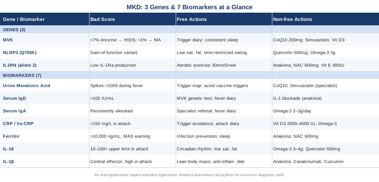
7 Biomarkers to Track in Mevalonate Kinase Deficiency
Biomarker tracking in MKD serves two distinct purposes: confirming disease activity during attacks, and detecting low-grade ongoing inflammation between episodes that may indicate long-term complication risk. The challenge is that many markers normalize between fever episodes, making timing critical. The most informative approach is to measure several of the biomarkers below both during and within a week after an attack, then again at stable baseline. That pattern — rather than any single abnormal result — tells the most complete diagnostic and monitoring story.
1. Urinary Mevalonic Acid
Why it matters: Urinary mevalonic acid is the most direct available biomarker for MKD. When mevalonate kinase activity is reduced, mevalonic acid accumulates and spills into the urine, particularly during fever episodes. In the severe form — mevalonic aciduria (MA) — excretion is massively elevated even between attacks, sometimes by a factor of 1000 or more. In HIDS (the milder phenotype), levels may be only mildly elevated at baseline but spike dramatically during fevers, providing a clear biochemical fingerprint of an active episode rather than an intercurrent infection.
How to measure it: This test is performed via urine organic acid analysis at specialized metabolic laboratories. A spot urine sample suffices; first-morning collection is preferred for consistency. Cost ranges from approximately $150 to $400 depending on the laboratory and whether it is bundled into a broader organic acids panel. The most diagnostically useful collection is obtained during or within 24 hours of peak fever, as levels approach normal between attacks in many HIDS patients and a normal result during a fever-free interval does not rule out MKD.
If the result is elevated — plan without supplements: Elevated urinary mevalonic acid during a documented fever episode establishes the biochemical basis for MKD diagnosis. The most effective non-pharmacological step that follows confirmation is systematic trigger mapping: maintaining a detailed diary that records attack onset, duration, symptom severity, and all exposures in the preceding five days (infections, vaccines, stress, surgery, dietary changes). Trigger avoidance — particularly around vaccinations, which are the most common pediatric trigger — is the highest-yield non-drug intervention for reducing attack frequency.
If the result is elevated — plan with supplements or medication: Persistently elevated mevalonic acid with frequent attacks is a primary indication for IL-1 blockade therapy under physician guidance. At the supplement level, CoQ10 (ubiquinol form, 200–300mg daily with a fatty meal) is sometimes considered because the mevalonate pathway normally produces coenzyme Q10 — a compromised pathway may produce less. Side effects are minimal and no cycling protocol is required. Simvastatin (20–40mg daily, prescription only) has been studied in small open-label trials as a way to paradoxically reduce substrate loading on the defective pathway; results are mixed but some patients show reduced attack frequency. Both must be discussed with a metabolic disease specialist before use.
2. Serum IgD
Why it matters: Elevated serum IgD was the original hallmark that gave HIDS its former name — Hyperimmunoglobulinemia D Syndrome. Values above 100 IU/mL (approximately 14 mg/L) are considered a classic diagnostic marker. However, IgD can be elevated in other inflammatory conditions and is normal in roughly 20–25% of genetically confirmed MKD cases, particularly in young children who have not yet mounted the cumulative immune response that drives IgD rise. It is a supporting marker and context-dependent, not a standalone diagnostic result.
How to measure it: Serum IgD is measured via a standard blood draw and immunoglobulin panel. It is available at most reference laboratories but is not always included in routine panels — a specific request is required. Cost: approximately $50–$150. Results are most informative when collected at stable baseline (not during or immediately after an attack) and when paired with IgA levels, as the combination carries greater diagnostic weight than either measure alone.
If the result is elevated — plan without supplements: A persistently elevated IgD in the setting of periodic fevers strengthens the clinical picture for MKD but does not require specific treatment on its own. The priority is confirming the diagnosis with MVK genetic testing and urinary mevalonic acid measurement. IgD levels tend to decrease gradually with age and with effective disease control, so serial measurements over years serve as an indirect treatment response indicator rather than an acute management tool.
If the result is elevated — plan with supplements or medication: IgD levels are not directly targeted by any current supplement. Effective IL-1 blockade — particularly with anakinra or canakinumab — typically produces gradual IgD normalization over months to years in treatment responders. Pediatric HIDS cohort data supports this trend. No supplement is established to reduce IgD specifically in MKD; the focus here belongs on attack frequency reduction rather than IgD as a direct target.
3. Serum IgA
Why it matters: IgA is elevated in the majority of HIDS patients, often alongside IgD but with greater consistency between attacks. Unlike IgD, which fluctuates more substantially with disease activity, IgA elevation tends to persist and provides a more stable indicator of ongoing immune dysregulation. It also helps distinguish HIDS from other periodic fever syndromes — particularly PFAPA (periodic fever, aphthous stomatitis, pharyngitis, adenitis) and FMF (familial Mediterranean fever) — where IgA elevation is less pronounced.
How to measure it: IgA is part of a standard immunoglobulin panel (IgG, IgA, IgM). When ordering, request IgD simultaneously, since the combination adds diagnostic precision. Cost: approximately $30–$80 when included in a standard panel. Serial measurement every six to twelve months provides a reasonable disease-monitoring baseline for patients under active management.
If the result is elevated — plan without supplements: Elevated IgA in the context of periodic fever warrants referral to a specialist in autoinflammatory diseases and should prompt MVK genetic testing if not yet performed. A systematic fever diary — recording attack timing, duration, temperature peak, and associated symptoms — is the most immediately actionable non-pharmacological step. No dietary interventions are specifically proven to normalize IgA in MKD.
If the result is elevated — plan with supplements or medication: IgA normalization is a downstream consequence of successful disease control, not a direct treatment target. Omega-3 fatty acids (EPA+DHA, 2–3g daily with meals) have well-documented general anti-inflammatory properties and may modestly reduce baseline inflammatory tone over time. Side effects include mild GI discomfort and a fish aftertaste; blood-thinning considerations apply at doses above 3g daily. No specific MKD evidence exists, but the risk-benefit profile at moderate doses is favorable.
4. C-Reactive Protein (CRP)
Why it matters: CRP is a sensitive acute-phase reactant that rises sharply within hours of MKD fever episode onset. While nonspecific — elevated in any significant infection or inflammatory flare — it is one of the most accessible tools for objectively documenting attack severity, establishing a personal attack CRP signature, and evaluating treatment response over time. High-sensitivity CRP (hs-CRP) adds precision for detecting low-grade inflammation between attacks that may indicate background disease activity not yet producing symptoms.
How to measure it: Standard CRP is inexpensive ($10–$40) and universally available. High-sensitivity CRP adds precision at lower ranges and costs approximately $20–$80. Both require a simple blood draw. The most informative testing strategy in MKD is a three-point measurement: during peak fever, within 24–48 hours of resolution, and at two to four weeks post-attack at stable baseline. This pattern establishes your personal attack-and-recovery curve, which is far more useful than isolated readings.
If the result is elevated during attacks — plan without supplements: CRP can exceed 150–300 mg/L during active MKD episodes — values that reflect systemic inflammation far beyond routine intercurrent illness. The most direct non-pharmacological response is refining trigger identification based on the attack diary. When CRP patterns correlate consistently with specific exposures (a particular vaccine series, school-year infection cycles), targeted avoidance planning becomes possible. Tracking CRP over time also creates the objective documentation that helps physicians justify escalation to biological therapy.
If the result is persistently elevated between attacks — plan with supplements: Baseline CRP above 5–10 mg/L between attacks signals ongoing low-grade inflammation and warrants medical review for amyloidosis risk. Vitamin D3 (2000–4000 IU daily, adjusted to maintain serum 25-OH vitamin D at 50–80 ng/mL) has modest evidence for CRP reduction in inflammatory conditions; annual monitoring of vitamin D levels is appropriate; no cycling required. Omega-3 fatty acids as described above provide additive modest benefit. Neither replaces escalation to biological therapy when CRP is consistently elevated between episodes.
5. Ferritin
Why it matters: Ferritin is an acute-phase protein that can reach extreme elevations during MKD attacks — sometimes exceeding 10,000 ng/mL in severe episodes. It reflects macrophage activation intensity and the degree of systemic inflammatory surge. Very high ferritin during an unusually prolonged attack is also a critical warning sign for macrophage activation syndrome (MAS), a rare but life-threatening MKD complication that requires urgent medical escalation. Between attacks, mildly elevated ferritin (above 200–300 ng/mL) may reflect residual inflammatory activity that warrants monitoring for secondary AA amyloidosis.
How to measure it: Serum ferritin is part of most standard metabolic or iron panels. Cost: approximately $20–$60. During an active attack, ferritin can rise dramatically within 24–48 hours. Any attack producing ferritin above 3000–5000 ng/mL that is not resolving within 7–10 days should prompt urgent physician consultation to rule out MAS. Measuring simultaneously with CRP and LDH adds important context.
If the result is elevated — plan without supplements: Persistently high ferritin above 500 ng/mL between attacks warrants urgent medical review to assess for amyloid protein A (SAA) accumulation and renal function. Non-pharmacological contributions include infection prevention (vaccines are paradoxically important despite being attack triggers — timing and prophylactic coverage matter), consistent sleep, and stress reduction, all of which reduce ferritin elevation from inflammatory triggers. None replaces medical management when ferritin is chronically elevated.
If the result is elevated — plan with supplements or medication: IL-1 blockade with anakinra (1–2 mg/kg per day, subcutaneous injection) is the most evidence-supported intervention for reducing ferritin during attacks and normalizing baseline levels over time. For patients not yet on biological therapy, N-acetyl cysteine (NAC, 600mg twice daily) has demonstrated ability to reduce oxidative inflammatory load in other conditions; evidence in MKD is absent but the safety profile is favorable. Avoid iron supplementation unless confirmed iron deficiency is present alongside elevated ferritin — the coexistence requires specific clinical interpretation.
6. Interleukin-18 (IL-18)
Why it matters: IL-18 has emerged as one of the most discriminating biomarkers in MKD. Multiple studies have documented that MKD patients carry markedly elevated IL-18 levels — often 10 to 100 times the upper limit of normal during attacks — values that exceed what is typically seen in other autoinflammatory syndromes including systemic juvenile idiopathic arthritis, PFAPA, and FMF. This extreme IL-18 elevation reflects the degree to which the NLRP3 inflammasome is activated when mevalonate kinase is deficient and geranylgeranyl pyrophosphate (GGPP) production falls. IL-18 testing may also help resolve diagnostically ambiguous cases where genetic results are uncertain.
How to measure it: IL-18 measurement requires a specialized immunology or research laboratory. It is not a standard clinical test at most hospitals. Cost: approximately $200–$600. Samples should be collected during active fever for maximum diagnostic yield and processed promptly due to the molecule's short stability in circulation. Some academic medical centers with autoinflammatory disease programs run this in-house; otherwise samples go to international reference labs.
If the result is markedly elevated — plan without supplements: Confirmed extreme IL-18 elevation points directly to inflammasome activation as the driver of disease. Non-pharmacological interventions that reduce NLRP3 inflammasome priming include: consistent circadian rhythm (circadian disruption independently upregulates NLRP3 transcription), reducing infectious exposures, and limiting dietary saturated fats and refined carbohydrates, which are known NLRP3 activators. These reduce the baseline from which attacks are launching, even if they cannot prevent all episodes.
If the result is markedly elevated — plan with supplements or medication: IL-18 itself is not yet directly targeted by approved biological therapies in MKD, but IL-1 blockade (which targets a parallel arm of the same inflammasome cascade) often produces secondary IL-18 reductions in treatment responders. Omega-3 fatty acids (EPA+DHA, 3–4g daily with meals) have demonstrated NLRP3 inflammasome-dampening effects in human cell research and some clinical data. Quercetin (500mg twice daily, 8 weeks on / 2 weeks off) inhibits NLRP3 in multiple human cell studies; human trial evidence for autoinflammatory conditions is preliminary; side effects are minimal. Discuss both with your treating immunologist before starting.
7. Interleukin-1β (IL-1β)
Why it matters: IL-1β is the central effector cytokine in MKD. Reduced geranylgeranyl pyrophosphate (GGPP) — a downstream product of the mevalonate pathway — triggers caspase-1 activation, which cleaves the IL-1β precursor into its biologically active form. This mechanism is why IL-1 inhibitors are the most effective treatment for MKD and why agents targeting other cytokines such as TNF or IL-6 are generally far less useful in this condition. Direct measurement of IL-1β during attacks can confirm pathway involvement and guide treatment escalation decisions.
How to measure it: Serum IL-1β can be measured via ELISA at specialized immunology laboratories. Reliability has important caveats: IL-1β has a very short serum half-life, levels fluctuate rapidly, and results vary by assay method. Plasma collected during peak fever and processed within one hour provides the most reliable readings. Cost: approximately $150–$400. This test is most valuable for establishing the diagnosis in atypical cases or confirming treatment response rather than as a routine monitoring tool.
If the result is elevated — plan without supplements: Confirmed IL-1β elevation provides the biological rationale for escalating to IL-1 inhibitor therapy. Before initiating biologics, non-pharmacological measures include reducing infectious exposures (the most common trigger for IL-1β surge in MKD), maintaining lean body mass (adipose tissue is an independent IL-1β source), and following a structured anti-inflammatory dietary pattern. These can modestly reduce baseline IL-1β tone between attacks without addressing the genetic root cause.
If the result is elevated — plan with supplements or medication: Anakinra (1–2 mg/kg/day, subcutaneous injection) is the most evidence-supported IL-1β blocker in MKD and is particularly effective as rescue therapy during attacks. Canakinumab (150–300mg subcutaneous every 8 weeks) provides longer-acting IL-1β blockade and is more practical for continuous prophylactic use in patients with frequent episodes. Both are prescription medications requiring specialist management. At the supplement level, magnesium glycinate (300–400mg nightly) has modest anti-inflammatory properties and is well-tolerated long-term; no cycling required. Curcumin (BCM-95 form, 500–1000mg twice daily with meals) has some human evidence for IL-1β modulation; avoid combining with anticoagulants or blood-thinning medications.
With these seven biomarkers mapped against one another — each measuring a different dimension of MKD activity — it becomes possible to track disease progression and treatment response with genuine precision. The pattern across biomarkers over time tells a richer story than any single number, and carrying this data into specialist appointments fundamentally changes the quality of the clinical conversation.
The Genetics Behind MKD — What 3 Key Genes Reveal
Understanding the genetics of MKD goes beyond confirming a diagnosis. It helps predict who will have more severe disease, explains why two patients with identical diagnoses can have dramatically different attack frequencies and treatment responses, and points toward specific biological levers that are most relevant for each individual. The three genes below form the core genetic framework of MKD: the causative mutation, the inflammasome amplifier, and the natural inflammatory brake.
Gene 1: MVK — The Root of the Problem
What the gene does: The MVK gene on chromosome 12q24.11 encodes mevalonate kinase, the enzyme that phosphorylates mevalonic acid into mevalonate-5-phosphate in the mevalonate pathway. This pathway produces cholesterol, coenzyme Q10, isoprenoids, and other molecules essential for cellular function. Pathogenic variants in MVK reduce enzyme activity, and the degree of residual activity determines clinical phenotype: below 1% of normal activity produces Mevalonic Aciduria (MA), the severe form with developmental delays and near-continuous inflammation; 1–7% residual activity produces HIDS, the milder periodic fever form. Confirmed variant information is catalogued in the NCBI Gene entry for MVK.
Key variants and what they mean: Over 200 pathogenic variants have been described. The most common — p.Val377Ile (V377I) — is present in approximately 85% of European HIDS patients, typically in compound heterozygosity with a second variant. V377I alone produces roughly 3–6% residual enzyme activity, placing homozygous V377I patients firmly in the HIDS range. The p.Ile268Thr (I268T) variant confers lower residual activity and is associated with more frequent and severe episodes. Null variants (nonsense, frameshift, or splice-site mutations) virtually eliminate enzyme function and are characteristic of MA. Knowing your specific genotype allows a reasonable prediction of which severity tier applies.
If the variants are moderate (V377I/V377I or similar) — plan without supplements: Systematic trigger identification and avoidance is the highest-yield non-pharmacological strategy. Work with your specialist to build a comprehensive trigger map. Develop a pre-treatment plan for unavoidable high-risk triggers (scheduled surgeries, vaccination series). Maintain consistent sleep and stable physiological routines between attacks, as circadian and metabolic stress lower the attack threshold. Moderate aerobic exercise 3–5 times per week is generally beneficial between attacks and reduces baseline inflammatory tone; avoid vigorous exertion during prodromes.
If the variants are moderate — plan with supplements or medication: CoQ10 in ubiquinol form (200–300mg daily with a fatty meal) addresses potential pathway-related CoQ10 underproduction; well-tolerated long-term; no cycling required. Simvastatin 20–40mg daily (prescription only) has been studied in open-label HIDS trials as a paradoxical substrate-reducer; discuss specifically with a metabolic disease specialist. Vitamin D3 maintenance (as described in the CRP section) supports immune regulation across all MVK variant groups.
If the variants are severe (null mutations or I268T compound heterozygous) — plan: Severe genetic combinations warrant continuous rather than attack-rescue IL-1 blockade. Canakinumab is preferred for its every-eight-week dosing schedule and established safety record. Monitoring for secondary AA amyloidosis via serum amyloid A (SAA) levels and urinalysis is essential in patients with severe genotypes and frequent uncontrolled attacks. Developmental and neurological monitoring is required for pediatric MA patients.
Gene 2: NLRP3 — The Inflammasome Amplifier
What the gene does: NLRP3 encodes a central component of the NLRP3 inflammasome — the multiprotein complex responsible for activating caspase-1, which then processes both pro-IL-1β and pro-IL-18 into their active inflammatory forms. In MKD, the reduction in GGPP production abnormally activates this inflammasome, driving the cytokine surge that causes fever episodes. Common gain-of-function NLRP3 variants in the general population can amplify this already-dysregulated response, potentially explaining why some HIDS patients with identical MVK mutations experience significantly more severe or more frequent disease than others with the same genotype.
Key variants and modifying role: The Q705K variant (rs35829419) is the most studied common population-level NLRP3 variant with functional consequences. It is associated with increased IL-1β production in response to inflammatory triggers across multiple conditions. The R262W variant carries stronger gain-of-function effects. These are not the rare mutations causing Cryopyrin-Associated Periodic Syndrome (CAPS) — they are modifier variants that lower the inflammatory threshold for patients who already carry primary autoinflammatory disease mutations like those in MVK.
If the NLRP3 variant score is unfavorable — plan without supplements: Dietary strategies that directly reduce NLRP3 activation have accumulating human evidence. Limiting saturated fat, refined carbohydrates, and processed sugar reduces NLRP3 transcriptional activity and inflammasome priming. Time-restricted eating (eating within an 8–10 hour window) and ketogenic dietary periods have shown NLRP3-suppressing effects in clinical research, though direct evidence in MKD is not yet available. Consistent, high-quality sleep (7–9 hours, fixed schedule) reduces the nocturnal inflammasome priming that amplifies attacks triggered by daytime stressors.
If the NLRP3 variant score is unfavorable — plan with supplements: Quercetin (500mg twice daily, 8 weeks on / 2 weeks off) inhibits NLRP3 inflammasome assembly in human cell models. Omega-3 fatty acids (EPA+DHA, 3–4g daily with meals) reduce NLRP3 transcription through PPAR-γ activation; no cycling required; monitor for blood-thinning effects above 3g. Resveratrol (250–500mg daily with meals) activates SIRT1, a negative regulator of NLRP3 signaling; evidence remains primarily preclinical; generally well-tolerated at these doses. Discuss all three with your treating immunologist before adding to an existing treatment plan.
Gene 3: IL1RN — The Natural Brake
What the gene does: IL1RN encodes the interleukin-1 receptor antagonist (IL-1Ra) — the body's primary endogenous inhibitor of IL-1 signaling. IL-1Ra competes with both IL-1α and IL-1β for binding to the IL-1 receptor without activating it, functioning as a molecular off-switch for the IL-1 inflammatory cascade. In MKD, the ratio of IL-1β to IL-1Ra during attacks is critically and pathologically imbalanced — excessive IL-1β production meets an already-stressed IL-1Ra system. Genetic variants in IL1RN that reduce IL-1Ra production can worsen disease expression in patients who already have elevated IL-1β from MVK loss-of-function mutations.
Relevant variants: The most studied IL1RN variant is the intron 2 variable number tandem repeat (VNTR). Allele 2 (two repeats) is associated with lower circulating IL-1Ra and increased inflammatory susceptibility across multiple autoimmune and autoinflammatory conditions. In an MKD patient who also carries IL1RN allele 2, the natural brake on IL-1β is structurally weaker. Direct evidence linking this allele to MKD severity specifically is preliminary, but the biological pathway is well-established.
If the IL1RN variant score is unfavorable — plan without supplements: Since the IL-1Ra system represents the endogenous brake on IL-1β, everything that upregulates natural IL-1Ra production is directly relevant. Regular moderate aerobic exercise — 30 minutes, four to five times per week — is one of the most consistently documented upregulators of IL-1Ra in human trials. Anti-inflammatory dietary patterns high in polyphenol-rich vegetables and soluble fiber support the conditions under which IL-1Ra is produced in adequate amounts. Avoid prolonged fasting and high-intensity exercise during attack prodromes, as acute physiological stress temporarily suppresses IL-1Ra relative to IL-1β.
If the IL1RN variant score is unfavorable — plan with supplements or medication: Anakinra (100mg subcutaneous daily, or 1–2 mg/kg in children) is essentially a recombinant replacement for the natural IL-1Ra that a reduced-function IL1RN allele produces less of — making it the most logically targeted and evidence-supported intervention in this genetic context. Injection site reactions are the most common side effect; infection risk is elevated and should be monitored. NAC (600mg twice daily) and natural vitamin E tocopherols (400 IU daily) may support the anti-inflammatory environment; no cycling required for either; avoid high vitamin E doses with concurrent anticoagulants. Both are adjunctive at best and should be paired with medical management for IL1RN-compromised patients.
Understanding all three layers — the causative MVK mutation, the NLRP3 amplifier, and the IL1RN brake — gives a substantially more complete picture of why MKD expresses differently in different people. Carrying this genetic context into conversations with your autoinflammatory disease specialist enables more targeted treatment decisions and more realistic expectations about disease trajectory.
What Recent Research Is Changing About MKD Management
MKD has been studied for roughly four decades, but the past decade has produced a quiet but meaningful revolution in how the condition is understood and managed. Several findings that are now foundational were actively debated as recently as the early 2010s. The ten insights below represent a shift from viewing MKD as a periodic fever syndrome toward recognizing it as a mechanistically precise metabolic-inflammatory disease with specific, targetable vulnerabilities.
1. GGPP Deficiency, Not Mevalonic Acid Accumulation, Is the Actual Inflammatory Driver
The earlier explanatory model assumed that accumulated mevalonic acid was inherently toxic and proinflammatory. Research by Simon and colleagues demonstrated that the actual driver is deficiency of geranylgeranyl pyrophosphate (GGPP), a downstream isoprenoid. When GGPP falls, caspase-1 activates and processes IL-1β independent of infection. This reframing explains why reducing mevalonic acid substrate via statins does not reliably suppress inflammation in all patients — the problem is downstream.
2. The NLRP3 Inflammasome Is Central to MKD Pathology, Not Incidental
Early treatments relied on nonspecific anti-inflammatories. The identification of the NLRP3-caspase-1-IL-1β axis as the specific cascade activated by GGPP deficiency provided the mechanistic foundation for IL-1 blockade as targeted therapy. It also explained why TNF inhibitors and IL-6 inhibitors, effective in other autoinflammatory conditions, show minimal benefit in MKD. The mechanistic clarity made the treatment choice not just empirical but rationally derived.
3. IL-18 Distinguishes MKD From Other Fever Syndromes More Precisely Than IgD
IL-18 is produced downstream of the same caspase-1 activation that generates IL-1β. In MKD, IL-18 can reach 10–100 times the upper limit of normal — values higher than those observed in systemic JIA, PFAPA, or FMF. Researchers including Ter Haar and colleagues have highlighted IL-18's discriminating value in diagnostically ambiguous cases, suggesting it may be more diagnostically specific than IgD, which was historically considered the signature marker.
4. Amyloidosis Is a Real Long-Term Consequence of Uncontrolled MKD
Long-term follow-up studies of HIDS patients have documented secondary AA amyloidosis as a genuine complication in patients with persistent uncontrolled inflammation — identifiable through elevated serum amyloid A (SAA) measured serially between attacks. This finding shifted clinical thinking from reactive attack management to proactive continuous treatment in patients with high attack burden, establishing amyloidosis prevention as a primary treatment goal rather than a theoretical concern.
5. Trigger Mapping Is Therapeutically Underused and Demonstrably Effective
Clinical registry data — including Eurofever/PRINTO cohort data — have shown that most MKD patients can identify consistent, reproducible triggers: vaccination is most common in children, followed by minor infections, physiological stress, and surgical procedures. Yet formal trigger mapping is rarely offered to patients as a structured intervention. Systematic attack diary methodology, combined with specialist-guided trigger avoidance planning, reduces attack frequency without any pharmacological escalation.
6. Simvastatin Works in Some Patients Through a Counterintuitive Pathway
Statins block HMGCR, the step upstream of MVK, reducing overall flux through the mevalonate pathway. In two open-label studies in HIDS patients, simvastatin at 20–40mg daily reduced attack frequency in a subset. The mechanism — reducing substrate loading on an already-compromised enzyme — is paradoxical but coherent. Response variability may be related to individual differences in NLRP3 or IL1RN modifier genetics. This is a specialist discussion, not a self-management option.
7. Canakinumab Has Shown Sustained, Objective Efficacy Specifically in MKD
The CLUSTER trial and subsequent MKD-specific cohort studies demonstrated that canakinumab (anti-IL-1β monoclonal antibody) does not merely reduce subjective attack symptoms — it produces objective, measurable reductions in CRP, SAA, and ferritin over sustained periods. This is directly relevant to amyloidosis prevention. Its every-eight-weeks subcutaneous dosing schedule represents a significant quality-of-life improvement over daily anakinra injections.
8. Residual Enzyme Activity Is More Predictive Than Phenotype for Long-Term Prognosis
Two clinically similar HIDS patients — same attack frequency, same symptom severity — can have significantly different residual enzyme activities and correspondingly different long-term trajectories. Biochemical measurement of mevalonate kinase activity in lymphocytes provides prognostic information that genotyping alone cannot supply, particularly for compound heterozygous patients where the functional impact of one variant is uncertain. This test, available at specialized metabolic labs, changes treatment intensity decisions in meaningful ways.
9. Gut Microbiome Status May Modulate Attack Thresholds in MKD
While MKD-specific microbiome studies are limited, evidence in related autoinflammatory conditions consistently shows that gut dysbiosis raises innate immune reactivity — elevating baseline NLRP3 priming and IL-1β responsiveness. Clinical observations in HIDS cohorts suggest that periods of gut dysbiosis (antibiotic courses, gut infections, dietary disruption) often precede fever attacks, suggesting the microbiome may act as a modifiable upstream modulator. Maintaining microbiome diversity through dietary means is mechanistically justified even without condition-specific trials.
10. Psychological Stress Activates the Exact Pathway Compromised in MKD
Psychological stress drives the HPA axis to increase cholesterol synthesis demand — placing additional flux pressure on a pathway already limited by MVK deficiency. Stress is among the most commonly reported adult triggers in MKD registries, and the mechanism connecting chronic psychological stress to mevalonate pathway dysregulation is now biologically coherent. Structured stress reduction is therefore not generic lifestyle advice for MKD patients — it is mechanistically targeted intervention.
Complementary Approaches Backed by Human Evidence
The three modalities below were selected for having the most meaningful combination of biological rationale and human clinical evidence directly relevant to MKD's specific drivers: autoinflammatory activation, stress-mediated trigger cascades, and gut-immune dysregulation.
The Autoimmune Protocol (AIP) — Reducing Baseline Inflammatory Load
Developed by Dr. Sarah Ballantyne and detailed in The Paleo Approach, the Autoimmune Protocol is a structured dietary elimination-and-reintroduction framework designed for conditions driven by immune dysregulation and intestinal permeability. While MKD is autoinflammatory rather than strictly autoimmune, the underlying biology overlaps substantially: innate immune activation, systemic cytokine excess, and potential gut-barrier involvement are common to both. The AIP systematically removes immunologically stimulating foods — grains, legumes, nightshades, dairy, eggs, seed oils, and alcohol — while emphasizing nutrient-dense proteins, diverse vegetables, and anti-inflammatory fats.
A pilot trial of the AIP in inflammatory bowel disease (Konijeti et al., 2017, published in Inflammatory Bowel Diseases) showed significant clinical improvement alongside reduced fecal calprotectin, an objective mucosal inflammation marker, in a predominantly female cohort. While this is not MKD-specific, the principle is directly applicable: reducing dietary inflammatory triggers lowers baseline innate immune activation. For MKD patients, a lower baseline inflammatory tone may elevate the stimulus required to trigger a full fever episode.
To apply the AIP in MKD: initiate the full elimination phase for 4–6 weeks during a stable, attack-free period. Track attack frequency, urinary mevalonic acid, and CRP before and after the elimination phase. The structured reintroduction phase — one food category reintroduced at a time, every 5–7 days — can identify which specific dietary factors, if any, correlate with increased attack tendency. Involve a registered dietitian familiar with the AIP protocol, particularly for children, to ensure nutritional adequacy during both phases.
Mindfulness-Based Stress Reduction (MBSR) — Interrupting Stress-Triggered Attacks
Given the established role of psychological stress in triggering MKD fever episodes through HPA axis activation and NLRP3 inflammasome priming, structured stress reduction is mechanistically justified rather than merely supportive. Mindfulness-Based Stress Reduction (MBSR) — the standardized 8-week protocol developed by Jon Kabat-Zinn — has been shown in randomized controlled trials to reduce circulating inflammatory cytokines including IL-6 and CRP in chronic inflammatory populations. A 2014 meta-analysis by Sanada and colleagues published in PLOS ONE found statistically significant CRP reductions across MBSR studies in clinical populations with elevated baseline inflammation.
The MBSR protocol involves 2.5 hours of weekly group instruction, 45 minutes of daily home practice (body scan, mindful movement, sitting meditation), and a full-day retreat at week six. Programs are widely available in both in-person and online formats. No MKD-specific MBSR trials have been conducted — the evidence base is from comparable inflammatory conditions — but the mechanistic link between stress-driven HPA activation and mevalonate pathway overload in MKD makes the connection more than theoretical.
For MKD patients, the most immediately practical application is a consistent daily practice of 10–20 minutes rather than beginning with a full formal MBSR program. Body-scan meditation during prodromal periods — when early warning signs appear before fever develops — may interrupt the stress-amplification component of the cascade. Insight Timer, the Calm app, and accredited online MBSR programs provide structured guidance without requiring in-person attendance. The intervention carries no side effects and no drug interactions, making it one of the most accessible adjunctive strategies available to any MKD patient.
Microbiome-Directed Approaches — Supporting the Gut-Immune Interface
The gut microbiome exerts substantial influence over innate immune signaling, including NLRP3 inflammasome activity and IL-1β production. Dysbiosis — reduced microbial diversity, altered microbial composition — increases intestinal permeability, elevates systemic endotoxin exposure, and primes innate immune cells toward exaggerated responses. In MKD patients, whose inflammatory threshold is already compromised by MVK deficiency, a dysbiotic gut microbiome may be contributing to attack frequency without ever being identified as a modifiable trigger.
Human clinical evidence supports microbiome-directed interventions in inflammation-driven conditions. Research consistently shows that increased short-chain fatty acid (SCFA) production — driven by fermentable fiber intake — downregulates NLRP3 activation and IL-1β secretion through GPR43 signaling and histone deacetylase inhibition. This mechanism is now well-established in human gut biology research, providing a credible pathway through which dietary fiber intake modifies the very inflammatory cascade that drives MKD episodes.
For MKD patients, practical microbiome support begins with a high-fiber dietary pattern built around prebiotic-rich foods — chicory root, leeks, garlic, onions, asparagus, slightly resistant starch sources — and regularly consumed fermented foods such as kefir, kimchi, and natural yogurt (unless in AIP elimination phase). A daily diverse fiber intake of 25–35g is a practical and achievable target. Following any antibiotic course required for MKD-triggering bacterial infections, a clinically researched probiotic strain — Lactobacillus rhamnosus GG or Saccharomyces boulardii — for four weeks post-treatment can support restoration of microbial diversity. Discuss any probiotic use with your immunologist, as certain immune-compromised states require additional caution before starting.
Conclusion
Mevalonate kinase deficiency is rare, but the precision available for measuring and managing it has improved substantially. Seven biomarkers — from urinary mevalonic acid to IL-18 — offer a detailed, trackable map of where inflammation is originating and whether it is being controlled. Three key genes explain not just why MKD exists in a given patient but why it varies in severity, and how the natural regulatory systems around IL-1β may be working with or against the primary enzyme defect. The research insights of the past decade have moved MKD from a poorly understood fever syndrome to a mechanistically clear condition with specific, targetable biological levers.
The next smart step is not more reading — it is action. Bring a biomarker tracking plan to your next specialist appointment. Request genetic results that include NLRP3 and IL1RN alongside MVK. Begin systematic trigger mapping if you have not done so. If complementary approaches are of interest, start with MBSR and a dietary anti-inflammatory framework — both are accessible, low-risk, and mechanistically grounded in MKD biology. Better information, consistently applied and shared with a qualified specialist, is the clearest path toward meaningful improvement in quality of life with this condition.