This article contains AI generated content.
· UpdatedMilwaukee Shoulder Knee Syndrome — 5 Genes And 7 Biomarkers To Track
Understanding Milwaukee Shoulder Knee Syndrome Beyond the Diagnosis
If you or someone you care about has been diagnosed with Milwaukee shoulder knee syndrome, the standard clinical response often looks predictable: manage the pain, consider corticosteroid injections, and eventually discuss joint replacement. That path isn't wrong, but it leaves an enormous gap — it doesn't address why the crystals are forming in the first place, why joint destruction is progressing, or why some people with the same diagnosis deteriorate far more quickly than others.
Milwaukee shoulder knee syndrome (MSKS) — also called basic calcium phosphate crystal deposition disease or hydroxyapatite deposition disease — involves the abnormal accumulation of BCP crystals in joint spaces. These crystals aren't random. They form because of disrupted calcium-phosphate metabolism, impaired natural crystal inhibitors, and an inflammatory response that amplifies tissue destruction. Understanding these mechanisms opens real possibilities for upstream intervention.
Generic recommendations around rest, anti-inflammatories, and physical therapy address downstream symptoms. They rarely touch the actual drivers: mineral dysregulation, genetic predispositions affecting crystal inhibition pathways, or inflammatory signaling that turns a minor crystal deposit into a cascade of cartilage loss. Two people with MSKS can have very different biochemical profiles fueling their condition — which is why a one-size-fits-all approach so often disappoints.
This article takes a more precise approach. It covers the most actionable biomarkers to track — including several that most clinicians don't routinely order for this condition — and explores the genetic variants that research has linked to crystal deposition and inflammation pathways. For each marker and gene, you'll find a practical protocol grounded in mechanism, not just symptom management. Better information doesn't guarantee better outcomes, but it changes the quality of the decisions you're able to make.
Summary
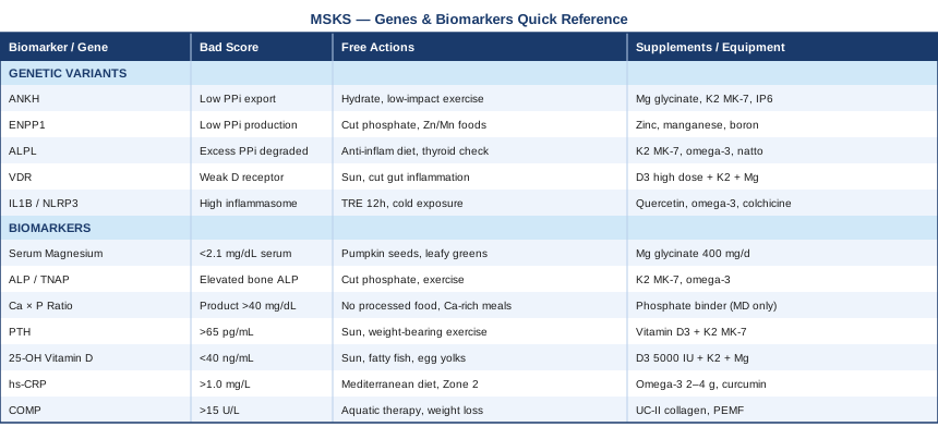
This article maps Milwaukee shoulder knee syndrome through two lenses most clinical visits never reach: 7 biomarkers that reveal the metabolic drivers of crystal formation and joint destruction, and 5 genetic variants that shape the biological machinery behind calcium phosphate balance and inflammation. For each, you'll find a practical plan — with and without supplementation — grounded in mechanism rather than guesswork. Beyond that, the article draws on Peter Attia's longevity medicine framework to show how systemic metabolic health connects to joint decline, and it covers three complementary approaches — tai chi, photobiomodulation, and mindfulness — that carry enough clinical evidence to be worth considering. The goal is not to replace medical care but to give you tools to track what's actually happening upstream, and act on it.
7 Biomarkers to Track If You Have Milwaukee Shoulder Knee Syndrome
Why Standard Blood Tests Often Miss the Picture
Most people with MSKS come away from a blood draw with a basic metabolic panel and perhaps a CRP result. That's a starting point, but the core drivers of BCP crystal formation — dysregulation of pyrophosphate metabolism, calcium-phosphate imbalance, and cartilage matrix breakdown — require a more targeted panel. The seven markers below collectively cover the upstream and downstream drivers of this condition. Several are inexpensive and widely available; others require more specialized ordering. All of them have a mechanistic reason to be tracked in MSKS, and each gives you something actionable to work with.
Biomarker 1: Serum Magnesium
Why it matters
Magnesium is the most underappreciated mineral in the context of ectopic calcification. It competes directly with calcium for crystal nucleation sites in cartilage and synovial tissue. When intracellular and extracellular magnesium are low, calcium crystals — including BCP and calcium pyrophosphate — form more readily. Research in rheumatology literature has observed that patients with BCP crystal deposition tend to have lower serum and intracellular magnesium compared to matched controls, though this finding remains underemphasized in routine clinical care.
Magnesium also plays a role in modulating alkaline phosphatase activity and in dampening inflammatory signaling pathways. Its deficiency is extremely common in Western populations due to soil depletion of dietary sources, chronic stress, and widespread proton pump inhibitor use — three factors that frequently overlap with the older female demographic most affected by MSKS. This makes magnesium both a meaningful therapeutic target and an easy place to start.
How to measure it
Standard serum magnesium is available through any basic blood panel: $10–$30. However, serum magnesium reflects only about 1% of total body magnesium and can appear normal even with significant intracellular depletion. Red blood cell (RBC) magnesium is more sensitive and costs $40–$80 through specialty labs. Optimal target: above 2.1 mg/dL for serum; above 5.5 mg/dL for RBC magnesium. If serum is borderline, request the RBC version for a more complete picture.
If the score is low — the plan without supplements
Focus on magnesium-dense whole foods: pumpkin seeds, dark leafy greens (spinach, Swiss chard), almonds, black beans, and dark chocolate above 70% cacao. Reduce alcohol intake, which substantially increases urinary magnesium loss. Eliminate processed foods and refined grains, which have minimal magnesium content and increase metabolic demand for it. Chronic psychological stress elevates cortisol, which increases renal magnesium excretion — stress reduction is therefore a direct magnesium-preserving strategy. Aim for at least 400 mg daily from food sources consistently.
If the score is low — the plan with supplements or equipment
Magnesium glycinate or magnesium malate are the best-tolerated forms for systemic repletion. Begin at 200 mg elemental magnesium at night and increase to 400 mg over two weeks. Magnesium bisglycinate is well-absorbed and causes far less GI discomfort than oxide or citrate forms. Frequency: daily, continuous — no cycling needed for maintenance. Side effect to watch: loose stools at high doses; reduce the dose if this occurs. Topical magnesium oil applied to the skin is an alternative if gut tolerance is poor. Avoid magnesium oxide entirely — poor bioavailability and high risk of GI side effects. Recheck RBC magnesium every 3–4 months initially, then every 6 months once stable.
Biomarker 2: Tissue-Nonspecific Alkaline Phosphatase (ALP)
Why it matters
This biomarker sits at the biochemical heart of MSKS. Tissue-nonspecific alkaline phosphatase (TNAP, encoded by the ALPL gene) is the enzyme responsible for degrading inorganic pyrophosphate (PPi). PPi is the body's primary endogenous inhibitor of calcium crystal formation — when PPi levels are adequate, crystals don't nucleate easily. When TNAP activity is excessive, PPi is consumed too rapidly, removing the natural brake on BCP crystal formation. This mechanism is central to the pathophysiology of MSKS.
Elevated ALP is usually interpreted clinically as a liver marker. But the bone and tissue ALP isoforms tell a completely different story about joint mineral metabolism. Fractionated ALP, which distinguishes the liver isoform from the bone/tissue isoform, is far more informative in the MSKS context. It's one of the most mechanistically direct biomarkers available for this condition and is rarely discussed in standard rheumatology consultations.
How to measure it
Total ALP is included in standard comprehensive metabolic panels: $10–$20. Fractionated ALP with bone-specific ALP adds isoform specificity: $50–$120. Normal total ALP: 44–147 U/L, though lab ranges vary. Elevated bone-specific ALP with normal liver ALP points specifically toward abnormal bone and joint mineral metabolism. If total ALP is elevated without liver pathology (normal GGT, normal bilirubin), request fractionation before concluding it's bone-related.
If the score is elevated — the plan without supplements
Reduce high-phosphate processed food intake: phosphate additives in packaged meats, fast food, and carbonated soft drinks (phosphoric acid) drive excess phosphate absorption, which shifts calcium-phosphate balance in ways that promote crystal formation. Maintain adequate hydration to support renal phosphate clearance. Evaluate thyroid function — both hypothyroidism and hyperthyroidism alter bone ALP activity significantly. Regular low-impact movement (swimming, walking, cycling) supports healthy bone remodeling without mechanical overloading of affected joints.
If the score is elevated — the plan with supplements or equipment
Vitamin K2 (MK-7 form) activates matrix Gla protein (MGP), which is an independent and potent inhibitor of soft tissue calcification that works downstream of the PPi pathway. Dose: 100–200 mcg MK-7 daily, continuous. Side effects: minimal at these doses; important caution with warfarin (K2 interaction). Omega-3 fatty acids (2–4 g EPA+DHA daily): reduce inflammatory cytokines that secondarily induce TNAP expression. Avoid calcium supplementation in excess of dietary needs — supplemental calcium in patients with active BCP crystal burden should be reviewed with a physician. Monitor fractionated ALP every 4–6 months alongside the full mineral panel.
Biomarker 3: Serum Calcium and Phosphate Ratio
Why it matters
The calcium-phosphate product (Ca × PO₄, abbreviated Ca×P) is a key determinant of whether BCP crystals will nucleate in soft tissue and joint spaces. When this product exceeds a critical threshold in joint fluid or tissue microenvironments, crystal formation becomes thermodynamically favorable. While this threshold is most aggressively managed in renal disease, the same biophysical principle governs crystal deposition in MSKS. Both values need to be tracked together — isolated hypercalcemia or isolated hyperphosphatemia each tell part of the story, but the product reveals the systemic crystallization risk.
Hyperphosphatemia is often subclinical and diet-driven in otherwise healthy individuals. The proliferation of phosphate additives in processed food has increased average dietary phosphate intake substantially over the past four decades — a trend that maps onto increased prevalence of ectopic calcification conditions. This is an environmental driver that is largely invisible in routine care.
How to measure it
Serum calcium and serum phosphate are both included in basic metabolic panels: $10–$30. Calculate the Ca×P product in mg/dL units. A product above 55 is definitively concerning in renal disease; for joint tissue crystallization risk in MSKS, lower thresholds in the 40–45 range remain informative. Target: calcium 8.5–10.2 mg/dL; phosphate 2.5–4.5 mg/dL; product below 40. Ionized calcium (unbound fraction) adds precision for borderline total calcium results: $30–$60.
If values are out of range — the plan without supplements
Reduce dietary phosphate load by eliminating processed meats, sodas, packaged snacks, and fast food. Increase calcium from whole food sources such as dairy, bok choy, and broccoli rabe — paradoxically, adequate dietary calcium reduces intestinal phosphate absorption by binding phosphate in the gut. Timing matters: eat calcium-rich foods with phosphate-rich meals to reduce net phosphate absorption. For elevated calcium, always evaluate vitamin D and PTH (see below) before concluding it's dietary — pathological causes need to be ruled out.
If values are out of range — the plan with supplements or equipment
If phosphate is chronically elevated despite dietary changes and confirmed through repeated testing, low-dose calcium carbonate with meals (under medical supervision only) can act as a phosphate binder. This is not a self-prescribing strategy — the balance between reducing phosphate and avoiding excess calcium in BCP crystal conditions requires physician oversight. For abnormal serum calcium, vitamin D optimization (Biomarker 5) and PTH assessment (Biomarker 4) are the first interventions. Self-supplementing calcium when BCP crystal burden is already confirmed is contraindicated without explicit physician guidance.
Biomarker 4: Parathyroid Hormone (PTH)
Why it matters
PTH is the master regulator of calcium homeostasis. Secondary hyperparathyroidism — elevated PTH driven by vitamin D deficiency or declining renal function — promotes calcium mobilization from bone and increases the total calcium load available for ectopic crystal deposition in joint tissue. Chronic low-grade PTH elevation creates precisely the biochemical environment that accelerates BCP crystal formation in MSKS.
In older patients, where MSKS is most prevalent, secondary hyperparathyroidism due to vitamin D insufficiency and age-related renal decline is frequently present and under-recognized. PTH also stimulates osteoclast-mediated bone resorption, compounding the structural damage to periarticular bone that worsens joint stability in advanced MSKS. Tracking PTH gives you a direct window into the calcium-regulatory axis that underlies crystal formation risk.
How to measure it
Intact PTH (iPTH) assay: $30–$80. Standard normal range: 10–65 pg/mL. Functional optimal range, as applied in Peter Attia's clinical practice and discussed in longevity medicine contexts: 20–55 pg/mL. Values above 65 with concurrent low vitamin D strongly suggest secondary hyperparathyroidism. Always interpret PTH alongside vitamin D (Biomarker 5) and serum calcium simultaneously — isolated PTH values are easily misread without the full context.
If PTH is elevated — the plan without supplements
Midday sun exposure is the most effective non-supplemental intervention: 15–30 minutes of direct skin exposure (arms, legs, torso) on most days drives vitamin D3 synthesis, which then suppresses excess PTH production via a direct feedback loop. Weight-bearing physical activity supports bone mineral density and reduces the stimulus for compensatory PTH elevation. Address hydration and potential nephrotoxic exposures (chronic NSAID use, excessive protein in compromised kidneys, dehydration) to preserve renal function. Dietary calcium from whole food sources — not supplements — reduces PTH stimulation without adding free calcium to circulation.
If PTH is elevated — the plan with supplements or equipment
Vitamin D3 optimization (see Biomarker 5) is the primary intervention when secondary hyperparathyroidism is vitamin D-driven. Ensure adequate combined calcium intake from food plus any supplementation (1000–1200 mg daily for postmenopausal women). Vitamin K2 (MK-7, 100–200 mcg daily) is the essential co-factor — it activates osteocalcin and MGP, directing calcium toward bone tissue and away from soft tissue and joint spaces. Continuous use, no cycling required. If PTH remains elevated after vitamin D optimization over 3–4 months, evaluate renal function comprehensively and consider endocrinology referral — autonomous hyperparathyroidism needs to be excluded.
Biomarker 5: 25-OH Vitamin D
Why it matters
Vitamin D deficiency is one of the most prevalent and modifiable upstream factors in crystal arthropathy. It drives secondary hyperparathyroidism (Biomarker 4), impairs immune regulation, and increases inflammatory cytokine production — including IL-1β and TNF-α, which are key drivers of the synovial inflammation triggered by BCP crystals. Research has also documented that vitamin D influences the expression of genes involved in calcium transport and matrix calcification inhibitors, placing it upstream of multiple MSKS mechanisms simultaneously.
Peter Attia treats 25-OH vitamin D as a foundational biomarker — one he monitors in essentially every patient because its downstream effects span immune function, bone health, cardiometabolic risk, and inflammation. Thomas Dayspring has similarly emphasized optimal D levels, not merely "normal" range levels, as clinically relevant to inflammatory conditions. The gap between population-normal (above 30 ng/mL) and functionally optimal is large enough to matter significantly for MSKS patients.
How to measure it
25-OH vitamin D (calcidiol) blood test: $30–$60, widely available. Standard normal range: 30–100 ng/mL. Functional optimal range: 40–60 ng/mL (Attia's target for most patients; some researchers recommend 50–70 ng/mL for patients with active inflammatory conditions). Levels below 30 are clearly deficient; 30–40 is insufficient. Test seasonally — autumn and winter levels are typically lowest, spring and summer levels highest. A single annual test often misses the seasonal trough.
If levels are low — the plan without supplements
Midday sun exposure remains the most efficient source. In northern latitudes or during winter months, dietary sources become critical: fatty fish (salmon, mackerel, sardines, herring), egg yolks, and full-fat fortified dairy. Gut health significantly affects vitamin D absorption — fat malabsorption from low-fat diets, gallbladder dysfunction, or gut dysbiosis reduces dietary vitamin D uptake. Reducing adiposity, where applicable, improves functional vitamin D availability, since adipose tissue sequesters circulating vitamin D and reduces its bioavailability.
If levels are low — the plan with supplements or equipment
Vitamin D3 (cholecalciferol) supplementation: for deficiency below 30 ng/mL, 5,000 IU daily for 8–12 weeks, then recheck. For maintenance at 30–40 ng/mL: 2,000–4,000 IU daily. Always pair with vitamin K2 (MK-7, 100–200 mcg) — vitamin D increases calcium absorption and K2 ensures that calcium is directed to bone rather than soft tissues. Magnesium is a required cofactor for both enzymatic steps of vitamin D activation in the liver and kidney — ensure adequate magnesium (Biomarker 1) before loading vitamin D, or the conversion will be limited. Side effects: toxicity at sustained high doses above 10,000 IU without monitoring. Recheck 25-OH D at 3 months. Continuous use is appropriate for most patients in temperate climates.
Biomarker 6: High-Sensitivity C-Reactive Protein (hs-CRP)
Why it matters
BCP crystals don't sit passively in joint tissue. They are actively phagocytosed by synovial macrophages, which triggers the NLRP3 inflammasome and the release of IL-1β and IL-18. This drives local synovial inflammation and systemic inflammatory signaling. Hs-CRP is the most practical systemic proxy for this inflammatory cascade. In MSKS, persistently elevated hs-CRP indicates that the inflammatory component of the disease is active — and that joint destruction is likely progressing at an accelerated rate.
Thomas Dayspring and Peter Attia have both emphasized hs-CRP as one of the most informative and underutilized routine markers in medicine. At levels above 3 mg/L, cardiovascular risk increases substantially — and the same inflammatory state driving cardiovascular risk is actively destroying joint architecture in MSKS. This overlap is not coincidental; it reflects a shared chronic inflammatory biology.
How to measure it
High-sensitivity CRP assay: $15–$40. Note that standard CRP and hs-CRP use different assay sensitivities — request hs-CRP specifically. Optimal functional target: below 1.0 mg/L. Values of 1–3 mg/L indicate moderate chronic inflammation; above 3 mg/L indicates high systemic inflammation. Ensure no acute infection or illness within two weeks of testing — CRP can spike 100-fold with even minor acute inflammation, making the result uninterpretable for chronic inflammation assessment.
If hs-CRP is elevated — the plan without supplements
Diet is the most powerful lever: a Mediterranean-style dietary pattern reduces hs-CRP by 20–30% in well-controlled trials. Eliminate seed oils rich in linoleic acid (soybean, corn, sunflower oils), reduce refined carbohydrates, and increase omega-3-rich whole foods (fatty fish, walnuts, flaxseed). Sleep quality is equally critical — fragmented or insufficient sleep below 7 hours measurably increases hs-CRP and IL-6 the following day. Regular moderate-intensity aerobic exercise (Zone 2, see Strategy 3) lowers CRP over time in a dose-dependent way. Chronic psychological stress drives HPA-axis dysregulation and cortisol-mediated inflammatory amplification — structured stress management directly reduces hs-CRP.
If hs-CRP is elevated — the plan with supplements or equipment
Omega-3 fatty acids (EPA+DHA): 2–4 g of combined EPA+DHA daily from high-quality fish oil or algae-based supplements. This is the best-evidenced supplemental intervention for reducing hs-CRP across multiple systematic reviews and meta-analyses. Daily use, no cycling; side effects minimal (fishy burps — mitigated by enteric-coated forms or refrigerating the capsules). Curcumin with phospholipid complex or piperine for bioavailability: 500–1000 mg daily in divided doses. Multiple meta-analyses confirm hs-CRP reduction. Cycle 8–12 weeks on, 4 weeks off. Boswellia serrata (AKBA-standardized extract): 100–250 mg AKBA twice daily — evidence specifically for joint-tissue inflammation reduction. Combine with monitoring every 3–6 months to confirm response.
Biomarker 7: COMP (Cartilage Oligomeric Matrix Protein)
Why it matters
COMP is a structural glycoprotein that is a major component of cartilage extracellular matrix. When cartilage is actively breaking down — as occurs in the destructive arthropathy of MSKS — COMP is released into the bloodstream in proportion to the rate of cartilage degradation. Elevated serum COMP directly reflects active structural damage to the joint, not just inflammation or crystal burden. It is one of the few biomarkers that captures the tissue-destruction component of MSKS with reasonable specificity.
In MSKS, cartilage destruction can be rapid and is frequently underestimated clinically until radiological changes are advanced. BCP crystals activate matrix metalloproteinases (MMPs) in synovial cells, and these enzymes directly degrade the cartilage matrix including COMP. Tracking serum COMP over time provides a biochemical early warning signal for accelerating joint destruction, independent of whether symptoms have obviously changed. For research on COMP as a cartilage degradation marker, PubMed lists extensive literature in inflammatory arthropathies.
How to measure it
Serum COMP: $80–$200, available through specialty reference labs including Quest Diagnostics and LabCorp. Normal range is approximately 8–13 U/L, though values vary by assay and lab. Elevated levels above 15–17 U/L indicate significant cartilage turnover. This test is most valuable for longitudinal tracking — the trend over 6–12 months tells you far more than any single value. Establish a baseline early, before deciding on interventions, to have a reference point.
If COMP is elevated — the plan without supplements
Mechanical load reduction on affected joints is the most direct non-pharmacological intervention. Use assistive devices (canes, ergonomic joint supports), avoid repetitive overhead motion for shoulder involvement, and eliminate high-impact activities. Aquatic therapy is particularly valuable: warm-water exercise allows joint loading and muscle activation without compressive impact forces. Weight normalization significantly reduces compressive load on knee joints — each pound of body weight reduction translates to approximately four pounds less force per step at the knee. Cold therapy (ice packs, 15–20 minutes post-activity) reduces synovial inflammation acutely without the long-term risks of chronic NSAID use.
If COMP is elevated — the plan with supplements or equipment
Undenatured type II collagen (UC-II): 40 mg daily of the specific patented form (not standard hydrolyzed collagen) — meta-analyses have shown reduction in joint pain and, in some trials, biomarkers of cartilage degradation. Daily, continuous use. Glucosamine sulfate (pharmaceutical-grade Rotta preparation, 1500 mg/day): carries the strongest structural evidence among OA supplements, with several DMOAD (disease-modifying OA drug) trials showing reduced joint space narrowing. Evidence is mixed but the signal exists for this specific form. PEMF therapy (pulsed electromagnetic field devices): home devices have FDA clearance for musculoskeletal pain and have shown modest evidence for cartilage health in OA trials. Use 20–30 minutes daily on affected joints. Monitor serum COMP every 4–6 months to track whether interventions are changing the trajectory.
With these seven biomarkers tracked consistently, a meaningful picture of MSKS biology emerges — one that allows for informed, targeted interventions rather than trial-and-error management. The next layer is genetic, and it reveals why some individuals face higher baseline risk regardless of lifestyle.
5 Genetic Variants That Shape Individual Risk
The Pyrophosphate Pathway: Where the Genetics Concentrate
The genetics of Milwaukee shoulder knee syndrome converge on a central theme: the regulation of inorganic pyrophosphate (PPi). PPi is the body's primary endogenous brake on calcium crystal nucleation. When the genes controlling PPi production, transport, or degradation carry risk variants, the brake weakens and BCP crystals form more readily. Three of the five genes below directly regulate this pathway; the other two govern inflammation and vitamin D response — the amplifying factors once crystals are present.
Consumer genetic panels (23andMe, AncestryDNA) capture many of the relevant common variants in these genes, though they don't always report them explicitly within joint disease contexts. More comprehensive assessment through clinical rheumatology genetics panels or whole-genome sequencing provides broader coverage. Knowing your variant status doesn't predict MSKS with certainty, but it does indicate where your physiological vulnerabilities likely lie.
Gene 1: ANKH — The Pyrophosphate Transporter
The ANKH gene (ankylosis, progressive homolog) encodes a membrane channel protein that transports intracellular PPi to the extracellular space, where it inhibits calcium crystal nucleation in cartilage and joint tissue. Loss-of-function variants in ANKH reduce extracellular PPi levels, creating a permissive biochemical environment for both BCP and calcium pyrophosphate crystal formation. ANKH mutations have been identified in familial chondrocalcinosis (CCAL2) and are increasingly studied in sporadic crystal arthropathy. Patients with reduced ANKH function essentially have a constitutionally weakened anti-calcification system in their joint tissues.
If the gene shows a risk variant — the plan without supplements
Hydration is foundational: adequate water intake (2–3 liters daily, adjusted for body weight and activity level) supports renal mineral clearance and overall mineral homeostasis. Prioritize PPi-supporting nutrient density from whole foods: organ meats, legumes, and fermented foods contain natural phosphate metabolism cofactors that support this pathway. Avoid excess calcium supplementation, which can worsen BCP crystal burden by flooding the system with the primary crystal-forming cation. Regular, low-impact joint movement — swimming, cycling, walking — maintains synovial fluid circulation, which disperses crystal precursors and promotes local PPi distribution through joint fluid dynamics.
If the gene shows a risk variant — the plan with supplements or equipment
Magnesium glycinate (300–400 mg elemental magnesium daily): magnesium competes with calcium at crystal nucleation sites and supports PPi-dependent mineral regulation. Vitamin K2 MK-7 (100–200 mcg daily): activates MGP independently of the PPi pathway, providing a parallel calcification-inhibiting mechanism. Inositol hexaphosphate (IP6): an emerging research area — IP6 is a phosphate-based compound with documented crystal-inhibitory properties in vitro and early human data. Dose: 1–2 g daily with water on an empty stomach. Limited long-term human safety data; cycle 8 weeks on, 4 weeks off. No significant side effects at these doses. Magnesium and K2 are continuous; IP6 cycling is precautionary given the early stage of evidence.
Gene 2: ENPP1 — The PPi Generator
ENPP1 (ectonucleotide pyrophosphatase/phosphodiesterase 1) encodes the enzyme responsible for generating extracellular PPi from ATP in the cartilage matrix. Where ANKH transports PPi out of cells, ENPP1 creates it in the extracellular space. Loss-of-function variants in ENPP1 reduce PPi production at the cartilage surface — removing the critical inhibitor of BCP crystal formation precisely where it is most needed.
ENPP1 variants have been implicated in conditions ranging from generalized arterial calcification to insulin signaling abnormalities, underscoring how broadly this enzyme's function extends. In the joint-specific context of MSKS, reduced ENPP1 activity shifts the local balance from crystal-inhibiting PPi toward crystal-promoting inorganic phosphate — the precise imbalance that triggers crystal nucleation.
If the gene shows a risk variant — the plan without supplements
Reduce dietary phosphate load systematically: eliminate ultra-processed foods (which use phosphate additives extensively) and phosphoric acid-containing beverages. Whole food sources of zinc and manganese support ENPP1 enzymatic activity as these are cofactor metals for the enzyme. Moderate resistance exercise maintains muscle mass and metabolic flexibility, which preserves mitochondrial ATP production — the upstream substrate from which ENPP1 generates PPi. Reducing chronic psychological stress decreases cortisol-driven magnesium loss, which in turn supports ENPP1 cofactor availability.
If the gene shows a risk variant — the plan with supplements or equipment
Zinc glycinate (15–25 mg daily): ENPP1 is a zinc-dependent metalloenzyme; adequate zinc is required for enzyme stability and function. Take with food to reduce nausea. Important: excess zinc competes with copper — consider a balanced zinc/copper formula or ensure dietary copper from organ meats and shellfish. Manganese (2–5 mg daily): another metalloenzyme cofactor for ENPP1. Food sources include pineapple, hazelnuts, and whole grains; supplement at low doses only. High manganese intake is neurotoxic — stay strictly within the range above and do not combine high dietary and supplemental sources. Boron (3–6 mg daily as sodium borate or boron glycinate): emerging evidence supports boron's role in pyrophosphate-related mineral pathways and bone health. Well-tolerated at these doses, continuous use appropriate.
Gene 3: ALPL — The PPi Consumer
ALPL encodes tissue-nonspecific alkaline phosphatase (TNAP), the enzyme that degrades PPi. This gene is the functional antagonist of both ANKH and ENPP1 in pyrophosphate regulation. Gain-of-function variants or elevated basal ALPL expression result in accelerated PPi degradation — the crystal brake is continuously consumed faster than it can be replenished. Loss-of-function mutations in ALPL cause hypophosphatasia, a rare metabolic bone disease, but in MSKS the concern runs in the opposite direction: elevated TNAP activity is problematic, and this can reflect both genetic predisposition and secondary induction by inflammatory cytokines.
This gene therefore bridges the genetics and biomarker sections: an elevated bone-specific ALP on your blood panel (Biomarker 2) may partially reflect ALPL genetic activity and should prompt thinking about crystal inhibitor depletion as a mechanism.
If the gene shows a risk variant (elevated activity) — the plan without supplements
Reduce dietary factors that drive TNAP induction: high-fat, high-sugar diets increase hepatic and bone ALP expression. An anti-inflammatory, whole-food dietary pattern demonstrably keeps TNAP activity lower in longitudinal dietary studies. Limit isolated vitamin A supplementation — high retinol intake can increase bone ALP activity independent of dietary vitamin D. Maintain optimal thyroid function through regular testing; both hypothyroidism and hyperthyroidism alter ALP dynamics and should be treated if present.
If the gene shows a risk variant — the plan with supplements or equipment
Vitamin K2 MK-7 (100–200 mcg daily): K2 activates MGP, which inhibits calcification downstream and partially compensates for TNAP over-activity by working through a parallel mechanism. Omega-3 fatty acids (2–4 g EPA+DHA daily): reduce the inflammatory cytokine load that secondarily induces TNAP expression. Dietary natto (fermented soy): the richest food source of MK-7, providing approximately 900 mcg per 100 g serving — far exceeding any supplement dose. Japanese longevity research has associated regular natto consumption with reduced vascular and soft tissue calcification. For those not on anticoagulants, 50–100 g of natto three to five times per week is a practical and evidence-adjacent strategy. Side effect note: monitor if taking warfarin — K2 interaction requires INR adjustment under physician supervision.
Gene 4: VDR — The Vitamin D Receptor
The VDR gene encodes the vitamin D receptor, which mediates virtually all of vitamin D's genomic effects — including calcium transport regulation, immune modulation, and anti-inflammatory signaling in synovial tissue. Common VDR polymorphisms (FokI, BsmI, ApaI, TaqI) alter receptor binding efficiency and downstream transcriptional activity in ways that are clinically measurable. Individuals with less efficient VDR variants need higher circulating vitamin D to achieve the same biological effect.
This creates a practical problem: a patient with a high-risk VDR variant who tests at 50 ng/mL 25-OH vitamin D may be functionally insufficient compared to someone with optimal VDR function at the same circulating level. In MSKS, VDR function shapes both the inflammatory tone of synovial tissue and the calcium handling that underlies crystal formation risk — making this gene relevant to multiple pathways simultaneously. VDR polymorphisms in inflammatory joint conditions have been studied extensively in rheumatology literature.
If the gene shows a risk variant — the plan without supplements
Increase sun exposure strategically: higher ligand availability can partially overcome receptor inefficiency. The FokI "f" allele, for example, is associated with reduced VDR transactivation capacity — more circulating vitamin D3 means more receptor occupancy despite reduced efficiency. Dietary factors that enhance VDR signaling include omega-3 fatty acids, which modulate VDR transcriptional complex formation, and reducing gut inflammation through a whole food dietary approach (gut VDR expression is among the highest in the body and is impaired by gut dysbiosis).
If the gene shows a risk variant — the plan with supplements or equipment
Target higher-end 25-OH vitamin D levels (60–70 ng/mL rather than the typical 40–60 ng/mL target) under physician supervision with periodic monitoring. Dose: 5,000–8,000 IU D3 daily with K2 (100–200 mcg MK-7) and magnesium (adequate levels confirmed before loading). Resveratrol (250–500 mg daily with food containing fat): some research suggests resveratrol enhances VDR transcriptional activity — evidence is currently primarily in vitro and early clinical studies, so this remains a speculative addition. Cycle 8 weeks on, 4 weeks off given limited long-term data at supplemental doses. Monitor 25-OH vitamin D and serum calcium every 3–4 months when targeting the higher range.
Gene 5: IL1B and NLRP3 — The Inflammation Amplifiers
IL1B encodes interleukin-1 beta, the primary pro-inflammatory cytokine released when BCP crystals are phagocytosed by macrophages and synovial fibroblasts. NLRP3 encodes the inflammasome complex that acts as the crystal sensor — activating in response to BCP crystal uptake and triggering the IL-1β cascade. Variants in both genes affect the magnitude of the inflammatory response to a given crystal burden, essentially determining whether crystals produce mild chronic irritation or aggressive destructive synovitis.
NLRP3 gain-of-function variants have been documented in gout, CPPD, and related crystal arthropathies, with the same mechanism applying to BCP crystal disease. IL1B promoter polymorphisms affecting transcription are associated with more severe inflammatory joint disease phenotypes generally. These variants don't cause MSKS, but they determine whether a moderate crystal burden will inflict disproportionate damage.
If the gene shows a risk variant — the plan without supplements
Time-restricted eating (10–12 hour eating window): reduces NLRP3 inflammasome activation consistently across multiple metabolic studies, likely mediated through beta-hydroxybutyrate (a ketone body produced during the fasting window that directly inhibits NLRP3) and AMPK activation. This is not a dramatic fasting protocol — simply avoiding eating for 12 hours between dinner and breakfast is sufficient to produce this effect. Cold water exposure (cold showers progressing to cold water immersion): emerging evidence from immunological research suggests acute cold reduces NLRP3 activity and pro-inflammatory cytokine production. Begin with 30-second cold finishes to showers; progress gradually. Chronic psychological stress persistently elevates IL-1β production through cortisol-NF-κB pathways — stress management is a direct anti-inflammatory strategy for high IL1B expressors, not merely a wellness nicety.
If the gene shows a risk variant — the plan with supplements or equipment
Quercetin (500–1000 mg daily with fat for absorption, or as quercetin phytosome for better bioavailability): well-documented NLRP3 inflammasome inhibitor across human and preclinical studies. Cycle 8–12 weeks on, 4 weeks off; generally well-tolerated. Omega-3 fatty acids (3–4 g EPA+DHA daily): EPA and DHA directly modulate NLRP3 activation through lipid mediator pathways. Resveratrol (250–500 mg daily): activates SIRT1, which downregulates NF-κB upstream of IL-1β production. Colchicine (low dose, 0.5 mg daily): a prescription option with robust evidence for reducing crystal-induced inflammation in BCP and CPPD arthropathy — colchicine disrupts inflammasome assembly and neutrophil-crystal interaction. This is worth an explicit conversation with your rheumatologist, particularly if inflammatory flares are frequent and severe. Side effects: GI discomfort at higher doses; low-dose is generally well-tolerated. Drug interactions exist — physician review required.
Genetics and biomarkers together provide the most complete picture of why MSKS behaves the way it does in any individual. The next section draws on a broader framework for metabolic and inflammatory health that connects these findings to systemic longevity medicine.
10 Insights From Peter Attia's Framework That Apply Directly to MSKS
When Longevity Medicine Meets Rheumatology
Peter Attia's book Outlive: The Science and Art of Longevity (2023) doesn't address Milwaukee shoulder knee syndrome by name, but it maps a metabolic and inflammatory framework that is directly applicable to its upstream mechanisms. Attia's core argument — that the major chronic diseases share common roots in systemic inflammation, insulin resistance, and mitochondrial dysfunction — applies precisely to the biochemical drivers of MSKS. The insights below represent the most translation-ready elements of his framework for understanding and managing this specific condition.
1. Chronic Low-Grade Inflammation Is the Common Denominator
Attia argues that atherosclerosis, cancer, neurodegeneration, and metabolic syndrome all share a foundation of chronic low-grade inflammation. The same hs-CRP elevation that marks cardiovascular risk is simultaneously driving synovial destruction in MSKS. Treating inflammation systemically — not merely managing the joint locally — is the strategic leverage point that most rheumatology visits never reach. The biomarker panel above gives you the tools to see this inflammation; Attia's framework gives you the rationale for treating it aggressively even when joints feel manageable.
2. Metabolic Health Directly Affects Cartilage Biology
Insulin resistance impairs mitochondrial function in chondrocytes — the cartilage-producing cells responsible for maintaining joint matrix. This reduces their repair capacity, increases local oxidative stress, and makes cartilage more vulnerable to crystal-induced MMP activation. MSKS appears on the surface to be a mineral and mechanical disease, but metabolic health underlies the tissue's ability to resist and recover from crystal-driven damage. Fasting glucose, insulin, and HbA1c belong in the MSKS monitoring panel for this reason.
3. hs-CRP Below 1.0 mg/L Is the Functional Target, Not Just "Normal"
Population-derived "normal" laboratory ranges and functional optimal ranges are not the same thing. For hs-CRP, the standard normal extends to 10 mg/L. The target that Attia's clinical practice — and that Thomas Dayspring's lipid-inflammation work — treats as the functional goal for disease prevention is below 1.0 mg/L. For MSKS patients with active crystal-driven inflammation, that gap between "normal" and "optimal" represents years of accelerated joint degradation. Aim for below 1.0, not just below 10.
4. Zone 2 Cardio Is the Most Accessible Anti-Inflammatory Tool
Sustained low-intensity aerobic exercise — Zone 2 at roughly 60–70% of maximum heart rate, the pace where you can hold a full sentence — improves mitochondrial efficiency, reduces systemic inflammatory markers, and improves insulin sensitivity through well-characterized mechanisms. The dose-response is linear up to several hours per week. For MSKS patients with significant joint involvement, aquatic Zone 2 training (pool walking, water cycling) and cycling provide this stimulus without loading the affected shoulder or knee. Four to five hours per week is Attia's clinical recommendation; even two hours produces meaningful benefit.
5. Sleep Is a Clinical Variable, Not a Lifestyle Preference
Attia treats sleep as a non-negotiable longevity pillar. A single night of poor sleep (under 6 hours or highly fragmented) measurably increases circulating IL-1β, IL-6, and CRP. For MSKS patients — where IL-1β is the central cytokine driving synovial destruction via NLRP3 — sleep quality is not separable from disease activity. Treating insomnia, sleep apnea, or poor sleep hygiene is a direct rheumatological intervention. Track sleep objectively if possible; subjective sleep assessment is notoriously unreliable.
6. Protein Intake Protects the Musculoskeletal Foundation
Attia recommends 1.6–2.2 g of protein per kilogram of body weight daily for lean mass preservation, and the evidence base for this target in older adults is robust. In MSKS — where the patient demographic is predominantly older women with existing sarcopenia risk — inadequate protein accelerates muscle loss, reduces joint stabilizing capacity, and increases fall risk. Leucine-rich protein sources (animal protein, whey protein) maximize muscle protein synthesis signaling. Distributing protein across three or four meals (rather than concentrating it in one) improves utilization in older adults specifically.
7. Strength Training Is Joint Protection, Not Just Fitness
Attia treats grip strength and lower limb strength as primary predictors of all-cause mortality and functional independence. For MSKS, rotator cuff strength (shoulder stabilizers) and quadriceps strength (knee stabilizers) are the primary mechanical defenses against progressive joint instability and accelerated cartilage wear. Structured resistance training — adapted for joint limitations through seated exercise, elastic resistance bands, or aquatic resistance — is not optional for long-term joint preservation. A physical therapist experienced in inflammatory arthropathy can design an appropriate load-management program.
8. Vitamin D at 40–60 ng/mL Is a Functional Minimum
Attia's clinical practice maintains 25-OH vitamin D in the 40–60 ng/mL range — not merely "above deficiency." This higher target is relevant for MSKS because vitamin D's anti-inflammatory and calcium-regulatory functions are dose-dependent within the physiological range. The commonly encountered clinical message that any level above 30 ng/mL is "fine" leaves many patients in a functionally insufficient state where PTH suppression and immune regulation are incomplete. Seasonal testing and active supplementation to maintain the 40–60 ng/mL range are the standard Attia applies to patients with inflammatory conditions.
9. ApoB Captures an Inflammatory Burden Relevant to MSKS
While lipid markers may seem disconnected from joint disease, Attia's framework emphasizes ApoB (a marker of atherogenic particle count) as central to cardiovascular risk assessment. MSKS patients have elevated cardiovascular comorbidity risk through shared inflammatory pathways, and elevated ApoB correlates with the same oxidized-LDL-driven endothelial inflammation that drives vascular disease and amplifies systemic cytokine load. Patients managing MSKS who also carry elevated ApoB are facing a double inflammatory burden. Tracking ApoB alongside hs-CRP gives a fuller systemic picture.
10. Track Trends, Not Single Values
Attia's approach emphasizes that a single biomarker measurement tells you almost nothing in isolation. The trend of hs-CRP over 12 months, the trajectory of 25-OH vitamin D before and after supplementation, the direction of serum COMP over two years — these trends are the actionable signals. Building a systematic tracking habit, using a simple spreadsheet or an app like Heads Up Health, converts isolated data points into interpretable patterns. One of the highest-leverage things any MSKS patient can do is establish baseline measurements and re-test at defined intervals so that interventions can be evaluated objectively.
These ten principles collectively represent an upstream framework for managing MSKS that extends well beyond what routine rheumatology visits address. The final section covers three complementary approaches with sufficient human clinical evidence to merit consideration.
Complementary Approaches With Meaningful Evidence
Tai Chi for Joint Function and Inflammatory Modulation
Tai chi is a slow, controlled movement practice originating in Chinese martial arts tradition and now among the most extensively studied mind-body interventions in rheumatology. For MSKS, its relevance spans three areas: maintaining joint range of motion without high mechanical load, improving proprioception and balance (critical for reducing fall risk in patients with shoulder and knee involvement), and reducing systemic inflammatory markers through its documented stress-reduction and autonomic regulation effects.
A landmark randomized controlled trial by Wang and colleagues, published in Arthritis Care and Research (2009), found that 12 weeks of Yang-style tai chi practice significantly reduced pain and improved physical function in knee osteoarthritis patients compared to a control group. While this trial addressed osteoarthritis rather than MSKS specifically, the mechanical and inflammatory overlap between the two conditions makes the findings applicable — both involve synovial inflammation, cartilage degradation, and muscle stabilizer weakness. The tai chi intervention used 60-minute sessions twice weekly. For the broader evidence base for tai chi in joint conditions, multiple systematic reviews confirm consistent effects on pain, stiffness, and balance.
For MSKS patients, begin with a modified or seated tai chi program if shoulder or knee involvement is severe. The Yang style (slow, large-circle movements) is most commonly studied and most accessible for older adults with joint limitations. Aim for 20–30 minutes daily, three to five days per week. Guided instruction — in-person group classes or structured video programs — significantly improves form and adherence during the first 4–6 weeks, which is when the learning curve is steepest. Once the basic forms are learned, home practice becomes straightforward and self-sustaining.
Photobiomodulation for Local Inflammation and Tissue Repair
Photobiomodulation (PBM), delivered through low-level laser or LED devices emitting red and near-infrared light (typically 630–850 nm), uses non-thermal light energy to stimulate mitochondrial cytochrome c oxidase in tissue cells, increasing ATP production, reducing oxidative stress, and modulating local inflammatory signaling. In musculoskeletal medicine, PBM has accumulated substantial evidence for reducing joint pain, decreasing local inflammatory cytokine expression, and supporting tissue repair processes. For MSKS, the mechanism is relevant at both the synovial and cartilage levels — reducing inflammatory amplification by BCP crystals and supporting chondrocyte metabolic function.
A 2022 meta-analysis in Lasers in Medical Science examined PBM for knee osteoarthritis across multiple randomized trials and found statistically significant improvements in pain and stiffness scores versus placebo, with effect sizes in the moderate range. A Cochrane review on low-level laser therapy for osteoarthritis (Brosseau et al.) found moderate evidence for short-term pain reduction, particularly for knee involvement. MSKS-specific PBM trials are scarce, but the pathophysiological overlap with inflammatory arthropathy — specifically the shared role of IL-1β and MMP activation — provides a mechanistic rationale for its use as a complementary approach. For relevant clinical evidence on PBM and joint inflammation, the PubMed literature is now extensive.
Home PBM devices (panels or targeted handheld units from reputable manufacturers) are available at $200–$800 and can deliver therapeutic wavelengths at adequate power densities for musculoskeletal use. For shoulder involvement: position the device 6–12 inches from the shoulder joint, targeting the anterior, posterior, and lateral aspects in rotation over 10–20 minutes per session. For knee: target medial, lateral, and patellar aspects. Three to five sessions per week is the typical protocol. Avoid use over active malignancies, photosensitized medications, or active skin lesions. Results typically require 4–8 weeks of consistent use to become noticeable, and benefit appears to be cumulative with continued use.
Mindfulness-Based Stress Reduction for Pain and Inflammatory Load
Mindfulness-Based Stress Reduction (MBSR), the structured 8-week program developed by Jon Kabat-Zinn at the University of Massachusetts, has accumulated a substantial and methodologically rigorous evidence base across chronic pain conditions. Its relevance to MSKS operates through two distinct pathways: reducing pain catastrophizing (which measurably amplifies perceived pain intensity in chronic joint conditions through central sensitization mechanisms), and modulating HPA-axis function in ways that reduce cortisol-driven inflammatory amplification of IL-1β and TNF-α.
A landmark meta-analysis by Goyal and colleagues, published in JAMA Internal Medicine (2014), found that mindfulness meditation programs showed moderate-strength evidence for improving anxiety, depression, and pain across chronic conditions compared to active control conditions. This meta-analysis reviewed 47 randomized trials. For inflammatory joint disease specifically, research published in Annals of the Rheumatic Diseases demonstrated that MBSR participants with inflammatory arthropathy showed measurable reductions in disease activity scores and self-reported pain ratings over the 8-week program, with effects persisting at follow-up. For the evidence base on MBSR and inflammatory pain conditions, meta-analyses consistently confirm the pain-modulatory effects.
The standard MBSR protocol involves weekly 2.5-hour group sessions for 8 weeks, a single day-long silent retreat, and 45 minutes of daily home practice. For MSKS patients, formal sitting meditation can be adapted to chair-based practice if floor sitting is not tolerable, and gentle movement components (walking meditation, adapted body scan) are already part of the core curriculum. Digital MBSR programs — including online courses based on the original Kabat-Zinn protocol — provide a flexible and accessible format. Apps including Insight Timer and Ten Percent Happier offer guided practices consistent with MBSR methodology. Begin with 10–15 minutes of formal practice daily and build to 30–45 minutes over 4–6 weeks. The mechanism is not abstract: documented reduction in cortisol variability, decreased NF-κB activation, and lower circulating inflammatory cytokines have been measured in MBSR participants across multiple trials.
Moving Forward With a More Complete Picture
Milwaukee shoulder knee syndrome is a condition where the visible symptoms — pain, restricted motion, progressive joint deterioration — sit at the far end of a chain of biological events that begins with mineral dysregulation, genetic predispositions, and low-grade chronic inflammation. The good news embedded in that picture is that many of those upstream factors are measurable, and several are meaningfully modifiable with the right information.
The seven biomarkers covered here — serum magnesium, alkaline phosphatase, calcium-phosphate product, PTH, vitamin D, hs-CRP, and COMP — give you a functional map of what's driving your specific case. The five genetic variants — ANKH, ENPP1, ALPL, VDR, and IL1B/NLRP3 — reveal the constitutional architecture that shapes individual risk and guides the choice of interventions. Neither layer alone is sufficient; together, they support a genuinely personalized approach in a condition that too often receives only generic care.
The most practical next step is not to implement everything simultaneously. Start with what is most accessible: order a comprehensive panel including RBC magnesium, fractionated ALP, 25-OH vitamin D, intact PTH, and hs-CRP. Share the results with a rheumatologist or integrative medicine physician willing to engage with these markers in the MSKS context. From there, build one targeted intervention at a time — tracking your biomarker trends to know what's actually shifting, not just how you feel on a given day.
Better information leads to better questions. Better questions lead to better care. That's where meaningful progress in managing MSKS begins.
Women's Health Endocrine & Metabolic
Musculoskeletal: Bone Conditions Joint Conditions
Autoimmune: Inflammatory Conditions