This article contains AI generated content.
· UpdatedMixed Connective Tissue Disease: 5 Genes and 7 Biomarkers to Track
Introduction
Mixed connective tissue disease occupies an uncomfortable diagnostic space. It borrows symptoms from lupus, systemic sclerosis, polymyositis, and rheumatoid arthritis — which means that for many people, the path to a confirmed diagnosis stretches across years, multiple specialists, and a frustrating string of "possible" or "overlap" diagnoses. Even once MCTD is confirmed, the clinical picture keeps shifting. Raynaud's episodes come and go. Fatigue rises and falls without obvious cause. Pulmonary changes develop slowly and silently. Joint involvement waxes in the same month that skin symptoms wane.
What makes this especially difficult is that the standard clinical toolkit is built around averages. Treatment guidelines, medication thresholds, and follow-up intervals are designed around what tends to work across populations — not around the specific biochemistry of any individual patient. For some people, that is close enough. For many others, it leaves significant room for avoidable flares, undetected organ progression, and uncertainty about whether current management is actually working.
The better approach starts with precision. Specific laboratory biomarkers can reveal the real-time state of immune activity, organ strain, and inflammatory load far more clearly than symptom tracking alone. Certain genetic variants, identified through commercial testing or research-grade panels, can show which immune signaling pathways are structurally more vulnerable — and what lifestyle, nutritional, or medical interventions might specifically offset those vulnerabilities.
This article takes both approaches seriously. The primary section covers the seven most actionable biomarkers for tracking MCTD activity and protecting against its most dangerous complications, with concrete plans for what to do when numbers fall outside optimal ranges. A second section examines five key genetic variants identified in MCTD research, including practical compensatory strategies. Beyond those two angles, this article also covers a landmark dietary protocol with direct relevance to autoimmune biology, evidence-based complementary approaches, and a summary of one book that challenges conventional management thinking in ways that most patients never hear from their physician. Better information does not guarantee better outcomes — but it substantially improves the odds of making the right decisions at the right time.
7 Biomarkers That Track What MCTD Is Actually Doing
Biomarkers are not just diagnostic tools. Used longitudinally — tracked at regular intervals and interpreted in trends rather than as isolated data points — they become a real-time window into immune activity, organ status, and treatment response. The seven below were selected for their direct relevance to MCTD pathophysiology, their availability at standard commercial labs, and their actionability: each one, when out of range, points toward specific interventions.
1. Anti-U1-RNP Antibodies: The Diagnostic Cornerstone
Anti-U1-ribonucleoprotein (anti-U1-RNP) antibodies are the immunological signature of MCTD. High titers — typically above 1:160 by immunofluorescence, or strongly positive on ELISA — are required for most diagnostic criteria and distinguish MCTD from pure lupus or other overlap syndromes. Beyond confirming the diagnosis, serial anti-U1-RNP titers carry real prognostic weight. Rising titers can precede clinical flares by weeks; persistently very high titers correlate with elevated risk of pulmonary arterial hypertension (PAH) and, less commonly, renal involvement.
How to measure it: Anti-U1-RNP is ordered as a standalone test or as part of an extended myositis or connective tissue disease panel. Cost ranges from approximately $100 to $300 depending on the panel and insurance coverage. Recommended frequency during active disease management is every 6 to 12 months, or more frequently during suspected flares.
If the titer is high, the plan without supplements
Stress — both physical and psychological — directly activates the HPA axis and amplifies immune dysregulation. Consistent, restorative sleep of 7 to 9 hours per night is non-negotiable. An anti-inflammatory dietary pattern (Mediterranean or whole-food based, eliminating ultra-processed foods, refined carbohydrates, and seed oils) reduces systemic immune load. UV exposure triggers flares in connective tissue diseases via type I interferon upregulation; covering up and using mineral sunscreen is a low-cost protective step. A symptom journal tracking fatigue, Raynaud's frequency, hand swelling, and breath capacity can catch early flare signals before the next lab visit.
If the titer is high, the plan with supplements or equipment
Omega-3 fatty acids (EPA + DHA, 2–4 g/day): consistently anti-inflammatory; integrates into immune cell membranes and reduces prostaglandin-driven immune activation; use ongoing, no mandatory cycling; monitor for blood-thinning interactions if on anticoagulants; mild GI side effects at higher doses. Vitamin D3 + K2 (2000–5000 IU D3 with 100–200 mcg MK-7 K2): vitamin D directly modulates regulatory T cell function and interferon pathways central to MCTD; take with a fat-containing meal; retest 25-OH vitamin D levels after 3 months to adjust dose; target 40–60 ng/mL. N-acetylcysteine (NAC, 600 mg twice daily): supports glutathione synthesis and reduces oxidative stress; evidence in SLE and inflammatory conditions; cycle 8–12 weeks on, then reassess; avoid if history of kidney stones. Discuss hydroxychloroquine with your rheumatologist — it directly reduces anti-U1-RNP titers over months and remains one of the most evidence-supported first-line interventions in MCTD.
2. Complement C3 and C4: The Consumption Signal
The complement system is activated during immune complex deposition — the core immunological event in MCTD's lupus-like features. When complement proteins are being consumed faster than they are replaced, serum C3 and C4 levels fall. Low C3 (below 90 mg/dL) or low C4 (below 16 mg/dL) during a symptomatic period indicates active immune complex-mediated inflammation and warrants prompt clinical review. Persistently low C4 can also reflect an inherited C4 deficiency, which is itself an independent risk factor for autoimmune disease development.
How to measure it: C3 and C4 are typically ordered as a pair. Cost ranges from $50 to $150 for both at commercial labs. Many rheumatology follow-up panels include complement alongside ANA and anti-dsDNA.
If levels are low, the plan without supplements
Anti-inflammatory dietary pattern with emphasis on whole foods and reduction of omega-6 heavy vegetable oils. Careful UV protection — sunlight exposure can trigger immune complex flares in connective tissue diseases. Aggressive infection management: infections activate complement and can tip a borderline situation into a full flare. Review medication adherence with your rheumatologist; many MCTD patients with falling complement need active disease reassessment, not just observation.
If levels are low, the plan with supplements or equipment
Omega-3s (as above). Curcumin (500 mg twice daily with black pepper extract for bioavailability): well-documented anti-inflammatory with evidence in SLE and RA; cycle 12 weeks on, 4 weeks off; generally safe; may interact with anticoagulants. Infrared sauna (15–20 minutes, 2–3 times per week): reduces systemic inflammatory markers in some chronic inflammatory conditions; avoid during active flares or if cardiovascular complications are present. Discuss escalation of immunosuppression with your rheumatologist if complement continues declining.
3. High-Sensitivity CRP: The General Inflammatory Thermostat
High-sensitivity C-reactive protein (hs-CRP) is a non-specific marker — it rises with infection, autoimmune activity, cardiovascular stress, and poor metabolic health. In MCTD, that apparent lack of specificity is actually part of its value: elevated hs-CRP during a period of apparent clinical stability is a reliable signal that something inflammatory is happening, even if it cannot pinpoint the source. Peter Attia, MD, consistently recommends hs-CRP as a core baseline marker; values above 3 mg/L indicate elevated systemic inflammation, while values between 1 and 3 mg/L are borderline and worth monitoring.
How to measure it: hs-CRP is available at most commercial labs and through direct-to-consumer services. Cost typically ranges from $20 to $50. It is not automatically included in a standard metabolic panel — request it specifically.
If hs-CRP is above 3 mg/L, the plan without supplements
Moderate aerobic exercise for 25–30 minutes most days is one of the most consistent hs-CRP reducers demonstrated in human trials — begin gently if fatigue is limiting. Prioritizing 7–9 hours of sleep has a measurable effect on hs-CRP within weeks. Reducing dietary sugar and refined carbohydrate intake lowers the glycemic driver of low-grade inflammation. Modest weight loss in those with excess body weight (even 5%) significantly reduces hs-CRP through reduced adipose-driven IL-6 production.
If hs-CRP is above 3 mg/L, the plan with supplements or equipment
Omega-3s (2–4 g EPA + DHA) reduce hs-CRP across multiple meta-analyses; ongoing use without mandatory cycling. Magnesium glycinate or malate (300–400 mg at night): supports sleep quality and is associated with lower inflammatory markers; well-tolerated; avoid oxide form. Quercetin (500 mg twice daily with meals): anti-inflammatory flavonoid; cycle 8–12 weeks; check for medication interactions. A continuous glucose monitor (CGM) — now available over the counter (e.g., Dexcom Stelo, Abbott Lingo) — identifies post-meal glucose spikes that chronically elevate hs-CRP; addressing these through meal composition is one of the most direct inflammatory interventions available without a prescription.
4. Complete Blood Count with Differential: The Immune Snapshot
A standard CBC with differential reveals multiple disease-related patterns in MCTD. Lymphopenia (lymphocyte count below 1,000/µL) is common and reflects underlying immune dysregulation. Mild thrombocytopenia (platelet count below 150,000/µL) can occur during active disease. Anemia — from chronic inflammation, autoimmune hemolytic anemia, or nutritional deficiency — is also possible. The differential ratio matters: elevated neutrophils alongside lymphopenia may indicate a concurrent infection complicating the autoimmune picture.
How to measure it: CBC with differential is among the least expensive lab tests available. Cost typically ranges from $15 to $40 and is often included in rheumatology follow-up panels. Recommend checking every 3 to 6 months during active disease management.
If the count is abnormal, the plan without supplements
Ensure adequate caloric and protein intake — lymphocyte production requires sufficient substrate. Reduce infection exposure during periods of lymphopenia (hand hygiene, indoor air quality, avoiding crowded environments during respiratory season). Review current medications with your rheumatologist: several DMARDs and immunosuppressants can directly suppress white cell counts as a side effect, requiring dose adjustment.
If the count is abnormal, the plan with supplements or equipment
Methylated B12 and folate (methylcobalamin + methylfolate): support hematopoiesis; particularly important if MTHFR gene variants are present; ongoing use; generally well-tolerated. Ferrous bisglycinate (25–50 mg elemental iron) if iron-deficiency anemia is confirmed: significantly better tolerated than ferrous sulfate; take with vitamin C; check ferritin separately before adding iron supplementation; recheck CBC and iron panel every 8–12 weeks. If thrombocytopenia persists, discuss adjustment of immunosuppressive therapy with your rheumatologist rather than attempting supplementation alone.
5. Ferritin: The Dual-Role Marker You Cannot Ignore
Ferritin is usually associated with iron stores, but in the context of inflammatory disease it has a second identity: it is an acute-phase reactant that rises dramatically with active inflammation, regardless of actual iron levels. In MCTD, very high ferritin (above 500 ng/mL) should immediately raise suspicion for a significant disease flare, and values above 10,000 ng/mL can signal macrophage activation syndrome (MAS), a rare but potentially life-threatening complication requiring emergency management. On the opposite end, low ferritin (below 30–50 ng/mL) indicates true iron deficiency — common in women and in those with chronic inflammatory states — and causes pronounced fatigue and reduced immune resilience. Thomas Dayspring, MD, emphasizes that ferritin should always be interpreted alongside serum iron, TIBC, and transferrin saturation to distinguish inflammatory elevation from depleted stores.
How to measure it: Ferritin alone costs $20 to $50. A full iron panel (ferritin + serum iron + TIBC + transferrin saturation) costs $50 to $120 and is strongly preferred for meaningful interpretation.
If ferritin is elevated, the plan without supplements
Assess whether the elevation is infection-driven or disease flare-driven through clinical context. Temporarily reduce red meat intake (heme iron raises ferritin). Intensify anti-inflammatory dietary practices. If ferritin is persistently above 300 ng/mL without obvious infection, escalate clinical review. If above 500 ng/mL, contact your rheumatologist promptly.
If ferritin is low, the plan with supplements or equipment
Increase dietary iron from liver, grass-fed beef, and dark leafy greens consumed with vitamin C-rich foods for improved absorption. Cook with cast iron cookware (contributes measurable dietary iron). Ferrous bisglycinate (25–50 mg elemental iron): gentler than ferrous sulfate; take on an empty stomach if tolerated, or with vitamin C; recheck every 8–12 weeks; do not continue beyond confirmed deficiency resolution; constipation is common.
6. Vitamin D (25-OH): The Immune Regulator Most MCTD Patients Are Missing
Vitamin D receptors are found on virtually every immune cell type — T cells, B cells, dendritic cells, macrophages. Adequate vitamin D helps maintain regulatory T cell function, suppress Th17-driven inflammation, and modulate the type I interferon signaling that is mechanistically central to MCTD. Multiple observational studies have found that patients with autoimmune connective tissue diseases have significantly lower 25-OH vitamin D levels than healthy controls, and that lower levels correlate with higher disease activity scores.
Most functional medicine practitioners and researchers including Peter Attia consider 40–60 ng/mL to be a more clinically meaningful target than the conventional "sufficient" threshold of 30 ng/mL, particularly for immune-related conditions.
How to measure it: The 25-hydroxy vitamin D blood test costs approximately $30 to $70 at commercial labs. It remains one of the most underordered tests relative to its clinical utility. Check annually at minimum, or every 3–6 months when actively adjusting intake.
If 25-OH vitamin D is below 40 ng/mL, the plan without supplements
20–30 minutes of midday sun exposure on large skin surface areas (arms, legs) four to five days per week; effectiveness varies substantially by latitude, season, and skin tone. Increase dietary sources: fatty fish (salmon, mackerel, sardines), egg yolks from pasture-raised chickens, and liver. A UV-B lamp (such as Sperti brand) provides measurable vitamin D photosynthesis during winter months when sun access is limited.
If 25-OH vitamin D is below 40 ng/mL, the plan with supplements or equipment
Vitamin D3 + K2 (MK-7 form): 2000–5000 IU D3 daily, taken with a fat-containing meal; combine with 100–200 mcg MK-7 K2 for optimal calcium partitioning; ensure adequate magnesium intake (required for vitamin D conversion); retest after 3 months to calibrate dose toward the 40–60 ng/mL target; monitor calcium if supplementing long-term at higher doses.
7. NT-proBNP and DLCO: The Pulmonary Sentinels
Pulmonary arterial hypertension is the leading cause of mortality in MCTD, making pulmonary monitoring not optional but essential. Two specific markers carry the most weight. NT-proBNP (N-terminal pro-B-type natriuretic peptide) is a cardiac stress marker released when the right heart is under elevated pressure — precisely what develops in early PAH. Values above 125 pg/mL in an MCTD patient warrant urgent cardiopulmonary evaluation. DLCO (diffusing capacity for carbon monoxide) is measured during pulmonary function testing and reflects functional lung parenchyma capacity. A DLCO drop below 75% of predicted is one of the earliest detectable signs of interstitial lung disease (ILD) and developing PAH — often appearing before any respiratory symptoms.
How to measure it: NT-proBNP is a blood test costing $50 to $150 at most clinical labs. Full pulmonary function testing including DLCO is performed at hospital pulmonology departments and costs approximately $200 to $500. If either is abnormal, echocardiography ($500–$1500) is the next step. European MCTD management guidelines recommend annual DLCO screening in all patients given the high PAH risk.
If NT-proBNP is rising or DLCO is declining, the plan without supplements
Low-to-moderate intensity cardiovascular conditioning (walking, swimming, cycling) to preserve cardiac and pulmonary reserve — discuss intensity limits with your cardiologist before starting. Daily diaphragmatic breathing and pursed-lip breathing exercises improve ventilation efficiency and reduce the work of breathing. Eliminate all tobacco and avoid secondhand smoke and high-pollution environments. Optimize sleep positioning for oxygenation. If DLCO drops below 70% or NT-proBNP climbs above 300 pg/mL, escalate urgently to pulmonology and cardiology.
If NT-proBNP is rising or DLCO is declining, the plan with supplements or equipment
Coenzyme Q10 (CoQ10, 200–400 mg/day as ubiquinol with a fatty meal): supports mitochondrial energy production in cardiac tissue; meaningful evidence in heart failure and cardiac stress; no mandatory cycling; generally safe. L-citrulline (3–6 g/day): nitric oxide precursor; improves endothelial function and may reduce pulmonary vascular resistance in early PAH; cycle 3 months on, assess response; mild GI effects at higher doses. A pulse oximeter ($20–$50) tracks oxygen saturation during everyday activity and exertion — escalate if readings drop below 94% during normal effort. A home spirometer ($40–$80) allows tracking of peak flow and inspiratory volume trends between clinic visits. Prescription phosphodiesterase-5 inhibitors (sildenafil, tadalafil) are evidence-supported treatments for PAH in MCTD — discuss eligibility with your rheumatologist and cardiologist.
What Your Genes Reveal: 5 Variants Worth Knowing About
Genetic testing for autoimmune disease susceptibility is not yet a standard clinical tool, but research in MCTD has identified several genetic variants that consistently appear in affected populations. Understanding these variants does not change your diagnosis — but it can clarify which immune pathways are structurally more active in your specific case, and it can direct compensatory lifestyle and supplementation strategies toward the mechanisms that matter most.
1. HLA-DRB1 (*04 and *15 Alleles): The Autoimmunity Gateway
The human leukocyte antigen system is the molecular basis of immune self-recognition, and HLA-DRB1 variants are among the most powerful genetic risk factors for autoimmune diseases. In MCTD, the alleles DRB1*04 (more common in RA) and DRB1*15 (associated with SLE and MS) both appear with elevated frequency. These alleles influence how the immune system presents self-peptides — including fragments of U1-RNP — to T cells, raising the probability that autoreactive T cell responses will be initiated and maintained.
If the HLA risk allele is present, the plan without supplements
Strict UV protection (mineral sunscreen, protective clothing): UV exposure activates keratinocyte type I interferon production and can initiate flares in HLA-DR risk carriers. Mediterranean-style anti-inflammatory diet. Consistent sleep of 7–9 hours per night. Regular moderate-intensity exercise (not exhausting) to maintain immune regulatory balance. Avoid smoking (smoking amplifies HLA-DR-associated autoimmune risk by impairing immune tolerance mechanisms). Annual pulmonary screening and rheumatology review.
If the HLA risk allele is present, the plan with supplements or equipment
Vitamin D3 + K2 (2000–5000 IU daily): directly downregulates HLA-DR-driven immune activation and supports regulatory T cells; ongoing use; recheck levels every 3–6 months. Omega-3s (2–4 g EPA + DHA): reduces the inflammatory milieu in which HLA-driven autoreactivity operates. Discuss hydroxychloroquine with your rheumatologist: it directly reduces the HLA-DR-mediated antigen-presentation cascade and has the strongest evidence base among DMARDs in MCTD. NAC (600 mg twice daily): supports glutathione, reducing oxidative damage that amplifies HLA-DR-mediated flare triggers; 8–12 week cycles.
2. STAT4 (rs7574865 T Allele): The Interferon Amplifier
STAT4 (signal transducer and activator of transcription 4) mediates Th1 immune responses and interferon-gamma signaling. The rs7574865 T allele is one of the best-replicated genetic associations in autoimmune disease — found in SLE, RA, primary Sjögren's syndrome, and MCTD. Carriers, particularly T/T homozygotes, show exaggerated responses to IL-12 stimulation, amplified interferon-gamma production, and a chronically more Th1-skewed immune environment. This directly promotes the high-titer anti-U1-RNP responses that define MCTD.
If the STAT4 variant is present, the plan without supplements
Sleep optimization is mechanistically important here: growth hormone released during deep sleep suppresses the Th1/Th17 axis and moderates STAT4-driven interferon signaling. An omega-3 rich dietary pattern (fatty fish 3–4 times per week alongside flaxseed and walnuts) reduces the arachidonic acid substrate that fuels Th1-polarized inflammation. Minimize viral illness (frequent handwashing, adequate ventilation) because viral triggers activate STAT4 signaling directly.
If the STAT4 variant is present, the plan with supplements or equipment
Vitamin D3 (at 40–60 ng/mL serum levels): directly downregulates STAT4 transcription and shifts the immune response toward regulatory phenotypes; as above. Melatonin (0.5–3 mg at bedtime): modulates the Th1/interferon axis and improves sleep quality in autoimmune conditions; start at the lowest effective dose; avoid high doses (>5 mg) long-term; not suitable during pregnancy. Discuss JAK inhibitors with your rheumatologist if disease activity remains high — JAK1/2 inhibition directly blocks the STAT4 signaling pathway and is being explored in MCTD and related overlap syndromes.
3. IRF5 (rs2070197): The Interferon Signature Driver
IRF5 (interferon regulatory factor 5) is a transcription factor that sits upstream of type I interferon production. Risk variants at rs2070197 and related IRF5 haplotypes increase IRF5 expression, amplifying the production of interferon-alpha and interferon-beta. The elevated "interferon signature" — measurable in peripheral blood — is directly linked to anti-U1-RNP antibody production and is a known driver of disease flares in MCTD, lupus, and primary Sjögren's syndrome. In practical terms, IRF5 risk carriers tend to have more interferon-driven disease activity and may be more sensitive to viral triggers.
If the IRF5 variant is present, the plan without supplements
Aggressive management of viral infections: even minor respiratory viruses activate IRF5-mediated interferon responses that can precipitate flares. Consistent UV protection. Maintain sleep quality as a primary priority (interferon-alpha suppresses sleep architecture; better sleep in turn reduces basal interferon production). Reduce oxidative stress sources: address air quality, chemical exposures, and dietary oxidants.
If the IRF5 variant is present, the plan with supplements or equipment
Hydroxychloroquine: directly reduces type I interferon production by blocking Toll-like receptor signaling in endolysosomes; discuss with rheumatologist; standard MCTD first-line therapy. Resveratrol (500 mg with food): modulates IRF5 transcriptional activity in vitro; human evidence is limited but mechanistically plausible; 8–12 week cycles; generally safe; may have mild interactions with blood thinners. If interferon signature testing (available at specialized labs) confirms markedly elevated type I interferon, discuss anifrolumab or belimumab with your rheumatologist — both target interferon pathway components and have active research in MCTD-related overlap conditions.
4. PTPN22 (rs2476601 T Allele): The Immune Off-Switch Gone Wrong
PTPN22 encodes a protein tyrosine phosphatase that acts as a negative regulator of T cell receptor signaling — essentially an "off switch" for T cell activation. The R620W variant (rs2476601 T allele) disrupts this off-switch function, leaving T cells with a chronically lower threshold for activation. This variant is one of the most widely replicated genetic risk factors for multiple autoimmune diseases including RA, lupus, and MCTD. Notably, PTPN22 is also expressed in gut-associated lymphoid tissue, and some evidence suggests that gut permeability and microbiome composition can directly modulate its functional impact.
If the PTPN22 variant is present, the plan without supplements
Gut microbiome support is mechanistically relevant here: prebiotic fiber (chicory root, Jerusalem artichokes, green bananas) and regular consumption of fermented foods (kefir, kimchi, yogurt) help maintain gut barrier integrity and immune tolerance. Elimination or significant reduction of gluten: gluten-derived zonulin increases intestinal permeability, which worsens PTPN22-related immune dysregulation at the mucosal level. Avoid smoking (independently amplifies PTPN22-related autoimmune risk). Regular moderate exercise has been shown to support gut microbiome diversity and immune regulation.
If the PTPN22 variant is present, the plan with supplements or equipment
Multi-strain probiotic (Lactobacillus + Bifidobacterium-rich formulations): supports gut barrier integrity and mucosal immune regulation; use 30 billion CFU or more per day; 12-week trials; some people require individual strain adjustment. L-glutamine (5 g/day in water): supports intestinal epithelial integrity; ongoing use as needed; generally well-tolerated. Zinc carnosine (75 mg/day): supports gut mucosal barrier; good tolerability; cycle 8–12 weeks. Comprehensive microbiome testing (e.g., Viome or similar platforms) can identify specific dysbiosis patterns and direct more targeted probiotic and prebiotic selection.
5. TNFAIP3 (A20): When the Inflammation Brake Fails
TNFAIP3 encodes the A20 protein, a critical negative regulator of NF-κB signaling. NF-κB is the master transcription factor controlling inflammatory cytokine production — TNF-alpha, IL-6, IL-1 beta, and others. When A20 function is impaired by TNFAIP3 risk variants (including rs2230926), NF-κB activity escapes its normal constraints, and inflammatory gene expression is chronically elevated. TNFAIP3 variants have been identified in SLE, RA, Sjögren's syndrome, and MCTD, and they help explain why some patients maintain elevated inflammatory markers even when disease activity appears stable by clinical assessment.
If the TNFAIP3 variant is present, the plan without supplements
Dietary sugar reduction is mechanistically important: refined sugars directly activate NF-κB via advanced glycation end products (AGEs) and oxidative stress signaling. Prioritize sleep quality (sleep deprivation is one of the most powerful NF-κB activators in human physiology). A low-glycemic, anti-inflammatory whole-food pattern reduces the biochemical substrate for chronic NF-κB activation. Stress reduction practices (mindfulness, nature exposure, social connection) have measurable effects on NF-κB pathway activity in human intervention studies.
If the TNFAIP3 variant is present, the plan with supplements or equipment
Curcumin (500 mg twice daily with piperine): one of the most studied natural NF-κB inhibitors; cycle 12 weeks on, 4 off; check anticoagulant interactions. Boswellic acid / AKBA (500 mg twice daily): inhibits NF-κB via a different pathway than curcumin and works synergistically; 12-week cycles; generally well-tolerated. For patients with persistently elevated TNF-alpha-driven symptoms, discuss TNF inhibitors or other targeted biologics with your rheumatologist — TNFAIP3 variants may specifically predict better response to anti-TNF strategies.
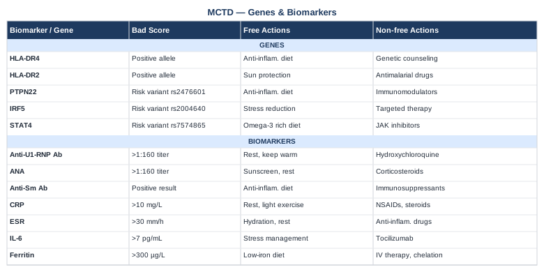
The following table summarizes the genes and biomarkers covered in this article alongside their optimal ranges and action options.
The Wahls Protocol: 10 Things That May Change How You Think About Autoimmune Disease
The Wahls Protocol by Dr. Terry Wahls (neurologist, University of Iowa) is arguably the most scientifically documented dietary intervention for autoimmune disease produced by a practicing physician in the last two decades. Wahls was herself diagnosed with secondary-progressive multiple sclerosis — another autoimmune condition with overlapping inflammatory mechanisms — and eventually became wheelchair-bound while on standard immunosuppressive therapy. After systematically reviewing the cellular and mitochondrial biology of neurodegeneration, she designed a dietary and lifestyle protocol that reversed her condition. Her clinical trial results in MS patients have been published in peer-reviewed literature. The protocol challenges the conventional model of autoimmune disease as purely a problem to be managed with immunosuppression, arguing instead that cellular nutrition is the upstream driver most physicians are not addressing.
Mitochondria are the real center of autoimmune disease, not just immune cells
Wahls' central thesis is that autoimmune disease reflects failing mitochondrial function across immune, neurological, and connective tissue cells. When mitochondria cannot produce adequate ATP, cells fail to regulate immune signaling, maintain epigenetic controls, or repair damage. This framing reorients the problem from "the immune system is attacking the body" to "the cells involved lack the biochemical substrate to regulate themselves." For MCTD patients, this is directly relevant: the connective tissue cells, vascular endothelium, and immune regulatory cells that malfunction in MCTD all depend on mitochondrial output.
You cannot out-medicate a nutritionally depleted immune system
Wahls argues — and supports with clinical trial data — that a body lacking the micronutrients needed to run immune regulatory chemistry will continue producing autoimmune activity regardless of how aggressively it is suppressed. Immunosuppression manages the downstream output of the problem but does not address what the immune cells are missing. This is not an argument against medication — it is an argument for addressing both layers simultaneously.
Nine cups of specific plants per day: why this is biochemistry, not ideology
The Wahls Protocol prescribes three cups of leafy greens, three cups of sulfur-rich vegetables, and three cups of deeply colored plants every day. This is not a general "eat your vegetables" recommendation. Each category targets specific biochemical pathways: leafy greens provide folate, B vitamins, and vitamins K and B2 for mitochondrial and methylation chemistry; sulfur vegetables drive glutathione production and detoxification; colored plants supply antioxidant polyphenols that directly protect mitochondria and immune regulatory cells. Together, these nine cups represent a systematic approach to micronutrient density, not merely dietary preference.
Sulfur vegetables reset the detoxification system
Cabbage, kale, broccoli, onions, garlic, leeks, and mushrooms are the primary sulfur-rich vegetables in the Wahls protocol. Sulfur compounds support the production of glutathione, the body's master antioxidant and a critical cofactor in the detoxification of inflammatory byproducts. Glutathione depletion is consistently documented in lupus and MCTD patients; supporting its synthesis through dietary sulfur is a direct, low-risk intervention with no cycling required.
Deeply colored plants protect the targets of MCTD-related damage
Anthocyanins (from berries, purple cabbage, beets), carotenoids (from orange and red vegetables), and polyphenols (from herbs, dark chocolate, green tea) protect mitochondrial membranes, support vascular endothelial integrity, and modulate NF-κB and interferon signaling. These are precisely the cellular systems under attack in MCTD — vascular endothelium, connective tissue, and immune regulatory circuits. Wahls frames colored plant compounds not as supplemental add-ons but as primary mitochondrial fuels.
The case against gluten and dairy in autoimmune disease
The Wahls Protocol requires elimination of gluten and dairy. Wahls explains this through gut permeability: both gluten and casein (dairy protein) have been shown to increase intestinal permeability in susceptible individuals, allowing partially digested peptides to enter systemic circulation and trigger immune responses. In patients with HLA risk alleles (especially DRB1 and DQ variants), these dietary peptides can directly activate autoreactive immune cascades. This is not a trend — it is a mechanistically coherent intervention for patients with autoimmune disease.
Grass-fed protein and omega-3-rich fish: rebuilding what autoimmunity is destroying
Wahls emphasizes quality protein from organ meats, grass-fed beef, and fatty cold-water fish for a specific reason: these foods provide the amino acids, fat-soluble vitamins, and long-chain omega-3 fatty acids needed to rebuild cellular membranes, support myelin and connective tissue, and maintain immune cell membrane composition. For MCTD patients experiencing progressive muscle and connective tissue involvement, this nutritional support is directly targeted at the affected structures.
Electrical muscle stimulation: the hidden recovery tool Wahls actually used
One of the less publicized elements of the Wahls Protocol is functional electrical stimulation (FES) of muscles — a technique used in rehabilitation medicine that Wahls used on her own leg muscles during her recovery. For MCTD patients with myositis or significant muscle weakness, neuromuscular electrical stimulation (NMES) devices provide a medically supported way to maintain or rebuild muscle mass when voluntary exercise is limited by pain or fatigue. Devices are available in clinical and consumer-grade formats.
The HPA axis is not a soft target — it is biochemically central
Wahls dedicates significant attention to the hypothalamic-pituitary-adrenal (HPA) axis as a direct driver of immune dysregulation. Chronic stress elevates cortisol, which initially suppresses inflammation but over time desensitizes immune cells to cortisol's anti-inflammatory signals — producing rebound inflammation and worsening autoimmune activity. Managing HPA axis tone through sleep, perceived safety, and stress reduction practices is framed not as wellness advice but as immune chemistry management.
Movement as cellular medicine: the discipline of exercising when the body resists
Wahls demonstrates in her clinical trials that progressive aerobic and resistance exercise — tailored to current capacity, even when that capacity is very limited — produces measurable improvements in fatigue, strength, and quality of life in autoimmune conditions. The mechanism includes improved mitochondrial density, anti-inflammatory myokine release (including IL-6 as an anti-inflammatory signal during exercise), and enhanced lymphatic drainage. The key is starting at genuinely appropriate intensity and building progressively rather than forcing effort during flares.
Complementary Approaches with Meaningful Human Evidence for MCTD
Medications and biomarker management are the backbone of MCTD treatment. But several evidence-supported complementary approaches can meaningfully improve symptom burden, functional capacity, and quality of life when applied appropriately. The five below were selected for having human clinical evidence — not just theoretical plausibility — relevant to the mechanisms of MCTD.
Mindfulness-Based Stress Reduction (MBSR)
MBSR is an eight-week structured program combining mindfulness meditation, body scan practices, and gentle movement. It was developed by Jon Kabat-Zinn at the University of Massachusetts Medical School and has since been studied in hundreds of clinical trials. Its relevance to MCTD is multi-layered: it directly reduces cortisol and HPA axis reactivity (which modulates autoimmune flare frequency), it reduces hs-CRP and inflammatory cytokine levels in controlled studies, and it significantly improves fatigue and pain perception in chronic autoimmune conditions.
A 2015 meta-analysis published in JAMA Internal Medicine reviewed 47 randomized controlled trials and found that mindfulness meditation programs produced moderate reductions in anxiety, depression, pain, and fatigue — all commonly experienced in MCTD. The same analysis found consistent effects on psychological well-being without evidence of harm.
Realistic application for MCTD: standard MBSR courses are available online through the University of Massachusetts Medical School and many hospital systems, usually at minimal cost. Starting with 10–15 minutes of daily body scan meditation is a reasonable entry point. During flares, active practices can be reduced to guided breath awareness. The critical factor is daily consistency over months rather than intensity in any single session.
Tai Chi
Tai chi is a slow-movement practice combining coordinated physical movement, breath control, and focused attention. Its combination of very low-intensity movement, parasympathetic activation, and circulatory stimulation makes it particularly suitable for MCTD patients who experience Raynaud's phenomenon, joint pain, and exercise intolerance. Unlike high-intensity exercise, tai chi can be performed during periods of moderate disease activity without triggering flares.
A 2013 systematic review in Rheumatology International examined tai chi across multiple rheumatic conditions and found consistent improvements in pain, physical function, balance, and fatigue. A separate review in PLOS ONE found that tai chi significantly reduced inflammatory markers including IL-6 compared to control groups in older adults with chronic inflammatory conditions.
For MCTD, 20–30 minutes of tai chi performed three to four times per week is a practical starting point. Beginner Yang-style sequences are widely available through video platforms and community programs. Raynaud's patients specifically benefit from tai chi's combination of whole-body circulatory stimulation and peripheral warming through movement, without the vascular stress of high-intensity exercise.
The Autoimmune Protocol (AIP) from Sarah Ballantyne
The Autoimmune Protocol (AIP) is a structured dietary elimination and reintroduction framework developed by Dr. Sarah Ballantyne (PhD, cell biology). It removes all dietary triggers most commonly associated with gut permeability and immune activation — including grains, legumes, dairy, eggs, nightshades, seeds, nuts, and all processed foods — while emphasizing nutrient-dense whole foods, fermented foods, and organ meats. After a minimum elimination period (typically 30–90 days), foods are systematically reintroduced to identify individual triggers. AIP directly addresses gut barrier integrity, microbiome composition, and the dietary drivers of MCTD's relevant immune pathways (especially PTPN22 and IRF5 variants discussed above).
A 2017 pilot study by Konijeti et al. published in Inflammatory Bowel Diseases examined AIP in Crohn's disease patients and found clinically meaningful reductions in disease activity and endoscopic inflammation within 6 weeks, with high adherence rates. While direct MCTD trials do not yet exist, the mechanistic overlaps — gut permeability, microbiome dysbiosis, and innate immune activation — make AIP one of the most structurally coherent dietary interventions for autoimmune connective tissue diseases.
Because MCTD is an autoimmune condition, AIP deserves serious consideration as an adjunct to medical management. Implementation is most successful with a structured plan and ideally dietitian support, as nutrient adequacy must be maintained throughout the elimination phase. The reintroduction phase is as important as the elimination phase — it is the only way to identify which specific foods trigger your individual response.
Breathing-Based Therapies
Breathing interventions are directly relevant to MCTD given the high prevalence of pulmonary involvement (ILD, PAH, pleuritis) and diaphragmatic dysfunction from polymyositis-like features. Diaphragmatic breathing training improves respiratory mechanics, ventilatory efficiency, and oxygen delivery at the tissue level. Separately, slow-paced breathing (4–6 breaths per minute) activates the vagal nerve, shifts the autonomic nervous system toward parasympathetic tone, and reduces sympathetic-driven immune activation — with documented reductions in inflammatory cytokines in controlled studies.
A review of breathing exercises in connective tissue diseases with pulmonary involvement found that supervised respiratory physiotherapy improved DLCO and exercise tolerance in patients with ILD-related restriction. The sessions emphasized diaphragmatic breathing, pursed-lip breathing, and inspiratory muscle training.
Practical application: 10–15 minutes of daily diaphragmatic breathing training (supine position, hand on belly, 5-second inhale, 7-second exhale) can be performed at any disease activity level. During stable periods, add inspiratory muscle training using a respiratory threshold device ($30–$60, widely available) to progressively strengthen ventilatory muscles. This is especially valuable for patients with declining DLCO where preserving respiratory reserve is a priority.
Microbiome-Directed Therapies
The gut microbiome is increasingly recognized as a modulator of systemic autoimmunity — not just gut disease. Dysbiosis (reduced microbial diversity, overgrowth of pro-inflammatory species, reduced short-chain fatty acid producers) drives increased gut permeability, systemic lipopolysaccharide exposure, and altered regulatory T cell development. In lupus, RA, and related conditions, specific microbiome patterns have been associated with disease activity. Several PTPN22 risk carriers (discussed above) show measurable gut permeability abnormalities that can be partially addressed through microbiome restoration.
Multiple randomized trials in SLE and inflammatory arthritis have demonstrated that high-dose, multi-strain probiotic supplementation significantly reduces inflammatory markers (hs-CRP, IL-6) and disease activity scores. A 2020 systematic review in Clinical Rheumatology found that probiotics reduced CRP by a mean of 1.8 mg/L across rheumatic diseases, with the strongest effects in patients with the highest baseline inflammation.
For MCTD, a realistic microbiome restoration protocol includes: prebiotic fiber diversification (varied plant foods, aiming for 30+ different plant species per week, as studied by the Sonnenburg Lab at Stanford); daily consumption of fermented foods (yogurt, kefir, sauerkraut, kimchi); a high-diversity probiotic formulation during and after antibiotic courses; and consideration of comprehensive microbiome testing to identify specific patterns worth targeting. This is not a replacement for medical management but an important complementary layer that addresses an upstream driver of immune dysregulation.
Conclusion
Mixed connective tissue disease does not lend itself to simple management frameworks. Its overlapping features, unpredictable course, and serious pulmonary complications demand both vigilance and precision. The seven biomarkers covered here — anti-U1-RNP, complement C3/C4, hs-CRP, CBC, ferritin, vitamin D, and NT-proBNP/DLCO — provide a longitudinal tracking system that goes well beyond a routine follow-up visit. The five genetic variants — HLA-DRB1, STAT4, IRF5, PTPN22, and TNFAIP3 — add a structural layer that can explain why certain pathways are more active and what may compensate for them. Add to these the nutritional and lifestyle frameworks from Wahls, Ballantyne, and the evidence-based complementary approaches, and the picture that emerges is not a cure but something genuinely useful: a coherent map for making better decisions.
The next smart step is straightforward. Review the biomarkers you have not tested recently — particularly DLCO, NT-proBNP, and vitamin D — and discuss adding them to your next rheumatology visit. Bring your genetic testing results if you have them. And begin with one dietary or lifestyle change that addresses the biomarker or pathway most out of range for you. Precision beats perfection. Starting with what is most measurable and most actionable is the right move.
Cardiovascular Respiratory Skin Autoimmune
Musculoskeletal: Joint Conditions Muscle Conditions
Autoimmune: Inflammatory Conditions Connective Tissue Conditions