This article contains AI generated content.
· UpdatedMorquio Syndrome Genes and Biomarkers — 2 Genes And 6 Biomarkers To Track
Introduction
Living with Morquio syndrome — or caring for someone who has it — places you in a position that most clinicians encounter only rarely, if ever. The diagnosis often comes slowly, the specialists are few, and the gap between what standard medical care offers and what daily life actually requires can be enormous. In a condition this rare, being well-informed is not optional; it shapes the quality of every decision made at every stage of the disease.
What compounds this reality is that most chronic-disease guidance simply does not apply. The skeletal changes, the progressive respiratory restriction, the cardiac complications — these are not problems addressed by advice to eat better and move more. Morquio syndrome is driven by a specific lysosomal enzyme deficiency that causes glycosaminoglycan accumulation throughout connective tissue. Understanding that biology is where useful guidance has to start.
This article focuses on what is measurable and actionable: the two genes that cause Morquio syndrome, and six biomarkers that together reflect the disease's impact on its major target systems. These are not abstract numbers — they reveal whether disease burden is increasing, whether enzyme replacement therapy is working, and where secondary complications are developing before they become emergencies.
Better information does not mean easy answers, and this article promises none. What it offers is a structured framework — starting with biomarkers as the most practical daily tracking tool, followed by the genetic picture that explains why those biomarkers behave as they do, a section on breathing as an underestimated but trainable dimension of Morquio management, and a look at complementary approaches with genuine supporting evidence. Each section is designed to raise the quality of decisions patients and families can bring to their care team.
Summary
This article covers 2 genes — GALNS and GLB1 — whose mutations account for all known forms of Morquio syndrome, and 6 biomarkers that together track disease burden, bone health, systemic inflammation, and cardiac risk. For each biomarker, you will find exactly how to measure it and what a bad result means biologically — plus what can realistically be done, both with and without supplements or equipment, including dosing, frequency, cycling, and side effects. The genetics section follows the same structure: what each gene mutation causes downstream, and where targeted support makes a measurable difference. A breathing science section translates the latest respiratory neuroscience — covering ten practical insights — into tools for the pulmonary complications that Morquio creates but that rarely receive systematic attention. The article closes with three evidence-supported complementary modalities adapted with the safety constraints that Morquio's cervical instability demands. If you have ever wondered what beyond enzyme replacement therapy deserves serious attention in Morquio syndrome, this article is a practical map.
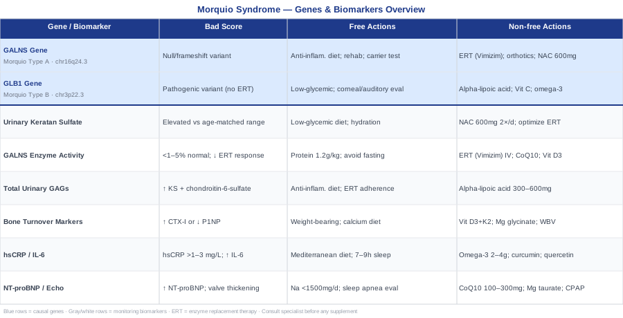
6 Biomarkers Worth Tracking in Morquio Syndrome
Biomarkers serve two distinct functions in Morquio syndrome: some are disease-specific, measuring the glycosaminoglycan accumulation that defines the condition; others are system-specific, capturing the secondary damage to bone, heart, and immune function that develops over time. Monitoring both categories provides a complete picture of where the disease stands at any given time — and where intervention will have the highest return.
1. Urinary Keratan Sulfate
Urinary keratan sulfate (KS) is the most disease-specific and diagnostically precise biomarker in Morquio Type A (MPS IVA). Because GALNS enzyme deficiency prevents the lysosomal breakdown of keratan sulfate and chondroitin-6-sulfate, these substrates accumulate in tissues and are excreted in urine in significantly elevated quantities. Urinary KS reflects total disease burden directly and, crucially, responds measurably to enzyme replacement therapy — making it the primary tool for assessing both disease severity and treatment response.
KS levels naturally decline with age even in unaffected individuals, so results must always be interpreted against age-matched reference ranges. A persistently elevated KS despite ERT may indicate inadequate dosing, antibody development against the enzyme, or insufficient infusion intervals.
How to Measure It
Spot urine or first-morning void sample; measured by tandem mass spectrometry (LC-MS/MS) or ELISA; normalized to urinary creatinine to correct for dilution. Requires a specialized metabolic laboratory — not available at standard hospital labs. Cost range: $150–$400. Results typically returned in 1–2 weeks. This test must be requested specifically as part of a metabolic disease panel.
If Keratan Sulfate Is High: The Plan Without Supplements
No lifestyle change has been shown to meaningfully normalize urinary KS in affected individuals independently of ERT. The supportive approach focuses on reducing upstream substrate availability: a low-glycemic, whole-food diet reduces circulating precursors for GAG synthesis; adequate hydration supports renal clearance; minimizing infectious illness reduces the inflammatory upregulation of GAG production. Work with the metabolic team to review ERT dosing frequency and antibody titer status if KS remains persistently elevated despite apparent ERT compliance.
If Keratan Sulfate Is High: The Plan With Supplements or Equipment
No over-the-counter supplement restores GALNS enzyme activity or directly lowers KS. However, N-acetylcysteine (NAC) at 600 mg twice daily supports glutathione production and may reduce the oxidative stress that lysosomal KS accumulation generates — evidence is early-stage from lysosomal storage disease models, not Morquio-specific human trials. Cycle for 8–12 weeks, then reassess. Take with food to reduce nausea. Side effects: GI discomfort, particularly on an empty stomach; inform the treating team before starting due to potential medication interactions. Enzyme replacement therapy (elosulfase alfa) remains the only medical intervention that directly reduces KS and should be optimized as the primary target before considering adjuncts.
2. GALNS Enzyme Activity
GALNS enzyme activity measured in leukocytes or dried blood spots is the diagnostic cornerstone of Morquio Type A. Quantifying N-acetylgalactosamine-6-sulfatase function confirms diagnosis when activity is severely reduced — typically below 1–5% of normal — distinguishes affected individuals from heterozygous carriers, and, when tracked in patients receiving ERT, can detect the development of neutralizing antibodies against the infused enzyme.
GALNS activity does not require frequent re-measurement in stable diagnosed patients, but it functions as a critical baseline reference and becomes important again if treatment response appears to diminish over time.
How to Measure It
Whole blood sample (purple-top tube, kept cold during transport) for leukocyte enzyme assay, or dried blood spot card for screening purposes. Cost range: $200–$600. Available through specialized metabolic laboratories and newborn screening programs in regions where MPS IVA panel testing is included. Antibody titer testing should be performed alongside enzyme activity in patients receiving elosulfase alfa.
If Enzyme Activity Is Very Low: The Plan Without Supplements
No dietary or lifestyle intervention increases GALNS enzyme production in affected individuals — the underlying genetic defect prevents it. Supportive steps focus on optimizing the cellular environment: adequate dietary protein (1.2–1.5 g/kg/day) supports cellular repair processes; avoiding prolonged fasting reduces catabolic stress on already-challenged cells; physical activity within safe orthopedic limits maintains overall metabolic function. These steps do not change enzyme levels but reduce the cumulative burden on a system already under significant stress.
If Enzyme Activity Is Very Low: The Plan With Supplements or Equipment
The only intervention that directly addresses GALNS deficiency is intravenous enzyme replacement therapy with elosulfase alfa (Vimizim), administered weekly under specialist prescription. Earlier initiation in the disease course is consistently associated with better functional outcomes. Research into pharmacological chaperone therapy — small molecules that stabilize misfolded GALNS protein and improve lysosomal targeting — is active for specific missense mutations but has no commercially available product at the time of writing. To support the cellular environment in which delivered enzyme must function, CoQ10 (ubiquinol form) at 100–200 mg daily with a fat-containing meal may support mitochondrial-lysosomal energy coupling; continuous use; side effects are rare but may include mild GI discomfort at higher doses. Pairing with vitamin D3 at 2000–4000 IU daily ensures enzymatic and cellular processes dependent on vitamin D receptor signaling are not additionally compromised.
3. Total Urinary Glycosaminoglycans
While keratan sulfate is Morquio-specific, total urinary glycosaminoglycans (GAGs) measure the broader picture of lysosomal storage disease burden. Total GAG testing is more widely available and less expensive than KS-specific assays, making it a practical screening and follow-up tool — particularly in settings where specialized metabolic testing access is limited. Qualitative GAG electrophoresis can also distinguish Morquio from other mucopolysaccharidoses by identifying which specific GAG fractions are elevated.
In Morquio Type A, the elevated fractions are keratan sulfate and chondroitin-6-sulfate. Think of total GAGs as the wide-angle lens and urinary KS as the close-up: together they provide both breadth and specificity.
How to Measure It
Spot urine or 24-hour urine collection; analyzed by spectrophotometric method (dimethylmethylene blue assay) or LC-MS/MS; normalized to creatinine. Cost range: $50–$200. More widely available than KS-specific testing — accessible at many reference laboratories and some hospital labs. Qualitative GAG electrophoresis adds $50–$100 and provides pattern information that is diagnostically useful when the MPS type has not been confirmed.
If Total GAGs Are Elevated: The Plan Without Supplements
There are no lifestyle interventions proven to normalize total GAG excretion in Morquio syndrome independently of ERT. An anti-inflammatory, low-glycemic dietary approach reduces systemic factors that upregulate GAG synthesis; staying well hydrated supports renal excretion; consistent ERT adherence is the primary lever. Monitoring trends over time provides more clinical value than any single measurement — a rising trajectory despite stable ERT warrants urgent discussion with the metabolic team.
If Total GAGs Are Elevated: The Plan With Supplements or Equipment
Alpha-lipoic acid at 300–600 mg daily with meals has early supportive evidence in lysosomal storage disease models for reducing oxidative consequences of GAG accumulation — no Morquio-specific human trial exists, and this application remains speculative. Apply cautiously and with specialist awareness. Cycle 8 weeks on, 4 weeks off to reduce the risk of B-vitamin depletion with long-term use. Side effects: nausea, skin rash at higher doses, potential interaction with thyroid medications. Pair with a nutrient-dense, antioxidant-rich dietary pattern — olive oil, berries, leafy greens — for a synergistic effect on oxidative burden.
4. Bone Turnover Markers
Skeletal involvement is the defining clinical feature of Morquio syndrome: the dysostosis multiplex, the spinal deformity, the joint laxity, the genu valgum, the progressive loss of height. Bone turnover markers — specifically bone-specific alkaline phosphatase (BAP), C-terminal telopeptide (CTX-I), and procollagen type 1 N-terminal propeptide (P1NP) — reflect the rate at which bone is being formed and resorbed. GAG accumulation in cartilage disrupts the extracellular matrix that skeletal remodeling depends on, creating a chronically abnormal pattern. Tracking these markers over time helps detect accelerated bone loss, estimate fracture risk, and evaluate whether physical and nutritional support is genuinely protective.
How to Measure It
Standard ALP: basic chemistry panel, $10–$30. Bone-specific ALP: $60–$150 at reference labs. CTX-I (bone resorption marker): $60–$150; requires a fasting blood draw before noon for reproducibility — CTX rises significantly with food intake, confounding comparison. P1NP (bone formation marker): $60–$150. Testing CTX and P1NP together every 6–12 months gives a longitudinal view of remodeling balance that standard ALP alone cannot provide.
If Bone Turnover Is Abnormal: The Plan Without Supplements
Weight-bearing activity within safe orthopedic limits stimulates osteoblast activity and bone formation — but this comes with significant caveats in Morquio syndrome given the cervical spine instability and joint hypermobility that characterize the condition. Any exercise or loading plan must be designed in collaboration with a physical therapist familiar with Morquio and cleared by the orthopedic or neurosurgical team before initiation. Low-impact weight-bearing — standing frames, supported walking, hydrotherapy — is often achievable and beneficial. Maximize sun exposure for endogenous vitamin D synthesis. Build calcium into the diet through dairy or fortified alternatives, leafy greens, and sardines with bones.
If Bone Turnover Is Abnormal: The Plan With Supplements or Equipment
Vitamin D3 at 2000–4000 IU daily with a fat-containing meal; target serum 25-OH vitamin D between 40–60 ng/mL; monitor blood levels every 3–6 months; at excessive doses (above 10,000 IU chronically), hypercalcemia is a real risk — never exceed tested and monitored doses. Pair with vitamin K2 (MK-7 form) at 100–200 mcg daily to direct calcium toward bone rather than vascular tissue; continuous daily use is appropriate; contraindicated with warfarin without physician coordination, as K2 directly interferes with warfarin dosing. Magnesium glycinate at 300–400 mg daily activates vitamin D and supports bone matrix synthesis; take in the evening for additional sleep benefit; loose stools are the most common side effect at higher doses. Low-intensity whole-body vibration (WBV) platforms at 0.3–0.6 g have preliminary evidence for improving bone mineral density in patients with limited mobility — consult a physical therapist familiar with Morquio before initiating, as not all positions are safe given spinal involvement.
5. Inflammatory Markers: hsCRP and IL-6
The accumulation of glycosaminoglycans in tissues is not metabolically passive. It activates lysosomal stress responses, generates reactive oxygen species, and triggers inflammatory signaling through toll-like receptors and the NF-kB pathway. High-sensitivity C-reactive protein (hsCRP) and interleukin-6 (IL-6) are practical proxies for this systemic inflammatory burden. In Morquio syndrome, chronically elevated inflammation accelerates tissue damage beyond what enzyme deficiency alone would cause — it contributes to the cardiac and pulmonary complications that develop over years, and it represents an additional modifiable target.
Thomas Dayspring and preventive cardiology researchers consistently flag hsCRP as among the most actionable inflammatory biomarkers given its responsiveness to lifestyle interventions — and that logic applies in Morquio as much as in cardiovascular disease.
How to Measure It
hsCRP: standard blood draw, widely available, $20–$50. Request high-sensitivity CRP specifically — the standard CRP test lacks the sensitivity needed to detect low-grade chronic inflammation. IL-6: blood draw, $100–$250 at reference labs; adds granularity when hsCRP is borderline or trending upward. Measure every 3–6 months to track trends — single-point elevations have limited interpretive value; patterns over time are what matter.
If Inflammatory Markers Are Elevated: The Plan Without Supplements
A Mediterranean-style anti-inflammatory dietary pattern — olive oil, fatty fish, vegetables, legumes, whole grains, with minimal ultra-processed foods and added sugars — consistently reduces hsCRP across diverse clinical populations. Sleep is one of the most powerful anti-inflammatory interventions available: 7–9 hours nightly significantly reduces IL-6 and TNF-α compared to chronic sleep restriction. Stress reduction, covered in the complementary approaches section below, directly modulates inflammatory cytokine production through the HPA axis. Each of these strategies is cumulative — combining them consistently produces greater hsCRP reduction than any single approach in isolation.
If Inflammatory Markers Are Elevated: The Plan With Supplements or Equipment
Omega-3 fatty acids (EPA + DHA) at 2–4 g daily from concentrated fish oil is among the best-studied interventions for hsCRP reduction; take with the largest meal to maximize absorption and minimize GI side effects; use continuously; at doses above 3 g/day, mild anticoagulant effects are possible — inform any prescribing physician managing antiplatelet or anticoagulant medications. Curcumin with piperine (500–1000 mg curcumin + 5–10 mg piperine daily) inhibits NF-kB signaling and has demonstrated IL-6 reduction in multiple trials; cycle 8 weeks on, 4 weeks off; GI upset is the most common side effect; avoid high-dose curcumin in patients with gallbladder obstruction or on blood thinners. Quercetin at 500 mg daily adds anti-inflammatory and mast cell-stabilizing effects; continuous use; generally well tolerated; take with vitamin C for improved bioavailability and additive antioxidant benefit.
6. Cardiac Biomarkers: NT-proBNP and Echocardiography
Cardiac involvement in Morquio syndrome is serious and frequently underappreciated. GAG deposition in cardiac valve leaflets and vessel walls causes progressive aortic valve thickening, mitral valve regurgitation, and over time, ventricular dysfunction. NT-proBNP (N-terminal pro-B-type natriuretic peptide) is a sensitive blood marker of cardiac wall stress: it rises when the heart is straining to compensate for valvular disease or ventricular impairment, often before clinical symptoms develop. Echocardiography provides the structural picture that NT-proBNP cannot — valve anatomy, regurgitation severity, ventricular dimensions, and systolic function.
Together, NT-proBNP and echocardiography create an early-warning system for cardiac decline, enabling proactive intervention rather than reactive crisis management.
How to Measure It
NT-proBNP: standard blood draw, $50–$120, widely available. Echocardiogram: $500–$1500 depending on facility; ideally performed at a center with MPS disease experience or reviewed by a cardiologist familiar with lysosomal storage disorders. Recommended surveillance: echocardiogram every 1–2 years for clinically stable patients; NT-proBNP as an interim check every 6–12 months. EKG adds rhythm monitoring for arrhythmia detection and costs $100–$300. Discuss with the cardiologist the specific NT-proBNP threshold that should trigger an expedited echocardiogram.
If Cardiac Markers Are Elevated: The Plan Without Supplements
Minimize sodium intake to under 1500 mg/day to reduce fluid retention and cardiac preload. Avoid isometric exercise and high-intensity aerobic exertion without explicit cardiologist clearance — in the setting of valvular disease, these create cardiac stress disproportionate to their cardiovascular benefit. Maintain appropriate body weight; even modest excess increases cardiac demand measurably. Prioritize evaluation for sleep-disordered breathing: obstructive sleep apnea is common in Morquio due to structural airway narrowing and is an independent driver of cardiac stress. Polysomnography should be performed if not recently completed, and treatment implemented if significant apnea is identified.
If Cardiac Markers Are Elevated: The Plan With Supplements or Equipment
CoQ10 (ubiquinol form) at 100–300 mg daily supports cardiac mitochondrial energy production and has been studied in heart failure and valvular disease contexts; take with a fat-containing meal for absorption; continuous use; side effects are rare at standard doses but can include mild GI discomfort at higher amounts. Magnesium taurate at 300 mg daily supports myocardial function and reduces arrhythmia risk; take at bedtime; generally well tolerated. Omega-3 fatty acids at 2–3 g EPA+DHA daily address both cardiac inflammation and triglycerides, which contribute to valvular dysfunction. For patients with confirmed sleep apnea, CPAP or BiPAP therapy is among the highest-impact cardiac-protective interventions available: nightly use reduces nocturnal hypoxia, cortisol surges, and inflammatory signaling that would otherwise chronically stress an already-compromised heart.
The biomarkers above all trace back to a common origin: the genotype of the affected individual. Understanding the specific gene mutation adds critical context to how these numbers should be interpreted — and what targeted approaches make the most sense.
The Genetic Foundation of Morquio Syndrome
Morquio syndrome is caused by mutations in one of two genes, depending on which enzyme is deficient and which glycosaminoglycans accumulate as a result. Both types are autosomal recessive — two pathogenic copies are required for disease expression — and both can vary considerably in severity depending on the specific nature of the variant. Knowing which gene is affected, and what category of mutation it carries, shapes the prognosis, the monitoring timeline, and the precision with which supplementary and medical interventions are applied.
The GALNS Gene — Morquio Type A
The GALNS gene on chromosome 16q24.3 encodes N-acetylgalactosamine-6-sulfatase, the lysosomal enzyme responsible for cleaving sulfate groups from two specific glycosaminoglycans: keratan sulfate and chondroitin-6-sulfate. When both gene copies carry pathogenic variants, these substrates cannot be degraded and accumulate progressively throughout the body — in skeletal cartilage, the cornea, cardiac valve leaflets, the upper airway, and indirectly in the nervous system through mechanical compression from skeletal deformity.
Over 250 distinct pathogenic variants in GALNS have been catalogued across affected populations. The genotype-phenotype correlation is imperfect but clinically real. Null mutations — nonsense variants, frameshift mutations, large deletions — that eliminate enzyme production entirely consistently produce the most severe phenotype: earlier onset, faster skeletal progression, and greater respiratory and cardiac involvement. Missense mutations that reduce but do not abolish enzyme activity often produce an attenuated phenotype with later onset and slower progression. Compound heterozygosity — one null variant and one missense variant — typically produces an intermediate clinical picture.
This genotype-informed picture matters for clinical timing: a patient with two null GALNS mutations warrants earlier initiation of ERT and more intensive multidisciplinary surveillance than a patient with two attenuated missense variants and a mild clinical presentation. Genetic counseling is essential for all families planning future pregnancies.
If GALNS Has a Pathogenic Variant: The Plan Without Supplements
No lifestyle or dietary intervention restores GALNS enzyme production or meaningfully alters gene expression in affected individuals. What can be addressed are the downstream consequences of the mutation: the oxidative stress from lysosomal dysfunction, the substrate availability for GAG synthesis, and the physical consequences of skeletal and respiratory involvement.
An anti-inflammatory, low-glycemic diet reduces circulating precursors available for GAG synthesis. Adequate dietary protein (1.2–1.5 g/kg/day) supports the cellular repair processes that are chronically challenged by lysosomal dysfunction. Physical rehabilitation — designed specifically around the cervical instability and joint hypermobility of Morquio — preserves functional capacity and slows secondary musculoskeletal deterioration. Respiratory muscle training (described in detail in the breathing section below) partially compensates for the thoracic restriction caused by skeletal involvement.
Carrier testing of first-degree family members is a high-yield, non-pharmacologic action with no side effects: it prevents delayed diagnosis in future family members and enables informed reproductive decision-making.
If GALNS Has a Pathogenic Variant: The Plan With Supplements or Equipment
Elosulfase alfa (Vimizim), administered intravenously every week, is the disease-modifying medical treatment for GALNS deficiency. Earlier initiation in the disease course is consistently associated with better functional outcomes. This decision requires specialist oversight and belongs entirely to the metabolic team.
Adjunct supplementation addresses the downstream effects of enzyme deficiency rather than the deficiency itself:
Vitamin D3 + K2 (2000–4000 IU D3 daily paired with 100–200 mcg MK-7 daily): protects against accelerated bone fragility driven by KS accumulation in cartilage and bone matrix. Daily use; blood monitoring every 3–6 months for vitamin D levels; warfarin interaction with K2 requires physician coordination.
NAC at 600 mg twice daily: supports glutathione production and reduces the oxidative burden from lysosomal KS accumulation. Cycle 8 weeks on, 4 weeks off. GI side effects possible, particularly on an empty stomach; take with food; inform the treating team before starting.
Orthotic and assistive equipment — cervical collars for atlantoaxial instability management, knee-ankle-foot orthoses, walking aids, adaptive seating, communication devices — represents some of the highest-impact non-pharmacologic interventions available. These do not address the enzyme deficiency directly but protect against its most disabling functional consequences.
For patients carrying specific missense mutations, emerging pharmacological chaperone research represents the most promising future therapeutic direction. Small molecules designed to stabilize misfolded GALNS protein and improve lysosomal trafficking are under active clinical development — no approved product exists at the time of writing, but this space is worth monitoring closely for relevant trial announcements.
The GLB1 Gene — Morquio Type B
Morquio Type B (MPS IVB) is caused by pathogenic variants in the GLB1 gene on chromosome 3p22.3, which encodes beta-galactosidase. This enzyme cleaves terminal galactose residues from multiple substrates, including keratan sulfate. When GLB1 function is severely reduced, keratan sulfate accumulates, producing a clinical picture that overlaps significantly with MPS IVA: skeletal dysplasia, short stature, joint laxity, and corneal clouding. Key differences include the absence of chondroitin-6-sulfate accumulation, a generally milder severity in most described cases, and — critically — the absence of an approved enzyme replacement therapy.
An important distinction that frequently confuses families: the GLB1 gene is also responsible for GM1 gangliosidosis, a neurologically devastating condition with a very different prognosis. The same gene, a different mutation affecting a different catalytic function of the same enzyme, produces a completely different disease. MPS IVB results from mutations that selectively impair keratan sulfate cleavage while preserving sufficient activity toward GM1 ganglioside — which is why neurological involvement is absent or minimal in MPS IVB. This distinction is essential for prognosis and for how families understand what the diagnosis means for long-term trajectory.
If GLB1 Has a Pathogenic Variant: The Plan Without Supplements
Because no ERT exists for beta-galactosidase deficiency in MPS IVB, the non-pharmacologic management strategy becomes even more central. Skeletal care, respiratory monitoring, cardiac surveillance, and functional rehabilitation parallel the MPS IVA approach closely, with the same safety constraints around cervical instability and joint hypermobility.
Corneal and auditory monitoring deserves particular emphasis in MPS IVB: beta-galactosidase deficiency commonly causes corneal clouding and sensorineural hearing impairment, both of which respond meaningfully to early detection. Protective eyewear, surgical options for corneal transplant, and hearing aids with communication support can substantially preserve quality of life when identified early. Regular ophthalmology and audiology appointments should be built explicitly into the care calendar — not left to symptom-driven scheduling.
A low-glycemic, anti-inflammatory diet reduces upstream substrate availability and supports overall metabolic function. A registered dietitian with metabolic disease experience can structure a practical eating pattern that achieves this without creating nutritional deficiencies in a growing child or an adult managing complex care needs.
If GLB1 Has a Pathogenic Variant: The Plan With Supplements or Equipment
Without an ERT option, the supplementation strategy focuses on cellular health, oxidative burden, and organ system protection:
Alpha-lipoic acid at 300 mg daily with meals: reduces oxidative stress from lysosomal dysfunction; cycle 8 weeks on, 4 weeks off; side effects include nausea and potential interaction with thyroid medications; avoid in thiamine-deficient states. Vitamin C at 500–1000 mg daily: supports collagen synthesis — particularly relevant given the connective tissue involvement — and antioxidant defense; continuous use; GI discomfort at higher doses; kidney stone risk with megadosing in susceptible individuals — stay well hydrated. Omega-3 fatty acids at 2–3 g EPA+DHA daily: anti-inflammatory support across all affected organ systems; continuous use; blood-thinning effects at higher doses as described above. Magnesium glycinate at 300 mg daily: musculoskeletal comfort, sleep quality, and bone matrix support; take at bedtime; generally well tolerated.
Assistive and adaptive equipment — mobility aids, orthotics, hearing aids, adaptive seating, communication technology — is the most impactful category of non-medical intervention for MPS IVB, particularly given the therapeutic gap left by the absence of ERT. Early physiotherapy and occupational therapy assessment should be considered mandatory components of care, not optional add-ons.
Understanding both the genetics and the biomarkers creates a solid foundation for monitoring and intervention. But one dimension of Morquio management rarely receives the systematic attention it deserves — and that is the way breathing mechanics can be deliberately trained and supported even within the structural constraints of the disease.
Breathing Science and Morquio Syndrome: What the Research Suggests
Respiratory complications — thoracic cage restriction, tracheal narrowing, recurrent pulmonary infections, obstructive sleep apnea — are among the leading causes of morbidity and early mortality in Morquio syndrome. What receives far less attention is that breathing mechanics and respiratory muscle function can be trained and optimized, at least partially, even within structural limitations. The neuroscience of breathing, covered extensively by Andrew Huberman on the Huberman Lab podcast and drawing on foundational research by neurobiologists including Jack Feldman, Mark Krasnow, and David Raichlen, has practical implications that translate directly into the respiratory challenges Morquio creates. The ten insights below represent the most relevant takeaways for this specific population.
1. CO2 Tolerance Is Trainable and Clinically Relevant
Perceived breathlessness is driven more by CO2 accumulation than by absolute oxygen depletion. In Morquio patients, altered breathing mechanics can dysregulate the CO2/O2 balance, triggering an earlier "air hunger" signal than the actual oxygen deficit warrants. Gradually improving CO2 tolerance through measured breath-hold training — starting with the BOLT test (Body Oxygen Level Test: hold breath after a normal exhale, measure seconds to the first involuntary urge to breathe) — trains brainstem chemoreceptors to tolerate higher CO2 before triggering the distress signal. This should be approached conservatively; patients with severe respiratory compromise must discuss breath-hold practice with a pulmonologist before initiating.
2. Nasal Breathing Improves Oxygen Uptake at Every Breath
Nasal breathing generates nitric oxide in the paranasal sinuses, which dilates pulmonary blood vessels and improves alveolar oxygen uptake by an estimated 10–15% compared to mouth breathing. For a Morquio patient with already-reduced thoracic volume, that efficiency gain at each breath cycle is significant. Establishing nasal breathing as the default during all non-maximal exertion is a zero-cost, no-equipment intervention. At night, mouth tape (Myotape or similar devices designed specifically for sleep) enforces nasal breathing — but only after polysomnography has ruled out significant obstructive sleep apnea. Using mouth tape with severe apnea is unsafe.
3. The Physiological Sigh Resets Respiratory Rhythm After Exertion
The physiological sigh — a double nasal inhale (short first breath to partial capacity, second breath to complete full inflation) followed by a long, complete exhale — is the body's most efficient mechanism for reinflating collapsed alveoli and restoring normal blood gas balance. Practicing 1–2 deliberate physiological sighs after physical effort helps Morquio patients recover respiratory rhythm more quickly and reduce post-exertional air hunger. No equipment, no training time, no meaningful side effects.
4. Diaphragmatic Breathing Expands Available Functional Capacity
In Morquio syndrome, thoracic cage expansion is structurally restricted. Diaphragmatic movement — downward piston motion that produces belly expansion rather than chest rise — is often less constrained and can partially compensate. 5–10 minutes of deliberate diaphragmatic breathing practice daily, with focused attention on belly expansion through each inhale, preserves and develops this compensatory capacity over weeks to months. Studies in restrictive lung conditions confirm that diaphragmatic breathing reduces respiratory rate, increases tidal volume, and reduces respiratory muscle fatigue across sessions.
5. Sleep-Disordered Breathing Must Be Actively and Repeatedly Investigated
Obstructive sleep apnea is significantly more prevalent in Morquio syndrome than in the general population due to macroglossia, tracheal narrowing, and midface hypoplasia. Huberman has highlighted consistently that untreated sleep-disordered breathing creates a cycle of hypoxia, inflammatory upregulation, cortisol elevation, and cardiac stress that amplifies every other disease process simultaneously. Polysomnography should be performed and repeated as the disease progresses — a single negative sleep study years ago does not remain reassuring as airway anatomy changes. Any significant apnea index warrants treatment matched to severity: CPAP, BiPAP, or positional therapy.
6. Inspiratory Muscle Training Builds Measurable Reserve
Devices such as the Threshold IMT or POWERbreathe provide calibrated resistance to inhalation, progressively strengthening the diaphragm and accessory inspiratory muscles in a manner analogous to resistance training for skeletal muscle. Studies in neuromuscular disease and chronic obstructive respiratory conditions show meaningful improvements in maximum inspiratory pressure and functional endurance capacity. Begin at the lowest resistance setting; increase gradually under physiotherapy supervision; 15–20 minute sessions, 5 days per week. Obtain pulmonologist clearance before starting.
7. Box Breathing Manages the Autonomic Amplification of Respiratory Symptoms
The autonomic nervous system and the respiratory system are bidirectionally linked: anxiety and sympathetic activation worsen respiratory efficiency, and respiratory distress amplifies anxiety. For Morquio patients managing chronic pain, procedural anxiety, and the ongoing psychological load of a progressive condition, box breathing — 4-count inhale, 4-count hold, 4-count exhale, 4-count hold — activates the parasympathetic branch, reducing cortisol and inflammatory cytokine release. Five minutes before sleep or before a stressful procedure represents a practical and evidence-supported entry point, with documented effects on heart rate variability and cortisol reduction.
8. CO2 Retainers Need Modified Protocols — This Is a Safety Point
Patients with advanced Morquio-related respiratory compromise who have developed CO2 retention (hypercapnia) cannot use standard CO2 tolerance training or any form of hyperventilation technique, as these can disturb an already-precarious respiratory drive. Any breathing protocol beyond the physiological sigh and basic diaphragmatic breathing requires pulmonologist review in patients with known hypercapnia or severely reduced forced vital capacity. This is not a caution for mild disease — it is a hard contraindication for advanced respiratory compromise.
9. Upright Positioning Maximizes Available Respiratory Reserve
Recumbent positioning further compresses an already-restricted thoracic cavity in Morquio patients. Strategic upright positioning — using wedge pillows, adjustable bed frames, or specifically tilted seating systems — during rest and sleep maximizes available respiratory excursion and measurably reduces nocturnal hypoxia in restrictive lung conditions. This is a zero-cost, zero-side-effect intervention that is frequently overlooked in care planning.
10. Spirometry Tracking Catches Decline Before Symptoms Develop
Regular spirometry — measuring forced vital capacity (FVC), FEV1, and the FVC/FEV1 ratio — tracks the restrictive lung disease trajectory in Morquio syndrome. Many patients experience slowly progressive loss of respiratory function that remains symptomatically silent until reserve is critically depleted. Annual spirometry — or every 6 months in patients with known respiratory involvement — creates the longitudinal data needed to make timely and proactive decisions about ventilatory support. The first spirometry measurement taken at diagnosis or at initial assessment serves as the most important personal baseline the patient will have.
Beyond monitoring and training the respiratory system, several complementary modalities address the broader quality-of-life challenges that Morquio syndrome creates — particularly chronic pain, fatigue, and the psychological weight of managing a complex progressive condition.
Complementary Approaches with Evidence Relevant to Morquio Syndrome
The modalities below have the most relevant supporting evidence for the specific challenges Morquio syndrome creates: chronic pain, musculoskeletal discomfort, fatigue, and psychological stress. All three have been selected for the combination of available clinical evidence and practical applicability within the safety constraints of Morquio syndrome. Given the cervical spine instability common in this condition, any hands-on or physically active approach must be cleared by the neurosurgical or orthopedic team before implementation.
Mindfulness-Based Stress Reduction
Chronic pain in Morquio syndrome is persistent and multidimensional — it arises from joint laxity, skeletal deformity, inflammatory tissue damage, and the secondary consequences of abnormal movement patterns that never fully resolve. Mindfulness-Based Stress Reduction (MBSR), developed by Jon Kabat-Zinn at the University of Massachusetts Medical School, addresses not the source of pain but the nervous system's amplification of the pain signal — a distinction that matters enormously when the source cannot be removed. By training attention and reducing emotional reactivity to physical discomfort, MBSR reduces pain intensity and the psychological distress that compounds physical suffering in chronic conditions.
A meta-analysis of 47 randomized trials by Goyal et al. (JAMA Internal Medicine, 2014) found that mindfulness meditation programs produced clinically significant reductions in anxiety, depression, and pain across diverse chronic disease populations. The evidence is not Morquio-specific — rare disease trials of this scale do not exist — but the underlying mechanisms (central pain sensitization, inflammatory cytokine modulation through stress reduction, HPA axis regulation) operate independently of the specific diagnosis.
MBSR is an 8-week structured program: 2–2.5 hours of group sessions weekly, plus a day-long retreat and 45 minutes of daily home practice. Adapted seated versions remove physical barriers for patients with limited mobility or postural constraints. Starting with a 10-minute daily guided breath-focused practice before committing to the full program builds the habit sustainably. Apps such as Insight Timer and Waking Up provide structured guided sessions, though a formally trained MBSR instructor delivers meaningfully better outcomes than self-directed app use alone, particularly in the initial learning phase.
Low-Level Laser Therapy / Photobiomodulation
Photobiomodulation (PBM) uses specific wavelengths of red (630–660 nm) and near-infrared (810–850 nm) light to stimulate cytochrome c oxidase activity in mitochondria, increasing cellular ATP production. In inflamed or painful joint tissue, this translates to reduced local inflammatory cytokine expression, reduced oxidative stress, and accelerated tissue repair. For Morquio patients dealing with chronic joint pain driven by laxity and abnormal mechanical loading, PBM offers a non-invasive, low-risk option that works through cellular mechanisms rather than requiring intact structural integrity — meaning the underlying skeletal dysplasia does not negate its mechanism.
A systematic review and meta-analysis by Bjordal et al. in Photomedicine and Laser Surgery found consistent short-term pain reduction from PBM compared to sham treatment across musculoskeletal conditions, with the strongest evidence at wavelengths of 630–850 nm delivered at appropriate irradiance levels of 3–5 J/cm² per session. The evidence is not Morquio-specific but targets the inflammatory and mitochondrial dysfunction mechanisms directly relevant to the condition.
At-home PBM panels delivering 660 nm and 850 nm wavelengths are safe for home use at recommended distances and durations: typically 10–20 minutes per session, 4–5 times per week, on affected joint areas. Maintain the manufacturer-recommended distance; avoid direct eye exposure; no additional precautions are needed for standard therapeutic intensities. Allow 3–4 weeks of consistent use before assessing benefit. Early improvements in joint comfort and morning stiffness are the most consistently reported outcomes. No significant adverse effects have been documented at standard therapeutic intensities in available literature.
Massage Therapy
Muscle guarding, myofascial restriction, and compensatory movement patterns are unavoidable consequences of Morquio syndrome's skeletal involvement. Muscles work harder and differently to protect hypermobile joints and maintain posture against a spine that is mechanically compromised. Over time, these compensation patterns generate significant soft tissue tension and fatigue that contribute substantially to daily pain. Therapeutic massage addresses these consequences directly — reducing tension, improving circulation, breaking the guarding cycle, and providing neurological pain relief through gate control mechanisms.
The safety constraint is non-negotiable: Morquio syndrome is strongly associated with atlantoaxial instability — laxity or malformation at the C1–C2 junction — which can cause catastrophic spinal cord compression under mechanical stress applied to the cervical spine. Any massage therapist treating a Morquio patient must be explicitly briefed on this before the first session, and work on the cervical spine must be entirely avoided. Deep tissue pressure, high-velocity techniques, and passive neck range-of-motion movements are absolutely contraindicated. A study published in Complementary Therapies in Medicine (Field, 2016) documented significant reductions in pain, anxiety, and inflammatory markers including cortisol and substance P following regular therapeutic massage in chronic pain populations, supporting its use as a safe adjunct within appropriately defined parameters.
Weekly or bi-weekly 45-minute sessions focused on the thoracic paraspinals, hips, and extremities — avoiding the cervical spine entirely — is a realistic starting frequency, with reduction to monthly maintenance once initial tissue tension has been addressed. Choose a licensed massage therapist with experience in hypermobility syndromes or connective tissue disorders; their technique will be appropriately gentle and targeted, avoiding the aggressive pressure that can destabilize lax joints. Bringing a written list of contraindications and safe zones to every session is a practical safety measure that does not require the therapist to fully understand the underlying rare disease.
Conclusion
Morquio syndrome is defined at the genetic level — by mutations in GALNS or GLB1 — but its trajectory is shaped at every stage by how thoroughly its downstream effects are monitored and managed. The six biomarkers covered here provide a concrete, measurable framework for doing exactly that: tracking disease-specific GAG accumulation, skeletal remodeling dynamics, systemic inflammation, and cardiac function in parallel, rather than waiting for symptoms to announce what a measurement would have caught months earlier.
The practical next step is clear: review which of these biomarkers have been measured recently, identify the gaps in the current monitoring picture, and bring that list to the next metabolic or multidisciplinary appointment. Whether the most urgent action is optimizing ERT dosing based on urinary keratan sulfate trends, scheduling an overdue echocardiogram, beginning a structured respiratory training program, or starting vitamin D3 and K2 to protect bone — each is a specific, evidence-grounded step that narrows the gap between what medicine currently offers and what living well with this condition actually requires. In a rare disease, taking systematic ownership of that monitoring picture is not a supplement to care; it is part of it.