This article contains AI generated content.
· UpdatedMuckle-Wells Syndrome: 3 Key Genes And 6 Biomarkers To Track
Introduction
Living with Muckle-Wells Syndrome (MWS) is a particular kind of exhaustion. The disease cycles between flares and quieter periods, but neither feels entirely safe. Flares bring fever, rash, joint pain, and sometimes progressive hearing loss; the quiet periods carry the unseen risk that chronic inflammation is silently damaging the kidneys and other organs. Most people diagnosed with MWS spent years in diagnostic limbo before a geneticist or autoinflammatory specialist finally recognized the pattern. That experience of being dismissed, or told it was something less serious, leaves a mark. If that resonates, this article is written with your experience in mind.
The challenge with MWS is that generic inflammation advice does not reach the molecular level where the disease actually operates. The driver is not a lifestyle imbalance — it is a gain-of-function mutation in NLRP3, a gene that controls the body's most powerful intracellular alarm system. No amount of stress reduction or anti-inflammatory diet will correct a constitutively overactive inflammasome protein. But that is not the full picture, because lifestyle, monitoring, and targeted interventions do matter — just in ways specific to this disease's biology, not generic wellness platitudes.
This article takes a more precise approach. The first major section covers six biomarkers that are the most clinically meaningful for MWS — not just broad inflammation markers, but the specific signals used to track disease activity, guide treatment adjustments, and catch the most dangerous complication (systemic amyloidosis) before it causes irreversible damage. For each biomarker, you will find a clear explanation of what it measures, how to get it tested and at what cost, what abnormal results mean, and what concrete steps can improve it — with and without supplementation.
The genetics section then unpacks the three most relevant genes and what they imply for disease severity, heritability, and the emerging science of inflammasome modulation. Following that, ten of the most actionable insights from Rhonda Patrick's synthesis of NLRP3 inflammasome research offer science-backed tools rarely discussed in clinical appointments. The article closes with three complementary approaches that have genuine mechanistic rationale and meaningful human evidence. Better data and clearer mechanistic understanding do not guarantee an easy path, but they make it possible to ask better questions and make smarter decisions.
Summary
This article identifies six clinically meaningful biomarkers for Muckle-Wells Syndrome — Serum Amyloid A, hsCRP, IL-18, urinary albumin, CBC with differential, and ESR — with precise guidance on measurement, interpretation, and what to do when results are abnormal, with and without supplements. The genetics section unpacks NLRP3, CASP1, and IL1B — three genes that together explain why two people with the same MWS diagnosis can have dramatically different disease courses, and what each gene's behavior implies for practical management. Ten research insights from Rhonda Patrick's NLRP3 inflammasome work follow, covering findings about ketone bodies, sulforaphane, uric acid, gut permeability, and sleep that are rarely discussed in clinical appointments but directly relevant to daily choices. The article closes with three complementary strategies — the Autoimmune Protocol, mindfulness-based stress reduction, and microbiome-directed therapies — each with mechanistic rationale specific to NLRP3-driven disease. If your current monitoring plan feels incomplete, or if you want to understand the molecular biology behind your diagnosis rather than just managing symptoms, read on.
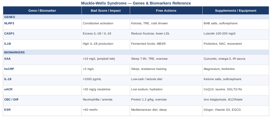
6 Biomarkers to Track in Muckle-Wells Syndrome
Monitoring inflammatory disease without the right biomarkers is like navigating without a map. For Muckle-Wells Syndrome, the stakes are especially high: the disease's most serious long-term complication — systemic AA amyloidosis — develops silently from chronically elevated inflammation, often without noticeable symptoms until significant kidney damage has already occurred. The six biomarkers below are the most clinically meaningful for tracking MWS disease activity, guiding treatment decisions, and catching complications before they become irreversible.
1. Serum Amyloid A (SAA)
Why it matters
Serum Amyloid A is the single most important biomarker in Muckle-Wells Syndrome. SAA is an acute-phase protein produced by the liver in response to IL-1β and IL-6 signaling — both massively amplified by the overactive NLRP3 inflammasome. Chronically elevated SAA is the direct biochemical precursor to AA amyloid fiber deposition in organs, most critically the kidneys. The risk of developing AA amyloidosis is not simply tied to peak levels during flares — it accumulates based on the time-averaged SAA over months and years. Even a modest but persistent elevation between episodes (above 10 mg/L) compounds silently into clinically significant amyloid deposition. MWS historically carries the highest amyloidosis risk within the CAPS spectrum, affecting up to 25% of patients before effective IL-1 inhibition was available.
How to measure it
SAA is measured from a standard blood draw but must be specifically requested — it is not included in routine inflammatory panels. Specialized reference laboratories and academic autoinflammatory disease centers include it in CAPS monitoring protocols; in other settings, the test may need to be sent to a reference lab. Cost in the US: approximately $50–$150 depending on insurance and laboratory. Should be measured during both active and quiescent periods to establish a meaningful individual baseline, and tracked as a trend rather than interpreted in isolation.
Target levels and red flags
Normal SAA: below 6–10 mg/L. European guidelines for CAPS management target SAA below 10 mg/L, with complete normalization (below 6 mg/L) as the ideal. During MWS flares, levels commonly reach hundreds to thousands of mg/L. Persistent elevation above 10 mg/L between flares — even in the absence of symptoms — is the clearest signal for amyloidosis risk and demands treatment review.
If the score is bad: the plan without supplements
Persistently elevated SAA in a treated patient demands immediate reassessment of the current IL-1 inhibitor regimen under specialist supervision. Both canakinumab (Ilaris) and anakinra (Kineret) are approved for CAPS/MWS, and dose or frequency adjustments may bring SAA into range. Beyond pharmacological optimization, non-supplement interventions that reduce upstream IL-1β and IL-6 signaling: reducing caloric excess (adipose tissue independently drives IL-6 and SAA through adipokine signaling), eliminating ultra-processed food (which primes NLRP3 through advanced glycation end products and oxidized seed oils), ensuring 7–9 hours of sleep consistently (sleep deprivation raises IL-6 acutely), and compressing the daily eating window to 12–14 hours through time-restricted eating, which reduces NLRP3 priming through multiple independent pathways. These steps will not override a gain-of-function mutation, but they meaningfully reduce the background inflammatory load that medication must work against.
If the score is bad: the plan with supplements or equipment
Curcumin (as BCM-95 or a liposomal formulation for bioavailability), 500–1000 mg twice daily with food, has demonstrated reductions in SAA and CRP in clinical studies of inflammatory conditions through NF-κB and direct NLRP3 pathway inhibition. MWS-specific RCT data are absent, but the mechanism is directly relevant. Cycle 8 weeks on, 2 weeks off; main side effect at high doses is loose stools. Omega-3 fatty acids (EPA + DHA combined, 3–4 g/day with food) shift eicosanoid production toward less inflammatory mediators and reduce IL-6 signaling; use continuously and monitor for bleeding tendency if combined with anticoagulants. Infrared sauna (3–4 sessions per week, 20 minutes at 55–60°C) has shown reductions in inflammatory markers including SAA in small human trials; a personal unit ranges from $2,000 upward, or wellness facilities typically charge $20–$30 per session.
2. High-Sensitivity C-Reactive Protein (hsCRP)
Why it matters
CRP is the most widely available and clinically familiar marker of systemic inflammation. In MWS, it rises sharply during flares and should fall toward normal during remission with adequate treatment. While less directly linked to amyloidosis risk than SAA, hsCRP is an excellent proxy for overall disease activity and a practical, affordable tool for monitoring treatment response at virtually any laboratory worldwide. Its rapid kinetics — rising within hours and falling within days of an inflammatory episode — make it particularly useful for detecting early flares and confirming treatment response in real time.
How to measure it
The high-sensitivity version (hsCRP) is preferred over standard CRP because it reliably detects lower levels of inflammation relevant to cardiovascular and inflammatory risk. Cost: $20–$60 in the US, frequently covered by insurance. Should be measured during both flares and stable periods to establish individual reference trends — a single result has substantially less value than a series of measurements over time.
Target levels and red flags
Optimal hsCRP is below 1 mg/L for cardiovascular and inflammatory purposes. In well-treated MWS between flares, hsCRP should approach this range. Persistent elevation above 3 mg/L during a supposedly quiescent period strongly suggests undertreated or inadequately controlled disease activity and should not be attributed to minor external causes without further investigation.
If the score is bad: the plan without supplements
Persistent hsCRP elevation between flares should prompt a full treatment review with the prescribing specialist. Non-supplement interventions with robust clinical evidence for reducing baseline CRP include: 7–9 hours of sleep consistently (even one week of poor sleep significantly raises CRP through IL-6 upregulation), resistance training 2–3 times per week (has among the strongest evidence for lowering baseline CRP independently of weight loss), moderate aerobic exercise 150+ minutes per week, reduction of refined carbohydrates and ultra-processed food, and reduction of visceral adiposity over time (which drives IL-6 and CRP through adipokine and free fatty acid signaling independently of NLRP3). Alcohol has a dose-dependent pro-inflammatory effect on CRP and should be minimized or eliminated entirely in MWS.
If the score is bad: the plan with supplements or equipment
Magnesium glycinate (300–400 mg before bed) has demonstrated modest CRP-lowering effects in clinical trials and simultaneously improves sleep quality, creating a compounding anti-inflammatory benefit; use continuously, well-tolerated at these doses. Berberine (500 mg twice daily with meals) has shown meaningful reductions in CRP in randomized controlled trials of metabolic and inflammatory conditions, likely through AMPK activation and gut microbiome modulation; cycle 8 weeks on, 4 weeks off to avoid adaptation. Cold water immersion (3–5 minutes at 10–15°C, 3–4 times per week) has human evidence for reducing CRP and IL-6 through autonomic and immune regulation; a dedicated cold plunge unit ranges from $2,000 to $8,000, or a cold shower finishing protocol achieves partial benefit at no cost.
3. Interleukin-18 (IL-18)
Why it matters
IL-18 is processed and released by Caspase-1 immediately downstream of NLRP3 activation — making it a direct readout of inflammasome activity rather than a downstream acute-phase marker. Research on CAPS/MWS patients has shown that IL-18 is dramatically elevated in MWS compared to other autoinflammatory diseases, often reaching 10–50 times normal values even between flares. Critically, IL-18 does not always normalize with IL-1β inhibition — canakinumab and anakinra primarily block IL-1β signaling downstream, not IL-18 release at the Caspase-1 level. This makes IL-18 a unique window into residual inflammasome activity that CRP and SAA alone cannot reveal. Persistently elevated IL-18 has been associated with progressive hearing loss and ongoing systemic tissue damage in CAPS, even in patients who appear clinically stable.
How to measure it
IL-18 is measured by ELISA from a blood sample and is primarily available through specialized autoinflammatory disease centers and reference laboratories (Mayo Clinic Laboratories and ARUP Laboratories in the US offer this test). Cost: approximately $100–$250; not routinely covered by insurance, and often ordered as part of specialist workup or research protocols. The sample requires careful handling — plasma should be separated quickly after the blood draw to prevent ex vivo cytokine release from degrading red cells from confounding the result.
Target levels and red flags
Normal serum IL-18 in healthy adults: below 200–250 pg/mL. In CAPS/MWS, levels frequently range from 1,000 to over 10,000 pg/mL even between flares. An IL-18 level dramatically exceeding what the CRP-based picture would predict implies residual NLRP3-Caspase-1 activity not captured by standard markers — and potentially not addressed by current treatment.
If the score is bad: the plan without supplements
Since IL-18 is not fully suppressed by IL-1 inhibitors, persistently elevated IL-18 warrants discussion of combination therapy or dose adjustment with an autoinflammatory specialist. Anti-IL-18 antibody therapy — including recombinant IL-18 binding protein (tadekinig alfa) — is under active investigation for IL-18-driven autoinflammatory diseases and represents an emerging pharmacological option. Without access to experimental therapies, the most accessible non-supplement approach is dietary or fasting-induced ketosis: beta-hydroxybutyrate (BHB), produced during carbohydrate restriction or extended fasting, directly inhibits NLRP3 inflammasome assembly at the protein level, reducing both IL-1β and IL-18 release. A modified low-carbohydrate diet achieving mild ketosis (0.5–1.5 mmol/L blood ketones) is feasible for most people and measurable with an inexpensive home ketone meter ($25–$50).
If the score is bad: the plan with supplements or equipment
Exogenous ketone salts or esters (10–15 g before meals, 1–2 times per day) raise blood BHB without requiring strict dietary ketosis; electrolyte monitoring (sodium, potassium, magnesium) is important with regular use, and GI tolerance should be established gradually. Sulforaphane (from fresh broccoli sprouts or concentrated supplements standardized to 40–60 mg/day sulforaphane) activates Nrf2, which reduces NLRP3 gene transcription and downstream IL-18 production; continuous use, minimal side effects. Quercetin (500–1000 mg/day combined with vitamin C for bioavailability) has preclinical evidence for direct NLRP3 protein complex inhibition and reduced IL-18; human clinical data are limited but the compound is safe at these doses; cycle 8 weeks on, 4 weeks off.
4. Urinary Albumin-to-Creatinine Ratio (uACR)
Why it matters
AA amyloidosis — the complication that transforms MWS from a manageable chronic disease into a life-threatening one — deposits amyloid fibers preferentially in the kidneys. The earliest detectable sign is microalbuminuria: small but abnormal amounts of albumin leaking into the urine well before macroscopic proteinuria develops. Standard urine dipsticks miss this early window almost entirely. By the time dipstick proteinuria is positive, significant amyloid deposition has already occurred and renal function may already be impaired. Sensitive urinary albumin testing is therefore not optional for MWS — it is the frontline screen for the disease's most serious and potentially irreversible consequence.
How to measure it
The preferred test is the urine albumin-to-creatinine ratio (uACR) from a first-morning urine sample, which corrects for urine concentration variability and gives a reliable result from a single collection. Cost: $20–$80, widely available in any laboratory setting. Should be performed at every routine clinical visit — every 6–12 months in stable disease, and more frequently when SAA is elevated or disease activity is suboptimal. A 24-hour urine collection for total protein may be added for higher precision when microalbuminuria is detected.
Target levels and red flags
Normal uACR: below 30 mg/g creatinine. Microalbuminuria: 30–300 mg/g (early warning signal requiring immediate treatment reassessment). Macroalbuminuria: above 300 mg/g (significant amyloid deposition is likely, nephrology involvement required urgently). Any result above 30 mg/g in an MWS patient should be treated as clinically urgent — it signals that SAA has been inadequately suppressed over a meaningful period.
If the score is bad: the plan without supplements
Abnormal proteinuria in MWS requires urgent escalation of anti-IL-1 therapy and immediate nephrology involvement. Non-supplement steps to protect kidney function and slow amyloid-related damage: reduce dietary sodium to below 2 g/day (reduces glomerular hypertension), maintain blood pressure below 120/80 mmHg through lifestyle and medication if necessary (glomerular filtration integrity is pressure-dependent), stay well hydrated at 2–3 liters of water per day, and eliminate NSAIDs entirely (nephrotoxic in the context of amyloid nephropathy). An ACE inhibitor or ARB prescribed by a nephrologist has specific evidence for kidney protection in proteinuric kidney disease and should be discussed urgently.
If the score is bad: the plan with supplements or equipment
SGLT2 inhibitors (dapagliflozin, empagliflozin) have robust clinical trial evidence for kidney protection in proteinuric kidney disease independent of diabetes status; this is a prescription intervention, but its use in non-diabetic inflammatory kidney disease is growing — discuss with a nephrologist. Coenzyme Q10 (200–400 mg/day with food, continuous use) supports mitochondrial function in renal tubular cells under inflammatory and oxidative stress; well-tolerated, no cycling required. Taurine (2–3 g/day, continuous) has early evidence for kidney-protective effects in inflammatory contexts through osmotic regulation and antioxidant mechanisms; minimal side effects, widely available.
5. Complete Blood Count with Neutrophil Differential
Why it matters
A full CBC with differential provides several clinically important layers of information in MWS. During flares, neutrophilia (elevated neutrophil count) is a hallmark, directly reflecting the proinflammatory output of NLRP3-driven IL-1β signaling, which rapidly recruits and primes neutrophils in the blood and tissues. The differential also provides essential treatment safety information: anakinra is associated with neutropenia in some patients, which requires periodic monitoring. Reactive thrombocytosis (elevated platelet count) accompanies active inflammation and serves as an indirect disease activity signal. Finally, anemia of chronic inflammation is common in inadequately treated MWS and independently contributes to fatigue, cognitive impairment, and cardiovascular risk — making its detection and context-appropriate management important.
How to measure it
A standard CBC with differential is one of the most accessible and affordable laboratory tests worldwide. Cost: $15–$50. No special preparation required. Should be performed at every clinical visit and during any suspected flare. Results gain their greatest value when interpreted alongside CRP, SAA, and clinical history rather than in isolation.
Target levels and red flags
Normal neutrophil count: 1.8–7.7 × 10³/μL. Persistent neutrophilia above 9–10 × 10³/μL between flares — especially with an elevated CRP — suggests ongoing disease activity. Hemoglobin below 12 g/dL (women) or 13 g/dL (men) with a normal or low reticulocyte count suggests anemia of chronic disease. Platelet count chronically above 450 × 10³/μL reflects sustained inflammatory signaling. If neutropenia develops on anakinra (below 1.0 × 10³/μL), dose adjustment is typically required.
If the score is bad: the plan without supplements
Persistent neutrophilia between flares warrants reassessment of treatment adequacy with the prescribing specialist. Anemia of chronic inflammation typically resolves with better disease control — iron supplementation is inappropriate unless true iron deficiency is confirmed separately (ferritin and transferrin saturation should be checked, since iron stores are typically normal in anemia of chronic disease). Adequate protein intake (1.2–1.6 g/kg body weight per day) supports red cell production, immune cell turnover, and lean mass maintenance. Consistent moderate exercise improves bone marrow function, red cell kinetics, and chronic neutrophil activation over weeks.
If the score is bad: the plan with supplements or equipment
For confirmed iron deficiency anemia (not anemia of chronic disease), iron bisglycinate (25–50 mg elemental iron with vitamin C, every other day based on recent evidence for improved absorption and reduced oxidative side effects) is the preferred form. Methylcobalamin B12 (1000 mcg/day sublingually) and methylfolate (400–800 mcg/day) support red cell maturation and are well-tolerated continuously. If neutropenia develops on a biologic, do not add immune-modulating supplements before discussing the situation with the prescribing specialist — the priority is medication adjustment, not supplementation.
6. Erythrocyte Sedimentation Rate (ESR)
Why it matters
The ESR is the oldest and most broadly accessible inflammation marker in clinical medicine. In MWS, it rises with systemic inflammation and tracks disease trends reliably, complementing the faster-reacting CRP. Its strength lies in its sensitivity to chronic, sustained inflammation — it tends to reflect longer-standing disease activity particularly well and provides a useful cross-check against CRP, which can be falsely reassuring in some clinical contexts. Where SAA and IL-18 testing is not readily available, ESR provides a meaningful alternative for monitoring baseline inflammatory tone and tracking treatment response over time.
How to measure it
ESR is measured from a blood draw using a simple sedimentation method, available in virtually every laboratory worldwide. Cost: $10–$40. Should be included at every routine visit alongside CRP and, where available, SAA. Results are affected by anemia, age, and sex — context matters and trends over multiple measurements are more informative than single values.
Target levels and red flags
Normal ESR: below 20 mm/hr in men, below 30 mm/hr in women (age-adjusted ranges apply). In active MWS, ESR commonly reaches 60–100+ mm/hr. Persistent elevation above 40 mm/hr in a treated patient suggests residual disease activity that merits investigation. A rising ESR trend over successive measurements, even within the normal range, is worth noting and correlating with clinical status.
If the score is bad: the plan without supplements
A persistently elevated ESR in treated MWS should prompt full reassessment of the treatment regimen, documentation of flare frequency, and review of recent lifestyle factors including infections, psychological stress, and sleep quality. Dietary pattern has robust evidence for ESR reduction across chronic inflammatory conditions: a Mediterranean-style diet (rich in extra-virgin olive oil, oily fish, colorful vegetables, legumes, and low in red meat and ultra-processed food) consistently lowers ESR in randomized trials. Sleep optimization — 7–9 hours, consistent timing, dark and cool room — reliably reduces baseline ESR when sleep is genuinely and consistently improved, not just on isolated nights.
If the score is bad: the plan with supplements or equipment
Ginger extract (600–1000 mg/day of standardized gingerol and shogaol) has randomized trial evidence for ESR reduction in inflammatory conditions including osteoarthritis and rheumatic disease; cycle 8 weeks on, 2 weeks off; mild GI effects are possible at higher doses. Vitamin D3 (2000–5000 IU/day with vitamin K2 MK-7, targeting serum 25-OH-D at 40–60 ng/mL) has immunomodulatory effects relevant to inflammatory dysregulation and NLRP3 transcription; monitor blood 25-OH-D every 6 months; continuous use is appropriate at these doses. Green tea extract (EGCG) (400 mg/day standardized to 45% EGCG) modestly reduces ESR and CRP through NF-κB inhibition; take with food to reduce nausea risk; continuous use is supported.
The six biomarkers above give a meaningful and layered picture of MWS disease activity, amyloidosis risk, treatment safety, and long-term organ protection. But to truly understand why these markers behave as they do — and why some people with the same diagnosis have a markedly more severe course — it helps to look upstream at the genes.
The Genetic Architecture of Muckle-Wells Syndrome: What NLRP3 and Its Pathway Reveal
Muckle-Wells Syndrome is one of the rare diseases where the molecular cause is known with near-certainty. Almost every case traces back to a single gene — NLRP3. But that gene does not act in isolation: its output is shaped by downstream effectors and pathway modifiers that account for much of the variability in disease severity, complication risk, and treatment response that clinicians observe in MWS patients who appear to share the same diagnosis. Understanding these three genetic layers helps explain the clinical picture far beyond what a mutation report alone conveys.
NLRP3 (CIAS1) — The Master Switch
What the gene does
NLRP3 (Nucleotide-binding domain, Leucine-rich Repeat, Pyrin domain-containing protein 3), also known by its historical name CIAS1, encodes the sensor subunit of the NLRP3 inflammasome — a large multi-protein complex that functions as an intracellular danger detector. Under normal conditions, NLRP3 assembles the inflammasome only in response to genuine biological threats (bacterial toxins, uric acid crystals, damaged mitochondria), triggering a controlled burst of IL-1β and IL-18 and then returning to baseline. In Muckle-Wells Syndrome, gain-of-function mutations in NLRP3 dramatically lower this activation threshold — or cause the inflammasome to assemble constitutively — leading to chronic, unregulated IL-1β and IL-18 production independent of any actual threat.
More than 200 distinct NLRP3 mutations have been identified across the CAPS spectrum. MWS is most commonly associated with mutations in the NACHT domain of the protein (the ATPase domain responsible for oligomerization and assembly), including p.Thr348Met, p.Arg260Trp, p.Val200Met, and others. The CAPS spectrum spans from the milder Familial Cold Autoinflammatory Syndrome (FCAS) through MWS to the severe Neonatal Onset Multisystem Inflammatory Disease (NOMID/CINCA), with the specific mutation's effect on protein conformation and ATPase activity determining where a patient falls. MWS-associated mutations typically produce moderate constitutive activation rather than the near-maximal activation characteristic of NOMID. The original identification of NLRP3/CIAS1 mutations in CAPS (Hoffman et al., Nature Genetics, 2001) transformed a mysterious familial fever syndrome into a molecularly understood and targetable disease.
An additional and clinically important consideration is somatic mosaicism: in an estimated 10–20% of clinically apparent MWS cases, the NLRP3 mutation is present in only a fraction of cells, having arisen as a post-zygotic event rather than being inherited or present in all tissues. Conventional genetic sequencing may miss low-frequency mosaic variants, and the percentage of affected cells directly influences disease severity and lab findings. Next-generation deep sequencing (allele fraction sensitivity down to 1–5%) should be requested when clinical features strongly suggest MWS but standard testing returns negative.
If the gene is bad: the plan without supplements
With a confirmed gain-of-function NLRP3 mutation, the fundamental pharmacological correction is IL-1 blockade — canakinumab or anakinra intercept the downstream IL-1β consequence of the mutant protein. Lifestyle alone cannot override a constitutively active NLRP3 protein. However, NLRP3 priming — the upstream signal step that determines how readily the mutant protein triggers full inflammasome assembly — is meaningfully modifiable without supplements or prescription changes. Key priming reducers include: time-restricted eating (12–14 hour fasting windows reduce TXNIP-mediated priming from glucose), avoidance of excess saturated fat (palmitate activates TLR4, one of the most potent known NLRP3 primers), consistent cold exposure (brief cold showers reduce inflammatory tone through autonomic regulation), and regular aerobic exercise (releases anti-inflammatory myokines that dampen NLRP3 signaling for hours). These strategies reduce the background signal that the mutant protein amplifies, even if they cannot correct the mutation itself.
If the gene is bad: the plan with supplements or equipment
Dietary or supplemental BHB directly inhibits NLRP3 inflammasome assembly at the protein level, preventing the NLRP3-ASC interaction required for Caspase-1 activation — a mechanism independent of IL-1 receptor blockade, demonstrated in a landmark 2015 Nature Medicine study (Youm et al., 2015). Target mild ketosis at 0.5–1.5 mmol/L blood ketones via a low-carbohydrate diet or exogenous ketone salts (10–15 g per dose, 1–2 times daily); measure with an inexpensive home ketone meter. Sulforaphane (standardized to 40–60 mg/day, or from 30–50 g daily fresh broccoli sprouts consumed raw) activates Nrf2, which reduces NLRP3 transcription before the protein is even made; continuous use, minimal side effects. Quercetin (500–1000 mg/day with vitamin C) has preclinical evidence for direct NLRP3 protein inhibition; human MWS-specific data are early; cycle 8 weeks on, 4 weeks off.
CASP1 — The Executioner of the Inflammasome
What the gene does
Caspase-1, encoded by CASP1, is not mutated in MWS, but it is the essential enzymatic effector that the overactive NLRP3 protein activates. Once the mutant NLRP3 oligomerizes and recruits the adaptor protein ASC, Caspase-1 is cleaved into its active form. This active Caspase-1 then processes pro-IL-1β and pro-IL-18 into their mature, secreted forms — the cytokines responsible for the symptoms, tissue damage, and biomarker elevations seen in MWS. Understanding Caspase-1 matters clinically because functional variants in CASP1 and neighboring pathway genes can modify the quantity of IL-1β and IL-18 produced once the inflammasome fires, helping to explain why two individuals with identically mutated NLRP3 can have different disease severity and different SAA levels.
Caspase-1 also initiates pyroptosis — a form of inflammatory cell death that amplifies the inflammatory signal by releasing the entire intracellular contents of activated immune cells into surrounding tissue. This pyroptotic amplification loop contributes meaningfully to the tissue damage and systemic spread of inflammation seen in severe or undertreated MWS, particularly in joints, the inner ear, and the kidney.
If the pathway is overactive: targeted approaches
All strategies that reduce NLRP3 activation also reduce downstream CASP1 activity. Additionally, minimizing known NLRP3 co-activators limits the Caspase-1 signal. Elevated serum uric acid (above 5.5 mg/dL) directly activates NLRP3 through monosodium urate crystal nucleation, amplifying the signal that reaches Caspase-1; reducing dietary fructose (the primary uric acid driver from diet) and high-purine foods (organ meats, certain seafood) is the most accessible non-prescription approach. Keep serum uric acid below 5.5 mg/dL and verify with a standard metabolic panel. Luteolin (100–200 mg/day, found naturally in parsley, chamomile, and celery, and available as a supplement) has preclinical evidence for Caspase-1 inhibitory effects; human clinical data in autoinflammatory disease are limited but the compound is well-tolerated and broadly safe. Avoiding cholesterol crystal formation — which occurs with high LDL in arterial walls — is another underappreciated Caspase-1 activator; optimizing LDL-C through diet and, where appropriate, pharmacological support reduces one additional co-stimulus.
IL1B — The Downstream Amplifier and Its Modifier Variants
What the gene does
IL1B encodes Interleukin-1 beta — the primary effector cytokine released by the NLRP3-Caspase-1 axis and the molecule that IL-1 inhibitors directly neutralize. While the causative mutation in MWS is upstream (in NLRP3), individual variants in IL1B itself — particularly in its promoter region — influence how much IL-1β is transcribed in response to the inflammasome signal and how strongly downstream target tissues respond. Promoter variants such as rs16944 and rs1143634 have been associated with higher baseline IL-1β production across multiple inflammatory conditions. A person with both an NLRP3 gain-of-function mutation and high-producing IL1B promoter variants may experience a more severe disease course, higher SAA levels, and a greater amyloidosis risk than someone with the same NLRP3 mutation but lower-producing IL1B variants.
This modifier dimension is not routinely assessed in standard CAPS genetic panels and is not yet part of clinical decision-making protocols, but it becomes a relevant consideration when disease severity appears disproportionate to the specific NLRP3 mutation, or when treatment response is unexpectedly incomplete.
If the gene is bad: the plan without supplements
High-producing IL1B variants respond to the same strategies that reduce NLRP3 priming and Caspase-1 activity described above. The gut microbiome is a particularly relevant modifier specifically for IL1B expression: dysbiotic gram-negative bacteria producing high levels of LPS drive systemic IL-1β elevation through TLR4-NF-κB signaling that is independent of NLRP3 itself. A diet rich in diverse fermented foods (yogurt, kefir, kimchi, tempeh), 30+ distinct plant foods per week for microbiome diversity, and low in dietary emulsifiers consistently reduces systemic IL-1β signaling in human intervention studies. Structured stress management (see the MBSR section below) also meaningfully reduces IL-1β production through cortisol pathway normalization.
If the gene is bad: the plan with supplements or equipment
Probiotics containing Lactobacillus rhamnosus GG and Bifidobacterium longum (10 billion CFU/day, continuous use) have randomized trial evidence for reducing circulating IL-1β in inflammatory conditions through microbiome-mediated immune modulation. Resveratrol (250–500 mg/day as trans-resveratrol) inhibits IL-1β transcription through SIRT1-NF-κB interactions; cycle 8 weeks on, 4 weeks off; avoid if on anticoagulant medications. N-acetylcysteine (NAC) (600–900 mg twice daily, continuous) replenishes glutathione and reduces reactive oxygen species — a key priming signal for IL1B transcription; well-tolerated, with supporting evidence across multiple inflammatory conditions.
The genetic architecture above explains what the biomarkers are measuring and why certain interventions work at the molecular level in MWS. The next section takes this deeper through the work of one of the most rigorous science communicators working on NLRP3 biology today.
The NLRP3 Inflammasome: 10 Research Insights Every MWS Patient Should Know
Dr. Rhonda Patrick is a biomedical scientist who has spent a significant portion of her career synthesizing the peer-reviewed literature on NLRP3 inflammasome biology, nutrition, and the interaction between genetics and lifestyle. Through her FoundMyFitness platform and podcast, she has made an unusually dense body of mechanistic research accessible and actionable for a non-specialist audience. The NLRP3 inflammasome — the molecule at the center of MWS — is one of her recurring subjects. The ten insights below represent the most clinically relevant takeaways from that body of work for someone living with an NLRP3-driven disease.
1. Ketone Bodies Physically Block Inflammasome Assembly
Beta-hydroxybutyrate (BHB), produced during fasting or carbohydrate restriction, prevents NLRP3 from oligomerizing and recruiting ASC — a step that must occur before Caspase-1 can be activated. This direct physical interference with the inflammasome assembly process was demonstrated in a 2015 Nature Medicine study (Youm et al.) and is now one of the best-characterized non-pharmacological NLRP3 inhibitory mechanisms known. Even mild ketosis at 0.5 mmol/L appears sufficient for meaningful effect. For MWS, this may be the most mechanistically grounded single dietary intervention available as a complement to medication.
2. Sulforaphane Acts Upstream of the Protein Itself
Sulforaphane from broccoli sprouts activates Nrf2, a transcription factor that reduces NLRP3 gene expression before the protein is made. This upstream action makes it different from almost all other food-based NLRP3 modulators, which act at the protein or cytokine level. Patrick has emphasized that fresh broccoli sprouts provide the highest sulforaphane yield of any accessible food source — typically 30–50 g per day consumed raw and chewed thoroughly — because chewing releases the myrosinase enzyme required for sulforaphane formation. Commercial sprout powders vary dramatically in quality; myrosinase activity in the supplement determines efficacy.
3. Time-Restricted Eating Reduces NLRP3 Priming Without Requiring Full Ketosis
Compressing the eating window to 8–10 hours reduces NLRP3 priming through at least three independent pathways: it lowers peak glucose (which normally activates TXNIP, a direct NLRP3 activator), reduces intestinal permeability and gut-derived LPS in the blood, and promotes autophagy — which clears damaged mitochondria, a primary danger signal that triggers NLRP3 assembly. A 12–14 hour fast offers meaningful inflammasome-suppressing benefits with substantially lower compliance demands than a full ketogenic diet, making it viable even for those who cannot or prefer not to pursue strict carbohydrate restriction.
4. The Omega-6 to Omega-3 Ratio Sets the Inflammatory Background Level
Arachidonic acid derived from omega-6 fatty acids — dominant in seed oils and ultra-processed food — competes with EPA and DHA for key enzymatic pathways and shifts eicosanoid production toward pro-inflammatory mediators. Patrick emphasizes that the modern Western dietary omega-6 to omega-3 ratio (often 15–20:1 versus the ancestral 4:1) creates a persistent inflammatory background that continuously amplifies NLRP3 sensitivity. Correcting this ratio through 3–4 g/day EPA + DHA supplementation and reduced seed oil consumption shifts the inflammatory tone meaningfully over 4–8 weeks.
5. Exercise Releases Anti-Inflammasome Signals Within Minutes
A single session of moderate aerobic exercise releases myokines from contracting muscle — including context-dependent anti-inflammatory IL-6 (which induces IL-10 in this setting) and irisin — that suppress NLRP3 activity for several hours. Consistent moderate aerobic exercise (150+ minutes/week) produces sustained reductions in baseline NLRP3-driven inflammation through multiple complementary pathways: improved insulin sensitivity, reduced visceral adipose signaling, and direct myokine effects. This is not exercise for general health in MWS — it is a specific anti-inflammasome strategy with a measurable molecular mechanism. HIIT sessions add metabolic benefits but should be approached cautiously during active flares.
6. Regular Sauna Use Is Associated With Lower Systemic Inflammatory Markers in Long-Term Human Data
Patrick has extensively documented the Finnish KIHD cohort findings showing that frequent sauna use (4–7 sessions per week at 80–100°C) is associated with dramatically lower CRP, IL-6, and long-term cardiovascular risk. The mechanism involves heat-shock protein induction — which directly inhibits NF-κB and NLRP3 signaling — along with autonomic nervous system recalibration that dampens inflammatory tone. For MWS patients who can tolerate heat and do not have active cardiac involvement, regular sauna sessions are among the few lifestyle interventions with long-term population-level evidence for reducing systemic inflammatory markers.
7. Even One Night of Poor Sleep Increases NLRP3 Gene Expression
Research referenced by Patrick shows that a single night of sleep deprivation (below 6 hours) measurably increases NLRP3 gene expression in peripheral blood mononuclear cells and raises circulating IL-1β and IL-18 within 24 hours. For someone with MWS, whose inflammasome is constitutively overactive due to a genetic mutation, sleep deprivation functions as an amplifier: it adds a transcriptional driver on top of an already dysregulated protein. Sleep hygiene in MWS is not optional self-care — it is a direct molecular intervention that reduces inflammasome output when practiced consistently.
8. Sub-Clinical Uric Acid Elevation Amplifies NLRP3 Activity Below the Gout Threshold
Serum uric acid above 5.5 mg/dL activates NLRP3 through monosodium urate crystal nucleation in tissues, even below the classic gout threshold of 6.8 mg/dL. Patrick has highlighted that standard laboratory reference ranges often accept values up to 7–8 mg/dL as normal, which obscures this sub-clinical inflammasome co-activation. For someone with MWS and an already hair-trigger NLRP3 protein, even modest uric acid elevation meaningfully lowers the flare threshold. The primary dietary driver is fructose — not purines as widely assumed — making reduction of sugary drinks, fruit juices, and processed food containing high-fructose corn syrup the most effective intervention.
9. Dietary Emulsifiers Increase Gut Permeability and Continuous LPS-Mediated Priming
Intestinal permeability allows lipopolysaccharide from gram-negative gut bacteria to translocate into the bloodstream, where it activates TLR4 — one of the most potent known primers of NLRP3 assembly. Patrick has focused particular attention on the role of food-grade emulsifiers (carrageenan, carboxymethylcellulose, polysorbate-80) found throughout ultra-processed food, which have been specifically demonstrated in human studies to increase gut permeability and systemic LPS levels. For MWS patients, eliminating these emulsifiers reduces one of the most consistent and modifiable NLRP3 priming signals from the daily environment.
10. Vitamin D Has a Direct Genomic Effect on NLRP3 Transcription
The vitamin D receptor (VDR) has binding sites in the promoter regions of genes controlling NLRP3 expression and IL-1β transcription. Vitamin D deficiency (25-OH-D below 20 ng/mL) is associated with higher baseline NLRP3 transcription and impaired regulatory T cell activity in human studies. Patrick recommends targeting 40–60 ng/mL serum 25-OH-D for optimal immune regulation — a level achievable through a combination of consistent sun exposure and supplementation (typically 2000–5000 IU/day vitamin D3 with K2 for cofactor support). Deficiency is extremely common, inexpensive to test ($25–$60), and straightforward to correct.
These ten insights connect directly to the biomarker and genetic layers covered earlier and provide a scientific rationale for daily choices that can genuinely complement medical treatment. The following section examines three additional approaches backed by human evidence for reducing autoinflammatory burden through different biological entry points.
Complementary Approaches with Meaningful Evidence
The Autoimmune Protocol (AIP)
Developed by Dr. Sarah Ballantyne, a medical biophysicist, the Autoimmune Protocol is a structured dietary elimination and reintroduction framework originally designed for autoimmune diseases and increasingly discussed for autoinflammatory conditions including CAPS. The AIP systematically removes foods associated with increased gut permeability and immune activation — including grains, legumes, nightshades, eggs, nuts, seeds, alcohol, refined sugars, and seed oils — while emphasizing nutrient-dense anti-inflammatory foods rich in omega-3 fatty acids, polyphenols, collagen, and prebiotic fiber. For MWS, the mechanistic rationale is direct: gut permeability, as covered extensively above, is one of the most modifiable and consistent NLRP3 priming signals, and the AIP directly targets the dietary contributors to intestinal barrier disruption. It is worth noting that MWS is classified as an autoinflammatory disease (driven by innate immune dysregulation) rather than a classical autoimmune disease — but the shared inflammatory pathway overlap makes the AIP's mechanisms highly relevant.
A randomized controlled trial published in Inflammatory Bowel Diseases (Konijeti et al., 2017) demonstrated significant improvements in clinical disease activity and inflammatory markers in IBD patients following the AIP — a condition sharing multiple inflammatory pathway features, including elevated IL-1β and intestinal permeability, with autoinflammatory syndromes. Human clinical trial evidence specific to MWS does not yet exist, but the mechanistic overlap and the IBD data provide the strongest available indirect support for this approach in NLRP3-driven conditions.
To apply the AIP for MWS: work with a registered dietitian experienced in elimination protocols to ensure nutritional adequacy throughout (iron, calcium, iodine, and omega-3 all require deliberate attention during the strict phase). Commit to 4–6 weeks of strict elimination before beginning systematic reintroductions, testing one food at a time over several months. Set realistic expectations — dietary change will not override an NLRP3 gain-of-function mutation, but meaningful reductions in flare frequency and background inflammatory marker elevation are plausible goals for motivated patients. Avoid the strict elimination phase during acute flares, as nutritional stress may worsen inflammatory burden; this protocol is most effective during stable periods.
Mindfulness-Based Stress Reduction (MBSR)
Chronic psychological stress activates the HPA axis and sympathetic nervous system, promoting NLRP3 priming through glucocorticoid receptor desensitization, elevated catecholamines, and bidirectional autonomic-immune crosstalk. For a person with MWS — whose inflammasome is already constitutively overactive — unmanaged psychological stress acts as a continuous amplifier that can increase flare frequency, worsen pain perception, impair sleep quality, and raise inflammatory marker baseline independently of disease treatment. This is not a peripheral concern: the mechanism is specific and measurable. MBSR, the structured 8-week program developed by Jon Kabat-Zinn at the University of Massachusetts Medical School, addresses this through formal seated meditation, body scan practice, and mindful movement, building a sustained autonomic and immune regulatory shift over weeks.
A systematic review and meta-analysis published in Brain, Behavior, and Immunity (Black and Slavich, 2016) found that mindfulness-based interventions significantly reduced circulating markers of inflammation including CRP and IL-6 in adults with chronic illness. Pain management outcomes and sleep quality — both significantly and independently impaired in MWS — showed reliable improvements across reviewed trials. Evidence specific to autoinflammatory diseases is limited by the rarity of these conditions, but the inflammatory mechanism overlap, the symptom burden MWS patients carry between flares, and the documented benefits across comparable chronic inflammatory diseases all justify MBSR as a low-risk, meaningful complement to standard care.
For MWS: complete a formal 8-week MBSR program — these are offered through university medical centers, hospital wellness programs, and yoga studios, and are also available through apps such as Insight Timer and 10% Happier. Aim for 20–30 minutes of formal practice daily, 5–6 days per week during stable periods. During active flares, shorter breath-focused sessions (5–10 minutes of slow diaphragmatic breathing) can meaningfully help manage acute pain without requiring the sustained attention capacity that full meditation demands. Combining MBSR with consistent sleep hygiene amplifies the autonomic regulatory benefits and reduces the overnight IL-1β spike associated with poor sleep.
Microbiome-Directed Therapies
The gut microbiome regulates systemic immune tone through multiple mechanisms that are directly relevant to MWS. Most critically, short-chain fatty acids (SCFAs) — particularly butyrate — produced by microbial fermentation of dietary fiber directly inhibit NLRP3 inflammasome activation through GPR109A receptor signaling and epigenetic mechanisms (HDAC inhibition reduces NLRP3-related gene expression). Conversely, a dysbiotic microbiome dominated by LPS-producing gram-negative species generates a continuous systemic NLRP3-priming signal through intestinal LPS translocation. Restoring microbiome diversity and SCFA-producing bacterial populations is therefore a mechanistically coherent and clinically accessible target for reducing inflammasome priming burden in MWS — one that can be pursued alongside medication without any conflict.
A landmark clinical trial published in Cell (Wastyk et al., 2021) demonstrated that a fermented food-rich diet — compared to a high-fiber diet — increased microbiome diversity and reduced 19 systemic inflammatory proteins including IL-1β over 10 weeks in healthy adults. This represents among the strongest direct human evidence for dietary modulation of systemic inflammatory tone through the gut microbiome, including specifically IL-1β signaling — the central effector cytokine in MWS pathology. The implication for MWS is that the microbiome is not a bystander; it is an active modulator of inflammasome priming that is accessible through daily dietary choices.
For MWS: aim for 30 or more distinct plant foods per week (diversity drives microbiome diversity more reliably than any single superfood), 30 g/day of dietary fiber from varied sources including both soluble and fermentable types, and daily fermented food consumption at meaningful amounts (150–200 g of live-culture yogurt, kefir, kimchi, or sauerkraut). A targeted probiotic containing Lactobacillus rhamnosus GG and Bifidobacterium longum at 10 billion CFU/day can supplement dietary approaches, particularly when dietary variety is limited. Prebiotic fibers — partially hydrolyzed guar gum, resistant starch (cooled and reheated potato or rice), and inulin-rich foods (chicory, leek, asparagus) — specifically promote SCFA-producing species; introduce at 2–4 g/day and increase gradually over weeks to minimize bloating and gas. Eliminate dietary emulsifiers from all packaged foods, as these selectively deplete the mucus-adhering commensal bacteria that maintain intestinal barrier function. This entire approach carries no serious side effects, requires no prescription, and has mechanistic rationale specific to NLRP3-driven autoinflammatory disease.
Conclusion
Muckle-Wells Syndrome is rare, molecularly understood, and genuinely monitorable in ways that most chronic inflammatory diseases are not. The six biomarkers covered in this article — particularly SAA and urinary albumin — are the difference between catching amyloidosis in its earliest stages and discovering it when kidney damage is already significant. Tracking them consistently, understanding what each number means biologically, and acting promptly on abnormal results gives a level of clinical agency that no amount of general health advice can match.
The most valuable immediate step is a direct conversation with your autoinflammatory specialist: which of these biomarkers are currently included in your monitoring panel, which are missing, and at what frequency should each be measured given your current disease activity? Bring these questions to the appointment. Then consider the complementary strategies — ketosis, sulforaphane, sleep, the Autoimmune Protocol, MBSR, and microbiome support — not as replacements for medical treatment, but as scientifically grounded tools for reducing the inflammatory background that medication cannot fully address alone. In a disease where cumulative inflammation is the real long-term enemy, better monitoring and smarter daily choices are meaningful and measurable leverage.
Musculoskeletal: Joint Conditions
Autoimmune: Inflammatory Conditions
Ear, Nose & Throat: Hearing & Balance Conditions
Urological: Kidney Conditions