This article contains AI generated content.
· UpdatedMucoid Degeneration of ACL — 4 Genes And 6 Biomarkers To Track
Introduction
If your MRI report mentions mucoid degeneration of the ACL and you've been sent home with a referral for physiotherapy and little else, you're not alone in feeling like the explanation was incomplete. The diagnosis describes what is visible on imaging — a diffuse increase in signal within the anterior cruciate ligament, often with cystic changes — but rarely explains why the ligament reached that state, or what specifically is driving the degeneration in your case. That gap between observation and mechanism is where most people get stuck.
Mucoid degeneration of the ACL is a condition in which the normal collagenous architecture of the ligament is progressively replaced by a gelatinous, mucin-rich substance. It is not a tear, but it is not simply normal aging either. The result is a ligament that can cause significant knee pain, stiffness at the extremes of flexion and extension, and a reduced capacity to repair itself without targeted support. The mechanisms involved span inflammation, connective tissue matrix remodeling, vascular limitation, and in many cases, an inherited structural vulnerability that no amount of generic stretching will address.
Generic advice — strengthen the surrounding muscles, reduce impact, manage your weight — is not wrong. But it is often insufficient for people with this condition because it treats the ligament as if it were a muscle responding to effort rather than a metabolically complex, collagen-dense, avascular tissue responding to signals most people have never been asked to track. Understanding which signals are dysregulated in your case is what changes the quality of your decisions.
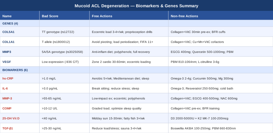
This article takes a more targeted approach. The first section covers the six most clinically informative biomarkers for this condition — measurable signals from blood and connective tissue that give you a window into the underlying biology. The second section examines the four genes most strongly linked to ACL structural vulnerability and what can be done when one or more of them is unfavorable. Together, these frameworks offer something that a standard orthopaedic consultation rarely provides: a personalized biological map for understanding why this is happening and where to intervene.
6 Biomarkers to Track for Mucoid Degeneration of the ACL
The six markers below reflect the key biological processes involved in ACL mucoid degeneration: systemic and local inflammation, matrix breakdown, connective tissue turnover, and nutritional sufficiency. No single marker tells the whole story — their value comes from reading them together. For each, you will find what it measures, what abnormal levels may mean for your ligament, and what evidence-backed strategies can move it in the right direction.
1. High-Sensitivity C-Reactive Protein (hs-CRP)
Why it matters
hs-CRP is the most widely available marker of systemic low-grade inflammation, and for a condition like mucoid ACL degeneration, chronic inflammation is not a side note — it is one of the central drivers. Even when levels are not dramatically elevated, values above 1.5–2.0 mg/L in the absence of acute illness indicate a pro-inflammatory systemic environment that steadily promotes matrix metalloproteinase activation, accelerates extracellular matrix breakdown, and impairs fibroblast function. In ligament tissue, this sustained catabolic signaling is exactly the kind of background condition that facilitates degenerative transformation. Peter Attia consistently targets hs-CRP below 0.5–1.0 mg/L in his patients for this reason.
How to measure it
Standard blood test, widely available. Usually covered by insurance when ordered with a metabolic panel. Out-of-pocket cost: $15–40. Target range for musculoskeletal health: below 1.0 mg/L. Values above 3.0 mg/L in the absence of acute infection or trauma warrant investigation into the source. Retest every 3–6 months when actively working to reduce it.
If the score is bad, the plan without supplements
The most evidence-supported non-supplement strategies for lowering hs-CRP are consistent moderate aerobic exercise (30 minutes, five days per week), an anti-inflammatory dietary pattern (Mediterranean-style, low in ultra-processed foods and refined sugars, high in polyphenols and omega-3-rich fish), sleep quality optimization targeting seven to nine hours nightly, and reduction of visceral adiposity, which is itself a source of chronic inflammatory cytokines. Time-restricted eating within a ten to twelve hour window has also shown measurable CRP reduction in several randomized trials, even without caloric restriction.
If the score is bad, the plan with supplements or equipment
Omega-3 fatty acids (combined EPA + DHA, 2–4 g/day with food) represent the intervention with the strongest and most replicated human evidence for hs-CRP reduction. Cycling: twelve weeks on, two to four weeks off, to avoid accumulation effects in those on anticoagulants. Curcumin with piperine (500 mg/day with food) is a reasonable adjunct with a modest but consistent evidence base. Magnesium glycinate (300–400 mg nightly) addresses a frequently missed deficiency that independently correlates with elevated CRP. Side effects at these doses are low; those on blood thinners should discuss omega-3 dosing with a physician before starting.
2. Interleukin-6 (IL-6)
Why it matters
IL-6 is a cytokine with a dual role that is particularly important to understand in the context of the ACL. Acutely, it promotes tissue repair. Chronically elevated, it does the opposite: it activates matrix metalloproteinases, disrupts collagen gene expression in fibroblasts, and sustains a catabolic signaling environment that works against ligament integrity. In degenerative knee conditions, elevated synovial IL-6 has been found to correlate with accelerated connective tissue breakdown. At the systemic level, resting serum IL-6 above 3.0 pg/mL in the absence of acute illness suggests a chronically dysregulated inflammatory state.
How to measure it
Serum IL-6 is not part of standard panels but is available through most major labs (LabCorp, Quest, and many sports medicine or functional medicine clinics). Cost: $30–80 out of pocket. Measurement requires same-day processing and is sensitive to exercise within 24 hours, so results should be taken under resting conditions. Interpret alongside hs-CRP for a fuller picture of inflammatory burden.
If the score is bad, the plan without supplements
Regular moderate aerobic exercise paradoxically lowers resting IL-6 despite raising it transiently during exertion — a well-established hormetic effect. Breaking up prolonged sitting with brief movement every 30–60 minutes has been shown in several trials to specifically reduce basal IL-6. Chronic psychological stress is a direct upstream driver of IL-6 production via cortisol, making stress management (sufficient recovery, social connection, reduced overtraining) a physiologically meaningful target. Sleep deprivation elevates IL-6 reliably; improving sleep architecture is not optional here.
If the score is bad, the plan with supplements or equipment
Omega-3 fatty acids (as above) reduce IL-6 alongside hs-CRP. Resveratrol (250–500 mg/day with food) has shown IL-6 suppression in clinical studies, particularly in metabolically compromised individuals. Cycling: eight weeks on, four weeks off. Cold water immersion (10–15 minutes at 10–15°C, three times per week) has emerging evidence for reducing chronic inflammatory cytokines, though it should not be used immediately post-exercise when a local anabolic response is desirable. Side effects are minimal at these doses; resveratrol may weakly inhibit CYP450 enzymes at high doses.
3. Matrix Metalloproteinase-3 (MMP-3)
Why it matters
MMP-3 (stromelysin-1) is an enzyme that degrades collagen types II, III, IV, IX, and X, fibronectin, laminin, and aggrecan — in other words, most of the structural components of a ligament's extracellular matrix. In the ACL, upregulated MMP-3 activity is directly implicated in the progressive breakdown of collagen architecture that characterizes degenerative conditions including mucoid change. Research has consistently found elevated MMP-3 in ACL tissue and synovial fluid in knees with degeneration, and serum MMP-3 may serve as a practical proxy for ongoing matrix catabolism. This is also a biomarker with a well-documented genetic component (discussed in the genetics section below), which makes it particularly important to measure in individuals who may carry the high-expression MMP3 promoter variant.
How to measure it
Serum MMP-3 is available as a specialty test through major reference labs and many rheumatology and functional medicine clinics. Cost: $50–120 out of pocket. Reference ranges vary by lab and sex (typically below 55 ng/mL in women, below 65 ng/mL in men), and elevation is most meaningful when persistent across serial measurements. Acute exercise or infection can transiently elevate MMP-3, so testing should be done under resting conditions.
If the score is bad, the plan without supplements
Reducing repetitive high-impact mechanical loading on the affected knee is the most direct lever for slowing MMP-3-driven matrix breakdown — not eliminating activity, but substituting toward low-impact modalities (swimming, cycling, resistance training within a pain-free range) during a repair phase. Controlled eccentric loading protocols, calibrated by a physiotherapist experienced in connective tissue rehabilitation, can promote organized collagen remodeling without triggering the excessive MMP activity that uncontrolled overloading generates. Dietary polyphenols (berries, green tea, olive oil) broadly inhibit MMP transcription pathways and are a low-friction lifestyle addition.
If the score is bad, the plan with supplements or equipment
Hydrolyzed collagen peptides (10–15 g/day, taken thirty to sixty minutes before exercise with 50–100 mg vitamin C) provide proline and hydroxyproline to support matrix anabolism as a counterbalance to elevated MMP-3-driven catabolism. Green tea extract standardized to EGCG (400–500 mg/day, cycled eight weeks on / four weeks off) has demonstrated direct MMP-3 inhibitory properties in preclinical and early clinical studies. N-acetylcysteine (600 mg/day) supports intracellular glutathione, reducing oxidative stress-driven upregulation of MMP transcription. Side effects are generally mild; EGCG in high doses warrants caution in those with liver conditions, and should be taken with food to reduce gastric irritation.
4. COMP (Cartilage Oligomeric Matrix Protein)
Why it matters
COMP is a structural protein found in cartilage, tendons, and ligaments. When these tissues undergo accelerated breakdown or abnormal turnover, COMP is released into the bloodstream at elevated levels. It is primarily used in rheumatology as a marker of cartilage damage progression, but its presence in ligament tissue makes it relevant for ACL conditions as well. Elevated serum COMP in the context of a known ACL degenerative process suggests active structural tissue catabolism — and tracking it serially (every three to six months) can help assess whether interventions are slowing or accelerating the process. Thomas Dayspring and Peter Attia have both noted that connective tissue turnover markers like COMP deserve wider clinical use outside of formal rheumatological settings.
How to measure it
Available through specialty and reference labs. Cost: $60–150 out of pocket. Some rheumatology panels include it. Reference ranges vary by assay (typically around 10–12 U/L upper limit), and single readings are less informative than trends. Elevated fasting levels that persist over multiple readings in a person with a documented connective tissue condition are clinically meaningful regardless of whether they exceed the formal reference range.
If the score is bad, the plan without supplements
Load management is again central: consistently overloading a degenerating ligament will produce persistently elevated COMP. A graded loading program — calibrated to stay within the repair zone rather than the damage zone — is the physiological foundation. Sleep quality deserves specific emphasis here: growth hormone and IGF-1 released during deep non-REM sleep are among the most potent endogenous drivers of connective tissue anabolic signaling. Chronic sleep deprivation reliably impairs this window and is associated with elevated connective tissue catabolism markers.
If the score is bad, the plan with supplements or equipment
The collagen peptide and vitamin C protocol described above applies directly here. Prolyl-hydroxyproline dipeptides, found in hydrolyzed collagen, have been shown in human clinical trials to stimulate collagen synthesis in ligament-derived fibroblasts by upregulating gene expression of collagen type I and type III. Blood flow restriction (BFR) training — using a tourniquet cuff to create metabolic stress at low loads — is an emerging tool for promoting connective tissue remodeling while minimizing compressive and tensile forces on the joint. BFR should initially be performed under professional supervision; home cuffs (B-Strong, Delfi) range from $150–600. Side effects are low when applied correctly; avoid in those with vascular disease or clotting disorders.
5. 25-Hydroxyvitamin D (25-OH Vitamin D)
Why it matters
Vitamin D is not simply a bone nutrient. Its receptor is expressed in ligament fibroblasts, immune cells, and muscle cells, and it directly modulates inflammatory gene expression, collagen synthesis pathways, and musculoskeletal coordination. In sports medicine and functional medicine contexts, vitamin D deficiency (below 20 ng/mL) and insufficiency (below 40 ng/mL) are independently associated with higher rates of musculoskeletal injury, elevated inflammatory markers, and impaired recovery. The presence of vitamin D receptors on ACL-derived fibroblasts suggests that this ligament may respond directly to vitamin D status — not just indirectly through systemic effects. Chronic insufficiency creates a permissive environment for the kind of low-grade inflammation and impaired repair signaling that characterizes mucoid degeneration.
How to measure it
Standard blood test included in most comprehensive wellness panels. Often covered by insurance. Out-of-pocket: $20–50. Optimal target for musculoskeletal health: 40–60 ng/mL — this is the range consistently cited by sports medicine specialists and longevity-focused clinicians, higher than the 20 ng/mL minimum set by general clinical guidelines. Test twice annually (once in winter and once in summer) to capture seasonal variation.
If the score is bad, the plan without supplements
Consistent midday sun exposure on the arms and legs (fifteen to thirty minutes between 10am and 2pm, without sunscreen on fair to medium skin tones; longer duration for darker skin) is the most physiologically natural route to raising vitamin D. At northern latitudes in winter, this is insufficient, but even partial solar exposure reduces the supplemental dose required. Dietary sources — fatty fish three times per week, egg yolks, fortified dairy, sun-exposed mushrooms — can contribute meaningfully alongside outdoor time.
If the score is bad, the plan with supplements or equipment
Vitamin D3 at 2,000–5,000 IU/day is the standard correction dose for most insufficient adults. Always co-administered with vitamin K2 (100–200 mcg MK-7 form) to direct calcium appropriately and avoid soft tissue calcification risks. Magnesium (discussed above) is required for vitamin D hydroxylation and activation — without adequate magnesium, supplemental vitamin D may remain partially inactive. Retest at three months and adjust based on results. Long-term doses above 10,000 IU/day carry hypercalcemia risk; stay below this without medical supervision.
6. TGF-β1 (Transforming Growth Factor Beta-1)
Why it matters
TGF-β1 is a cytokine with a context-dependent role that makes it particularly relevant — and particularly interesting — in mucoid ACL degeneration. When appropriately regulated, it is a primary driver of connective tissue repair, stimulating fibroblasts to produce collagen and organize the extracellular matrix. When chronically dysregulated, it promotes excessive fibrosis and abnormal matrix deposition — which is precisely what is happening in the mucoid transformation process. Elevated TGF-β1 in the context of ACL degeneration may reflect a repair process that has become pathologically sustained rather than completing normally. Measuring it alongside MMP-3 and COMP provides a more complete view of whether the tissue is in a catabolic-dominant, fibrotic-dominant, or balanced remodeling state.
How to measure it
Serum TGF-β1 is available through specialty labs and functional medicine clinics, though it requires careful blood handling and processing to avoid platelet contamination errors. Cost: $60–130. It is best interpreted as part of a panel alongside hs-CRP, IL-6, and COMP rather than as a standalone test. Values persistently above 25–30 ng/mL in the context of a known degenerative connective tissue condition warrant attention, though reference ranges vary by assay. This marker is not yet widely used in standard orthopaedic practice, but its clinical utility in degenerative soft tissue conditions is growing.
If the score is bad, the plan without supplements
TGF-β1 dysregulation is closely tied to overall inflammatory burden, mechanical load imbalance, and chronic psychological stress — all of which activate TGF-β upstream signaling. Reducing these inputs through the lifestyle strategies discussed in earlier sections is the foundational approach. Sauna use (three to four sessions per week at 80–90°C for fifteen to twenty minutes) has been associated with cytokine profile modulation in Finnish population studies and is a low-risk adjunct for those without contraindications. Sleep architecture optimization, particularly increasing slow-wave sleep, is important given GH's role in normalizing connective tissue cytokine balance.
If the score is bad, the plan with supplements or equipment
Boswellia serrata extract standardized to AKBA (100–250 mg/day, cycled every three months) has demonstrated anti-fibrotic properties and early TGF-β modulating effects in clinical data, with a reasonable safety profile at these doses. Vitamin D (already recommended above) independently suppresses dysregulated TGF-β signaling, providing an additional rationale for optimizing its levels. Photobiomodulation at 660–830 nm applied over the anterior knee has been shown in small clinical trials to modulate TGF-β expression in connective tissue, and is discussed in more depth in the complementary approaches section below.
Moving from biomarkers to genetics, it becomes clear why some of these values are harder to shift than others. Genetic variants can set the baseline expression level of enzymes like MMP-3, determine the quality of the collagen your body builds, and influence how well your ACL can repair itself. Understanding these variants does not predict an inevitable outcome — it shows you where targeted compensation is most needed.
What Genetics Research Suggests About ACL Structural Vulnerability
Genetic predisposition to ligament degeneration is not a sentence — it is a map. Knowing which genes may be working against you tells you which biological pathways need the most support, and which interventions will be most meaningful for your specific biology. The following four genes have the strongest research support for ACL integrity and degenerative risk.
COL5A1: The Collagen Organization Gene
COL5A1 encodes the alpha-1 chain of type V collagen, which regulates the diameter and spatial organization of collagen fibrils in tendons and ligaments. This is not simply a structural protein — it is a regulatory one. Without adequate and well-functioning type V collagen, the collagen fibrils of the ACL become irregular in diameter and less efficiently arranged, reducing both mechanical strength and resistance to degenerative change.
The BstUI RFLP polymorphism (rs12722) in the 3′ UTR of COL5A1 has been consistently associated with increased ACL injury risk across multiple independent studies. The TT genotype appears to confer the highest risk. Research by Posthumus and colleagues, published in the American Journal of Sports Medicine (2009) and replicated in subsequent cohorts, demonstrated that this variant is significantly overrepresented in individuals with ACL ruptures compared to matched controls. Later work from the same group extended the finding to chronic degenerative ACL conditions, suggesting that the structural vulnerability conferred by COL5A1 variants is not limited to acute injury events.
Evidence quality: moderate-to-strong for ACL injury risk; early but directionally consistent for chronic degenerative ACL conditions.
If the gene is bad, the plan without supplements
Since COL5A1 variants affect fibril geometry, the most relevant non-pharmacological strategy is collagen-stimulating mechanical loading: slow eccentric exercises targeting the posterior chain and ACL-surrounding structures, performed three to four times per week with forty-eight-hour recovery windows. Maintaining this consistency matters more than the absolute load. Proprioceptive training — balance board work, single-leg perturbation drills, neuromuscular control exercises — is equally important, because collagen organization in the ACL affects mechanoreceptor function as well as structural strength.
If the gene is bad, the plan with supplements or equipment
Hydrolyzed collagen peptides (10–15 g taken thirty to sixty minutes pre-exercise with 50 mg vitamin C) are the most targeted nutritional intervention. A small but well-designed trial by Shaw and colleagues, led by Keith Baar at UC Davis and published in the American Journal of Clinical Nutrition (2017), found that this protocol increased circulating collagen synthesis markers by approximately 65% over a control group. This suggests meaningful amplification of the collagen synthesis response to mechanical loading — precisely what a COL5A1-compromised ligament needs. Blood flow restriction training at twenty to thirty percent of one-repetition maximum (three to four sessions per week over eight to twelve weeks) is an adjunct that provides the collagen synthesis stimulus without the structural overload. Devices: $150–600 for home cuffs.
COL1A1: The Primary Structural Collagen Gene
COL1A1 encodes the alpha-1 chain of type I collagen — the dominant structural collagen of tendons and ligaments, comprising roughly seventy percent of the ACL's dry weight. A polymorphism in the Sp1 binding site of the COL1A1 promoter (rs1800012, G/T) has been associated with altered collagen content and reduced mechanical stiffness in connective tissue. The T allele, particularly in heterozygous (GT) and homozygous (TT) form, has been linked to higher rates of ACL and tendon injury across several study populations. From a biological mechanism standpoint, the Sp1 site variant appears to reduce transcriptional output of COL1A1, resulting in ligaments with lower absolute collagen content and consequently less structural resilience.
Evidence quality: moderate. The association is less consistently replicated than COL5A1, but the biological mechanism is well-supported.
If the gene is bad, the plan without supplements
Avoiding chronic high-volume impact loading patterns — particularly activities with repetitive decelerations, pivoting, and cutting — is the practical non-supplement recommendation for individuals with COL1A1 T-allele variants. This is not about eliminating sport or exercise, but about load management: periodizing training, monitoring weekly loading metrics, and ensuring adequate recovery between sessions. ACL injury prevention programs (such as the FIFA 11+ neuromuscular warm-up protocol) have Level I evidence for reducing ACL injury rates and are directly applicable to this population.
If the gene is bad, the plan with supplements or equipment
The collagen peptide protocol described for COL5A1 applies equally here. Additionally, lysyl oxidase cofactors — which govern the enzyme responsible for cross-linking type I collagen fibrils into mechanically stable bundles — deserve specific attention. Copper (1–2 mg/day from food or supplement), vitamin C (500 mg/day), and manganese (2–5 mg/day) are the three critical cofactors. Note: excess zinc supplementation above twenty-five milligrams per day competitively inhibits copper absorption and should be avoided without verified zinc deficiency. Silicon (as orthosilicic acid, 10 mg/day) has emerging evidence for supporting type I collagen gene expression.
MMP3: The Matrix Degradation Expression Gene
The MMP3 gene contains a well-characterized 5A/6A promoter polymorphism (rs3025058) that directly regulates MMP-3 transcription. Individuals carrying the 5A/5A genotype produce significantly higher baseline levels of MMP-3, meaning their matrix-degrading enzyme activity runs at a constitutively higher level. In a well-recovered state, this is not necessarily catastrophic — MMP-3 activity is needed for healthy tissue remodeling. But under conditions of chronic inflammation, mechanical overloading, or nutritional insufficiency, the 5A/5A genotype creates a catabolic bias that can tip connective tissue toward progressive breakdown.
This genetic finding is particularly important when read alongside the MMP-3 serum biomarker discussed earlier. If someone has elevated serum MMP-3 and carries the 5A/5A genotype, they have a compounded catabolic risk that justifies a more proactive intervention strategy than lifestyle modification alone.
Evidence quality: moderate for the promoter polymorphism's functional effect on MMP-3 expression; early but growing for musculoskeletal degeneration risk.
If the gene is bad, the plan without supplements
Anti-inflammatory dietary strategies are doubly important for this variant. Specifically, maximizing dietary polyphenol intake — blueberries, pomegranate, green tea, olive oil, dark chocolate, turmeric — provides broad MMP transcriptional inhibition through NF-κB and AP-1 pathway suppression. Eliminating ultra-processed foods, refined vegetable oils, and high-glycemic foods removes the primary dietary drivers of inflammatory MMP upregulation. Recovery between training sessions is not optional for 5A/5A individuals — it is a structural requirement for preventing matrix net catabolism.
If the gene is bad, the plan with supplements or equipment
EGCG from standardized green tea extract (400 mg/day, cycled eight weeks on / four weeks off) has direct MMP-3 inhibitory properties in preclinical and early clinical studies. Quercetin (500–1,000 mg/day with food, cycled similarly) has MMP-modulating properties alongside anti-inflammatory effects. Near-infrared photobiomodulation (810–1,064 nm, 10–15 minutes per session over the affected joint, three to five times per week) has shown MMP activity downregulation in soft tissue in pilot studies and is a low-risk adjunct particularly well-matched to this genetic profile. Side effects at these supplement doses are low; EGCG should be taken with food to avoid nausea.
VEGF: The Vascular Supply and Repair Gene
Vascular endothelial growth factor (VEGF) controls new blood vessel formation and is central to tissue healing. The ACL is one of the least vascular structures in the body — a characteristic that is part of why it heals so poorly after injury and degenerates without robust recovery capacity. VEGF gene polymorphisms, notably the -936 C/T variant (rs3025039), influence circulating VEGF levels and may affect the healing capacity of the ACL after degenerative insult. Individuals with lower VEGF expression may be further disadvantaged by the already-limited vascular environment of the ligament.
It is important to be honest about the evidence quality here: most VEGF studies in the ligament context are in animal models, with early associative human data emerging. This is a biologically plausible mechanism, not yet a firmly established clinical finding. It is worth knowing, particularly for individuals pursuing genetic testing and seeking to understand the full biological picture.
If the gene is bad, the plan without supplements
Exercise is the most potent physiological stimulus for systemic VEGF upregulation. Moderate-to-vigorous aerobic exercise (zone 2 cardiovascular training, sustained for thirty to sixty minutes) drives VEGF expression systemically. Eccentric loading of the affected tissue in combination with consistent cardiovascular conditioning is the practical prescription. Altitude exposure or simulated hypoxic training also upregulates VEGF, though this is less accessible for most people.
If the gene is bad, the plan with supplements or equipment
Red and near-infrared photobiomodulation (630–660 nm for superficial tissue, 810–1,064 nm for deeper penetration into the knee joint) has been shown to upregulate VEGF expression in soft tissue and is one of the most practically accessible tools for supporting neovascularization in a tissue as avascular as the ACL. L-citrulline (3–6 g/day) and dietary nitrates (beets, leafy greens) support nitric oxide production and vascular function, providing indirect support for VEGF-driven revascularization. These are low-risk, practical interventions appropriate for long-term use.
The Huberman Lab Episode That May Change How You Think About Ligament Recovery
The Huberman Lab episode featuring Dr. Keith Baar — professor of molecular exercise physiology at UC Davis and one of the world's leading researchers on connective tissue adaptation — stands out as one of the most practically actionable resources available for anyone dealing with a ligament condition. Baar has spent his career studying how tendons and ligaments adapt to mechanical and nutritional signals, and many of his findings directly contradict what most people with ACL conditions have been told. The episode runs through a framework built from peer-reviewed research, not anecdote, and several of the protocols discussed are directly applicable to mucoid degeneration.
The ten most impactful takeaways are summarized below.
1. Collagen Synthesis Occurs in a Narrow Post-Loading Window
Collagen synthesis in ligaments and tendons peaks roughly one to two hours after mechanical loading. Consuming collagen peptides or gelatin with vitamin C approximately thirty to sixty minutes before exercise places the precursor amino acids in circulation exactly when fibroblasts are primed to use them. Timing matters far more than total daily intake. This is not a marketing claim — it is a mechanism demonstrated by Baar's group in a clinical trial.
2. Vitamin C Is Structurally Required — Not Optional
The hydroxylation of proline and lysine — the step that creates the stable triple-helix structure of collagen — requires vitamin C as a cofactor. Without sufficient vitamin C present at the time of collagen synthesis, the resulting fibers are weaker and less stable. The threshold dose used in Baar's trials is modest: 50–200 mg taken alongside collagen, not gram-level megadosing.
3. Low-Load, Long-Duration Stimulus Is More Effective for Ligaments Than Heavy Loading
Tendons and ligaments adapt to lower loads held over longer durations more efficiently than muscles do. The optimal stimulus is slow, sustained isometric and eccentric contractions at twenty to forty percent of maximum load, held for thirty to sixty seconds per repetition. Heavy loading builds muscle effectively but does not generate the strain frequency that connective tissue fibroblasts respond to most strongly.
4. Gelatin Outperforms Whey Protein for Connective Tissue
In Baar's published clinical trial (Shaw G et al., American Journal of Clinical Nutrition, 2017), gelatin supplementation with vitamin C significantly increased circulating collagen synthesis markers compared to a placebo and more than a protein isolate control. Whey is effective for muscle hypertrophy — gelatin and collagen peptides are effective for ligament and tendon matrix. Both have a role in a connective tissue recovery program, but they are not interchangeable.
5. Mechanical Loading Is the Master Signal — Nutrition Amplifies It
Collagen supplementation cannot substitute for mechanical loading. A ligament that is completely unloaded will not respond meaningfully to nutritional support. The mechanical signal drives fibroblast activity; nutrition amplifies the magnitude of that response. The clinical implication is that rest without rehabilitation does not build a healthier ACL — it creates a less stimulated one.
6. The ACL's Avascular Nature Requires Compensatory Strategies
Baar discusses in detail how the ACL's limited blood supply restricts access to circulating nutrients and repair factors in ways that more vascular structures do not face. Strategies that address this — targeted local loading, photobiomodulation to improve microcirculation, supporting VEGF-driven revascularization — are disproportionately important for ACL health compared to structures like muscle or bone.
7. Estrogen Receptors on the ACL Have Clinical Implications
The ACL contains estrogen receptors, and fluctuating estrogen levels — particularly the pre-ovulatory drop — influence ACL mechanical properties and laxity. This is part of the explanation for why ACL injury rates are higher in female athletes, but it also has broader implications for understanding how hormonal health intersects with ACL integrity in anyone with hormonal dysregulation. Hormonal optimization is not separate from ligament health — they share biology.
8. Sleep Is a Non-Negotiable Connective Tissue Anabolic Window
Growth hormone — secreted primarily during slow-wave sleep — is one of the most potent endogenous drivers of collagen synthesis and connective tissue repair. Baar is explicit: chronic sleep deprivation actively undermines connective tissue recovery regardless of how well-designed the nutrition and training protocols are. Seven to nine hours of quality sleep is not a lifestyle preference in this context — it is a physiological requirement for ligament repair.
9. Chronic NSAIDs May Impair Long-Term Connective Tissue Healing
Counter to common clinical practice, regular NSAID use blocks prostaglandin pathways that are part of the physiological repair signaling cascade in tendons and ligaments. Short-term use for acute pain and swelling is likely acceptable. Using NSAIDs chronically as a management strategy for a degenerative condition is more problematic — it dampens the very signaling that connective tissue needs to remodel. Acetaminophen is less problematic in this regard, though it has its own limitations.
10. Myofibroblast Dysregulation Is the Hidden Engine of Fibrosis
When connective tissue repair goes wrong — as it appears to in mucoid degeneration — myofibroblasts (contractile fibroblasts) remain persistently active, driving aberrant matrix deposition rather than organized repair. Baar's framework suggests that well-calibrated loading protocols can normalize this process by providing the mechanical signals that allow myofibroblasts to exit the activated state. Broad, non-specific anti-inflammatory interventions that suppress all signaling without restoring mechanical cues may paradoxically sustain this dysregulation.
Complementary Approaches with Clinical Evidence Worth Considering
The following three modalities have meaningful human clinical evidence either directly for ACL or knee ligament conditions, or for closely related mechanisms (inflammation, soft tissue repair, chronic pain management) relevant enough to warrant inclusion here.
Low-Level Laser Therapy and Photobiomodulation
Photobiomodulation (PBM) is the application of red (630–680 nm) and near-infrared (810–1,064 nm) light to stimulate cellular repair. For mucoid ACL degeneration, its relevance is direct: PBM has been shown to increase mitochondrial ATP production in fibroblasts, reduce pro-inflammatory cytokines including IL-6 and MMP activity, promote collagen synthesis, and improve local microcirculation — all of which are precisely the pathways implicated in this condition. The near-infrared wavelengths penetrate deeply enough to reach intra-articular structures, making them applicable to the ACL specifically.
A systematic review published in Photomedicine and Laser Surgery examining low-level laser therapy for musculoskeletal tendon and ligament conditions found significant positive effects on pain and tissue repair markers compared to sham irradiation across multiple randomized trials. While studies targeting ACL mucoid degeneration specifically are not yet available, the cellular mechanisms are directly applicable, and the evidence base for soft tissue benefit is solid enough to justify inclusion.
For practical application, a protocol of 810–1,064 nm near-infrared irradiation over the anterior knee at 3–6 J/cm², three to five sessions per week for six to eight weeks, is consistent with protocols used in clinical trials. Consumer home devices (Joovv, Mito Red, Kineon Move+) range from $300 to $1,500. Results are cumulative and not immediate — a minimum of four to six weeks of consistent use is needed to assess response. No serious side effects have been reported at standard dosing; eye protection should be used and direct irradiation on any area of malignancy should be avoided.
Mindfulness-Based Stress Reduction (MBSR)
MBSR is an eight-week structured program developed at UMass Medical School that has accumulated robust clinical trial evidence for chronic musculoskeletal pain. Its relevance for mucoid ACL degeneration is not primarily mechanical — it is neuroimmunological. Chronic pain activates hypothalamic-pituitary-adrenal stress pathways that reliably upregulate IL-6, CRP, and cortisol, creating a feedback loop that simultaneously amplifies pain perception and sustains the inflammatory environment that impairs connective tissue repair.
A randomized controlled trial published in Pain Medicine (2014) by Garland and colleagues demonstrated that MBSR significantly reduced pain intensity, pain catastrophizing, and inflammatory cytokines including IL-6 in participants with chronic musculoskeletal conditions. These are not purely psychological outcomes — objective changes in neuroimmune regulation have been documented across multiple MBSR trials, and the IL-6 reductions are biologically meaningful for the conditions discussed in this article.
Practically, the standard MBSR program (twenty to thirty minutes of daily mindfulness practice over eight weeks) is available online through the UMASS Center for Mindfulness and through structured apps. For those whose knee pain limits floor-based postures, all practices can be done lying down or seated. Measurable effects on pain and stress biomarkers typically emerge by weeks four to six of consistent practice. There are no contraindications, and the intervention is cost-free if pursued through available online resources.
Massage Therapy and Soft Tissue Work
Therapeutic massage — particularly deep tissue, myofascial release, and connective tissue techniques — is relevant for mucoid ACL degeneration not because it directly treats the ligament but because it addresses the secondary tissue changes that amplify pain and restrict function. Chronic ligament conditions are rarely purely intra-articular: the surrounding quadriceps, hamstrings, hip flexors, iliotibial band, and posterior capsule all develop compensatory tension patterns that reduce range of motion, alter loading mechanics, and create their own pain contributions. Addressing these secondary changes is part of managing the condition.
A systematic review by Perlman and colleagues (referenced across rheumatology and physical therapy guidelines) examining therapeutic massage for musculoskeletal knee conditions found significant improvements in pain and physical function over eight-week massage protocols using Swedish and deep tissue techniques. The evidence for connective tissue-specific knee conditions is less voluminous than for osteoarthritis, but the mechanism of reducing compensatory muscular tension and improving lymphatic drainage within the joint is directly applicable.
A practical protocol for this condition is weekly forty-five to sixty minute sessions focused on the quadriceps, hamstrings, hip flexors, calf musculature, and iliotibial band over a six to ten week period. Self-massage with a firm foam roller (three minutes per muscle group, three to four times per week) is an accessible daily adjunct. Direct pressure over an acutely inflamed or post-surgical knee should be avoided; discuss timing and technique with your physiotherapist before beginning.
Conclusion
Mucoid degeneration of the ACL is not a straightforward condition, and it does not respond to straightforward interventions. The biology involved spans inflammation, matrix remodeling, connective tissue metabolism, vascular limitation, and in many cases an inherited predisposition that sets the baseline vulnerability. Understanding which of these processes is most active in your case is what makes targeted management possible.
The most accessible starting point is measurement: get your hs-CRP, vitamin D, and — if you can access it — your MMP-3 and COMP tested. Review your sleep, your loading patterns, and your dietary polyphenol and collagen intake. If genetic testing is available to you, the COL5A1 and MMP3 variants offer genuinely useful information for personalizing your approach. Then work from that data, not from generic protocols designed for average populations.
None of this replaces working with a sports medicine physician or physiotherapist who understands connective tissue biology. But arriving at those appointments with your own biomarker data, asking targeted questions, and understanding the mechanisms behind each recommendation you are given — that is the difference between being managed and being actively informed. Better information leads to better decisions. Start there.
Musculoskeletal: Joint Conditions Tendon & Ligament Conditions Sports Injuries
Autoimmune: Inflammatory Conditions Connective Tissue Conditions