This article contains AI generated content.
Mucolipidosis: 3 Genes and 7 Biomarkers to Track
If you're reading this, you've probably already sat through an appointment where a geneticist said a word like "GNPTAB" or "MCOLN1" and then moved on to the next topic before you could finish writing it down. Or maybe you're an adult who just learned that a milder, slower-moving version of this condition explains joints that never worked quite right. Either way, you're not looking for reassurance — you're looking for the actual mechanism, because the mechanism is what tells you what to expect and what, realistically, can be done.
General overviews of mucolipidosis tend to stop at symptom lists: skeletal problems, corneal clouding, developmental delay. That's true, but it's not usable. It doesn't tell you why a blood test can look "abnormal" in a reassuring way, why one gene produces a fatal infantile disease while a neighboring gene produces a manageable adult condition, or which lab values are actually worth asking your metabolic specialist to track between visits.
This article goes one level deeper. It walks through the three genes that cause the mucolipidoses, what each one actually does inside a cell, and what current, real-world management looks like when a child or adult carries a disease-causing variant — with and without medication or equipment involved. It then covers the laboratory values and imaging studies that clinicians use to follow disease activity over time, a genomics book that reframes how families should think about diagnosis itself, and a short review of complementary approaches with genuine — if limited — supporting evidence.
None of this reverses a gene mutation, and no supplement fixes a lysosomal trafficking defect. But precise information changes decisions: which specialists to see and how often, which questions to bring to an appointment, and which numbers on a lab report are worth watching. That's a realistic kind of hope, and it's the one this article is built around.
Summary
Mucolipidosis isn't one disease — it's a small family of conditions that all trace back to lysosomes failing to do their job, but for two completely different molecular reasons. Two of the three genes below break the delivery system that gets digestive enzymes into the lysosome in the first place; the third breaks a channel that the lysosome needs to communicate with the rest of the cell. That distinction is the single most useful thing to understand, because it explains why the severity, the organs affected, and the surveillance plan look so different across mucolipidosis II, III, and IV.
Below, you'll find what each gene does, what has actually been tried to manage its effects (from low-impact physical therapy to bisphosphonate infusions to experimental stem cell transplants), and the seven lab values and scans that give families and clinicians a way to track disease activity over time — including one surprisingly cheap blood test that can flag a specific mucolipidosis subtype before genetic results even come back. There's also a look at how the broader genomics field is rethinking rare-disease diagnosis in ways directly relevant to this condition, and a short, honest review of which complementary therapies have real supporting evidence for the pain, developmental, and comfort challenges that come with it.
The Three Genes Behind Mucolipidosis
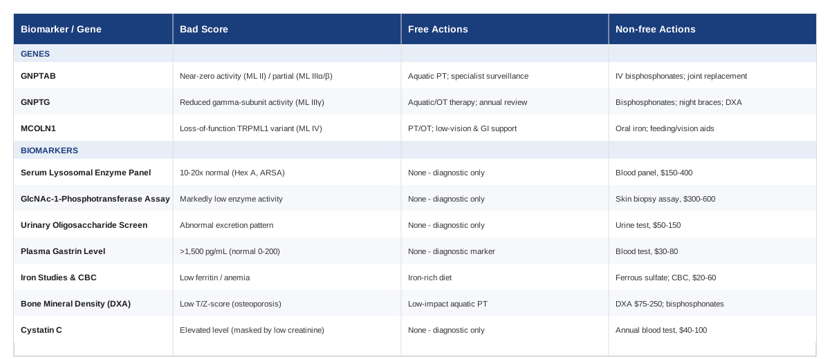
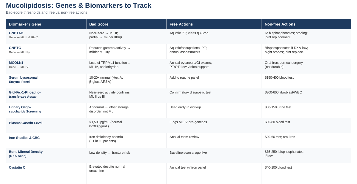
All three genes below cause autosomal recessive disease, meaning a child needs one altered copy from each parent to be affected — carriers with a single altered copy are generally unaffected. Two of the genes, GNPTAB and GNPTG, encode different subunits of the same enzyme complex, so it helps to think of them as two failure points on one assembly line. The third, MCOLN1, is mechanically unrelated — it doesn't affect enzyme delivery at all, but rather the lysosome's ability to function once enzymes are already inside it.
GNPTAB: The Gene Behind Mucolipidosis II and IIIα/β
GNPTAB encodes the alpha and beta subunits of an enzyme called N-acetylglucosamine-1-phosphotransferase. That enzyme's job is narrow but critical: it attaches a mannose-6-phosphate "shipping label" to dozens of digestive enzymes so they get routed into the lysosome, the cell's recycling compartment. Without that label, those enzymes get sent out of the cell entirely instead of into the lysosome. The lysosome, missing its enzymes, can't break down the materials it's supposed to, and those materials accumulate — while the enzymes themselves end up circulating at unusually high levels in the blood, which is actually how the condition is often first flagged.
The amount of leftover, functioning enzyme is what separates the two diseases this gene causes. Near-total loss of activity produces mucolipidosis II (I-cell disease), a severe, neonatal-onset condition with skeletal changes visible at birth, cardiorespiratory involvement, and a life expectancy usually measured in a small number of years. Partial, residual enzyme activity produces mucolipidosis IIIα/β, a considerably milder disease that presents in early childhood with joint stiffness, short stature, and skeletal changes, and that people can live with into adulthood. This genotype-severity relationship is well documented in the clinical genetics literature (LINKFIX url="https://pubmed.ncbi.nlm.nih.gov/30882951/" — Update on GNPTAB and GNPTG mutations) — actually formatted properly below.
According to the GeneReviews clinical summary on GNPTAB-related disorders, the amount of residual GlcNAc-1-phosphotransferase activity correlates directly with how early symptoms start and how quickly the disease progresses. A more detailed molecular breakdown of how specific mutation types predict this severity spectrum is available in the 2019 update on GNPTAB and GNPTG mutations, which remains one of the more useful genotype-phenotype references for families trying to understand where their specific variant falls on that spectrum.
If the Gene Is Altered: The Care Plan Without Supplements or Equipment
For both mucolipidosis II and IIIα/β, the foundation of care is non-pharmacologic and centers on protecting joints and organ systems rather than trying to alter enzyme activity, which currently isn't possible with lifestyle changes. Low-impact aquatic physical therapy is specifically recommended over land-based exercise because it strengthens muscles and preserves range of motion without loading already-strained joints and tendons — typically two to three sessions a week, adjusted as tolerance changes. Interactive cognitive stimulation programs are recommended for mucolipidosis II, where developmental involvement is more prominent.
Surveillance itself is the other half of this plan: outpatient visits every three months during infancy and toddlerhood, shifting to every six months as the child grows, with cardiac and respiratory monitoring intensified as those systems become more relevant. Any elective surgery or anesthesia should be deferred to tertiary centers experienced with the airway management challenges these patients present, since airway anatomy changes make routine intubation riskier than in the general population. None of this carries pharmacologic side effects, but the frequency matters: skipping the low-impact framing and pushing standard pediatric physical therapy protocols can accelerate joint damage rather than prevent it. These recommendations come directly from the GeneReviews management guidelines for GNPTAB-related disorders.
If the Gene Is Altered: The Plan With Supplements, Medication, or Equipment
Where bone pain and osteoporosis become significant — more common in mucolipidosis IIIα/β than in the more severe type II — bisphosphonate therapy, typically pamidronate given intravenously, is used to slow bone loss and reduce pain. Dosing generally follows an infusion cycle every three to four months, with bone mineral density reassessed by a DXA scan roughly every two to five years depending on severity, and calcium and vitamin D status monitored alongside it, since bisphosphonates can lower blood calcium. Side effects to expect include a flu-like reaction after the first infusion in many patients, and, with long-term high-dose use, a small risk of osteonecrosis of the jaw — which is why dental health is checked before starting.
Mechanical and surgical equipment plays a real role too: night bracing for hand and wrist stiffness, carpal or tarsal tunnel release surgery for nerve compression (which offers temporary rather than permanent relief), and hip or knee replacement, which the GeneReviews literature notes has been performed successfully in older adolescents and adults for pain relief. Hearing aids and mobility aids are added as needed rather than on a fixed schedule. Hematopoietic stem cell transplant has been attempted in a small number of mucolipidosis II cases; a detailed case analysis found biochemical improvement and preserved quality of life in one patient, but concluded that the actual benefit remains unclear due to the near-total absence of comparable untreated natural history data — this is not a routine option, and it carries real transplant-related risks that should be weighed carefully with a transplant specialist.
GNPTG: The Gene Behind Mucolipidosis IIIγ
GNPTG encodes the gamma subunit of the same GlcNAc-1-phosphotransferase enzyme complex disrupted by GNPTAB, but mutations here produce a generally milder, later-onset disease than either mucolipidosis II or IIIα/β. Mucolipidosis IIIγ mainly affects skeletal, joint, and connective tissue, with symptoms typically appearing around age three: slowed growth, short stature, joint stiffness, and dysostosis multiplex — a pattern of multiple bone abnormalities visible on imaging. Central nervous system involvement is far less prominent here than in mucolipidosis II, which is one reason cognitive development is often closer to typical in this subtype.
If the Gene Is Altered: The Care Plan Without Supplements or Equipment
The non-pharmacologic backbone looks similar to GNPTAB-related disease: physical and occupational therapy tailored to the individual, with low-impact aquatic therapy again favored for its ability to maintain function without joint strain. Annual assessment of musculoskeletal status, motor function, pain, growth, and cardiorespiratory function is standard, with pulmonary function testing typically done every five years rather than annually, reflecting the generally slower disease course. Clinic visits happen roughly twice a year in early childhood and can shift to annual after age six if the disease is stable.
One point worth being direct about: current guidance is explicit that no available intervention reverses the progressive limitation of joint motion in this condition — supportive therapies improve comfort and function, but they don't halt the underlying process. That's according to the GeneReviews summary on mucolipidosis III gamma.
If the Gene Is Altered: The Plan With Supplements, Medication, or Equipment
Bone health monitoring is more structured here than in some other subtypes: a baseline DXA scan is recommended for children over five and for adults at diagnosis, with follow-up scans every five years in children with normal results, every three years in adults with normal density, and every two years when density is already reduced. Bisphosphonate treatment is considered specifically when skeletal disease is significant and bone density is markedly low, following the same infusion-cycle and calcium-monitoring approach described for GNPTAB-related disease.
Night braces, particularly for the hands, are commonly used to preserve function, and joint replacement — hip and knee in particular — has been used successfully in older adolescents and adults for pain control. As with GNPTAB-related disease, these interventions manage symptoms and slow specific complications; they don't change the trajectory of the underlying enzyme defect.
MCOLN1: The Gene Behind Mucolipidosis IV
MCOLN1 works completely differently from the other two genes. It encodes mucolipin-1, more commonly known as TRPML1, a channel sitting in the membrane of the lysosome that controls calcium movement in and out of the compartment. This isn't a delivery problem — enzymes generally do reach the lysosome in mucolipidosis IV. Instead, the lysosome's internal signaling and recycling processes (autophagy) are disrupted, which particularly affects tissues with high turnover and high metabolic demand: neurons, the cornea, the retina, and the stomach lining.
Clinically, this produces a distinct pattern from the other two genes: psychomotor developmental delay and impaired vision from corneal clouding and retinal degeneration typically appear by the end of the first year of life, followed by a slow decline in neurological function through the second decade, and a shortened lifespan. A striking and clinically useful feature is constitutive achlorhydria — the stomach simply doesn't produce acid — which is present in essentially all affected individuals and drives several of the management points below. Corneal clouding is reported in roughly nine out of ten patients and is sometimes the very first symptom noticed, according to a long-standing clinical description of ocular findings in mucolipidosis IV, and the full clinical and management picture is laid out in the GeneReviews entry on mucolipidosis IV.
If the Gene Is Altered: The Care Plan Without Supplements or Equipment
Annual, structured surveillance is the backbone of care: comprehensive ophthalmology exams, neurologic assessment, and feeding and growth evaluation at minimum once a year, with more frequent review if regression is noted. Physical and occupational therapy address hypotonia and motor delay, and low-vision services and communication supports are introduced early rather than waiting for vision loss to become severe, since corneal clouding and retinal degeneration are present from infancy in most patients. Gastroenterology involvement is recommended for the constipation and bile reflux that come with the broader gastrointestinal involvement in this condition, and swallowing evaluation becomes more important as motor decline progresses, given aspiration risk.
None of this changes the underlying channel defect, and families should expect a surveillance-and-support model rather than a treatment aimed at the gene itself — that distinction is worth raising directly with a metabolic geneticist so expectations are calibrated correctly from the start.
If the Gene Is Altered: The Plan With Supplements, Medication, or Equipment
The single most concrete, evidence-backed intervention here relates to the achlorhydria mentioned above. Because stomach acid production is absent, iron absorption is impaired in roughly half of affected individuals, and about one in ten develops iron deficiency anemia. GeneReviews specifically recommends oral ferrous sulfate iron supplementation as needed, guided by annual complete blood count and iron studies — dosing is weight-based and typically continued as long as deficiency is present, then reassessed. Expected side effects include constipation, dark stools, and mild GI upset, which is one more reason gastroenterology follow-up matters in this group.
Kidney function monitoring uses cystatin C rather than standard creatinine specifically because muscle atrophy, common as the disease progresses, artificially lowers creatinine and can mask declining kidney function — this is tested annually alongside the iron panel. Elevated plasma gastrin, a direct consequence of the achlorhydria, is itself useful diagnostically and is checked as part of ongoing GI surveillance. On the equipment and procedural side, corneal surface intervention has been attempted historically — including conjunctival transplantation for corneal opacification — but results have generally been modest and non-durable, since the underlying storage process in corneal cells continues; this is worth discussing with a corneal specialist as a case-by-case option rather than a standard step.
Where Ali Torkamani and Gary Brecka Fit In, and Where They Don't
If you've spent time in consumer genomics or biohacking spaces, you've likely come across Ali Torkamani's work on polygenic risk scoring, or Gary Brecka's public advocacy for personalized biomarker panels and supplementation. Both are genuinely useful starting points — for common, complex conditions shaped by dozens or hundreds of small genetic effects layered on top of lifestyle factors, like cardiovascular disease risk or nutrient metabolism efficiency.
Mucolipidosis doesn't fit that model, and it's worth being direct about why. It's caused by a single gene, inherited in a straightforward recessive pattern, with a small number of well-characterized genes accounting for essentially the entire condition. A polygenic risk score has no meaningful role here, and a consumer raw-data genotyping panel (the kind used for ancestry or common-disease risk) is not built to reliably detect the specific structural variants, splice-site changes, and compound heterozygous mutations that cause mucolipidosis. The right tool is clinical-grade sequence analysis of GNPTAB, GNPTG, and MCOLN1 through a certified genetics laboratory, interpreted by a clinical geneticist — exactly the kind of testing described in the GeneReviews entries cited throughout this article.
Why "Epigenetics" Doesn't Explain Mucolipidosis Severity
It's worth addressing this directly because it comes up often: unlike many chronic diseases where epigenetic modification (methylation patterns, environmental gene-expression changes) meaningfully shapes outcomes, there is no established epigenetic biomarker that explains why one person with a GNPTAB mutation develops severe mucolipidosis II and another develops milder IIIα/β. The explanation that does hold up in the literature is allelic — the specific combination of mutations a person carries and how much functional enzyme activity those particular mutations leave behind, as detailed in the genotype-phenotype correlation research on GNPTAB and GNPTG. It's a less fashionable answer than "epigenetics," but it's the one supported by human data, and it's exactly the kind of distinction that matters when a family is trying to understand a specific diagnosis rather than a general category of disease.
Lab Values and Scans Worth Tracking Over Time
Genetic testing establishes the diagnosis once. The values below are what clinicians actually return to at follow-up visits to judge how the disease is behaving and whether an intervention is working. None of them are things a patient can improve through diet or exercise in the way a cholesterol panel might respond to lifestyle change — they're tracking a disease process, not a modifiable risk factor — but knowing what they mean turns a lab report from a wall of numbers into something you can actually follow.
Serum Lysosomal Enzyme Panel
In mucolipidosis II and III, several enzymes that should be inside the lysosome instead leak into the bloodstream, showing up at ten to twenty times normal levels. Hexosaminidase A is the most commonly used screening enzyme, alongside beta-glucuronidase and arylsulfatase A. This is a standard blood draw sent to a biochemical genetics laboratory, generally costing in the range of $150 to $400 depending on the specific panel and insurance coverage. It's primarily a diagnostic and severity marker rather than something that "improves" with treatment, though case reports after stem cell transplant have shown partial reductions.
GlcNAc-1-Phosphotransferase Activity Assay
This is the definitive confirmatory test for GNPTAB- and GNPTG-related disease, performed on cultured skin fibroblasts or white blood cells. It directly measures how much of the enzyme is still functioning, which is the single best predictor of whether a person's disease course will look more like severe mucolipidosis II or milder IIIα/β or IIIγ. Expect a skin biopsy, a specialized reference lab, several weeks' turnaround, and a cost typically in the $300 to $600 range.
Urinary Oligosaccharide and Mucopolysaccharide Screening
A relatively inexpensive urine test, usually $50 to $150, used early in a diagnostic workup to help distinguish mucolipidosis from related but distinct conditions like the mucopolysaccharidoses. Results in mucolipidosis are often normal or only mildly abnormal, which is itself diagnostically useful — a strongly abnormal result points toward a different storage disorder.
Plasma Gastrin Level
This one is specific to mucolipidosis IV and is a good example of a cheap test carrying outsized diagnostic value: because achlorhydria is essentially universal in this subtype, gastrin levels run far above the normal range (normal is roughly 0 to 200 pg/mL; affected individuals average over 1,500 pg/mL). It's a simple blood test, typically $30 to $80, and can support a clinical suspicion of mucolipidosis IV even before genetic confirmation comes back.
Iron Studies and Complete Blood Count
Also specific to mucolipidosis IV, this tracks the anemia risk created by impaired iron absorption from achlorhydria. It's routine, inexpensive bloodwork ($20 to $60), recommended annually, and directly guides whether and how much oral iron supplementation is needed.
Bone Mineral Density (DXA Scan)
Relevant across mucolipidosis II, IIIα/β, and IIIγ, this imaging study tracks osteoporosis and fracture risk from progressive skeletal involvement. Costs generally run $75 to $250 depending on setting and insurance. Baseline scans start around age five in the milder subtypes, with follow-up intervals of two to five years depending on severity — and results directly inform whether bisphosphonate therapy is worth starting.
Cystatin C
A kidney function marker preferred over standard creatinine in mucolipidosis IV specifically because muscle atrophy, common as the disease progresses, artificially lowers creatinine and can hide real declines in kidney function. It's a simple annual blood test, typically $40 to $100, with no meaningful downside beyond the blood draw itself.
The Genome Odyssey: Ten Ideas Reshaping Rare Disease Diagnosis
Stanford geneticist Euan Ashley's book The Genome Odyssey isn't about mucolipidosis specifically, but its central subject — families spending years chasing a diagnosis that genome sequencing could have delivered in days — maps almost exactly onto what many mucolipidosis families experience before a metabolic geneticist finally lands on the right gene. The ideas below are drawn from that book and the broader genomic medicine research it summarizes, and each one has a direct, practical parallel to navigating this specific condition.
The Diagnostic Odyssey Is a Measurable, Real Delay
Families of children with ultra-rare genetic diseases routinely spend years and see numerous specialists before getting a molecular diagnosis. Recognizing this as a systemic, fixable problem — not just bad luck — is what pushed genomic medicine toward faster, broader first-line testing rather than sequential single-gene tests.
Speed Changes Outcomes in Critically Ill Infants
The clearest demonstration of this came from a randomized trial testing rapid whole-genome sequencing in critically ill infants: among babies enrolled in the first 25 days of life, those who received rapid sequencing were diagnosed at a rate of 32%, compared to 0% in those who received standard genetic testing alone, according to the NSIGHT1 randomized controlled trial. For a condition like mucolipidosis II, where early recognition shapes airway and cardiac management from the first weeks of life, that speed has real clinical weight.
A Negative Genetic Test Isn't Necessarily the End of the Story
Genetic knowledge is updated constantly, and a gene's link to a disease sometimes isn't established yet at the time a family is first tested. Systematic reanalysis of existing genetic data — without a new sample — has been shown to raise diagnostic yield substantially over time, in some pediatric cohorts from roughly 31% to 53%, according to research on clinical exome reanalysis. If your family had inconclusive genetic testing more than two or three years ago, asking your genetics team about reanalysis is a reasonable, low-cost next step.
One Gene, Multiple Diseases, Depending on the Damage
A recurring theme in genomic medicine is that the same gene can produce wildly different diseases depending on exactly how a mutation disrupts protein function. This is precisely what happens with GNPTAB, where near-complete loss of function causes severe mucolipidosis II and partial loss of function causes the considerably milder mucolipidosis IIIα/β.
Sequencing Parents Alongside the Child Finds More Answers
Trio sequencing — testing both parents alongside the affected child — consistently outperforms testing the child alone, because it lets geneticists immediately see which variants were inherited versus newly arising, dramatically narrowing the search. For a recessive condition like mucolipidosis, where both parents are typically unaffected carriers, this approach is especially informative and worth confirming is part of any planned testing.
A Diagnosis Changes Management Even Without a Cure
Ashley's book repeatedly makes the point that a confirmed molecular diagnosis is valuable on its own, independent of whether a treatment exists — it stops unnecessary further testing, connects a family to the correct surveillance schedule, and opens the door to natural history studies and patient registries built around that exact gene.
Personalized, N-of-1 Therapies Are No Longer Theoretical
The most striking real-world precedent here is milasen, an antisense oligonucleotide drug custom-designed for a single child with a fatal genetic condition, developed and given to the patient within about a year of diagnosis, as described in the landmark New England Journal of Medicine report. It's not a template that currently exists for mucolipidosis, but it demonstrates that ultra-rare, single-patient genetic therapy has moved from theory to precedent — a meaningfully different landscape than existed a decade ago.
Natural History Data Is the Real Bottleneck, Not Sequencing Technology
Sequencing has gotten fast and cheap; understanding what happens to untreated patients over time has not kept pace. This exact gap was the central limitation identified in the review of stem cell transplant outcomes in mucolipidosis II, which explicitly called for better registries of untreated patients before transplant benefit could be properly judged.
Carrier Status Matters for Future Family Planning
Because mucolipidosis is recessive, a confirmed diagnosis in one child has direct implications for future pregnancies. Genetic counseling around carrier testing, and where relevant, prenatal or preimplantation testing options, is a standard next conversation once a causative gene is identified.
Understanding Your Own Genome Changes How You Advocate
The book's broader argument is that genomic literacy isn't just for clinicians — a family that understands its child's specific variant is better positioned to evaluate research trial eligibility, ask sharper questions at appointments, and connect with the right disease-specific advocacy and research organizations.
Complementary Approaches Worth Considering Alongside Medical Care
None of the approaches below touch the underlying genetic defect, and none have been studied in mucolipidosis patients specifically — the condition is too rare for dedicated trials to exist. What follows is evidence drawn from closely related populations: children with chronic joint pain, developmental disability, and pediatric chronic pain more broadly. That's a meaningful limitation worth stating plainly, and each option below should be discussed with your care team rather than substituted for medical surveillance.
Massage Therapy
For the joint pain and stiffness common in mucolipidosis IIIα/β and IIIγ, massage therapy is a reasonable, low-risk addition to standard physical therapy, since it targets the same muscular guarding and anxiety that amplify perceived pain in chronic joint conditions.
The most relevant human evidence comes from a randomized study in children with juvenile rheumatoid arthritis, where parent-administered massage for 15 minutes daily over 30 days reduced anxiety and stress hormone levels immediately, and reduced pain by self-report, parent report, and physician assessment over the study period, according to this study on massage therapy in juvenile rheumatoid arthritis.
In practice, this looks like short, gentle, parent-delivered massage of affected joints a few times a week, done alongside — never instead of — prescribed physical therapy, and avoided over any acutely inflamed or post-surgical joint without a clinician's sign-off.
Music Therapy
For children with mucolipidosis IV, where developmental delay and sensory impairment (particularly vision loss) are central features, music therapy offers a way to engage and support development through a channel that doesn't depend on vision.
A systematic review of randomized controlled trials of music therapy in children with autism spectrum disorder and intellectual disability — the closest comparable population studied — found consistent improvements in engagement, social communication, and initiation, as summarized in this review of music therapy RCTs. This isn't mucolipidosis-specific evidence, and results shouldn't be assumed to transfer directly, but the population and developmental profile overlap enough to make it a reasonable, low-risk option.
Realistically, this means structured sessions with a credentialed music therapist, typically weekly, focused on interactive singing or instrument play rather than passive listening, since the evidence favors active engagement over background music.
Guided Imagery and Progressive Muscle Relaxation
For the chronic pain that accompanies skeletal disease in mucolipidosis II, IIIα/β, and IIIγ, relaxation-based approaches address the anxiety and muscle tension that can amplify pain independent of the underlying joint damage.
A Cochrane review of psychological therapies for chronic and recurrent pain in children found that relaxation training alone, along with cognitive behavioral approaches, effectively reduces pain intensity across several chronic pediatric pain conditions, per this Cochrane summary. A specific randomized trial of audio-recorded guided imagery for children with recurrent abdominal pain also found meaningful symptom reduction, as reported in this guided imagery trial — notably relevant given the gastrointestinal symptoms common in mucolipidosis IV.
In practice, this means short, age-appropriate guided imagery or progressive muscle relaxation recordings used a few times a week, ideally introduced by a pediatric psychologist familiar with chronic illness, with no known physical side effects and no conflict with medical treatment.
Conclusion
Mucolipidosis comes down to two distinct mechanical failures: GNPTAB and GNPTG break the system that delivers enzymes into the lysosome, while MCOLN1 breaks a channel the lysosome needs to function once those enzymes arrive. That single distinction explains most of what differs across mucolipidosis II, III, and IV — severity, organs affected, and the surveillance plan your care team should be following. None of the management options described here reverse the underlying gene defect, but they meaningfully change comfort, function, and how early complications get caught: bisphosphonates for bone loss, iron for achlorhydria-driven anemia, low-impact therapy for joint preservation, and a defined schedule of lab values and scans to track it all.
The next useful step isn't a supplement or a device — it's a conversation. Bring this article's gene breakdown and biomarker list to your next appointment with a metabolic geneticist or genetic counselor, ask which of the seven values above are already being tracked and which aren't, and ask directly whether reanalysis of older genetic testing might be worthwhile. That single conversation is the most concrete, evidence-grounded move available right now.
Musculoskeletal: Bone Conditions Joint Conditions
Digestive: Stomach & Esophagus Conditions
Eye: Retinal Conditions
Urological: Kidney Conditions