This article contains AI generated content.
· UpdatedMucopolysaccharidosis Genes and Biomarkers — 9 Genes and 7 Biomarkers to Track
Introduction
Living with mucopolysaccharidosis, or caring for someone who has it, means navigating a condition that looks different depending on which enzyme is missing, how much residual activity remains, and which organs absorb the most damage over time. MPS is not one disease. It is a family of at least nine distinct lysosomal storage disorders, each caused by the failure of a single enzyme responsible for breaking down glycosaminoglycans — the long-chain sugar molecules that would otherwise accumulate in cells, tissues, and organs throughout the body. The result is a clinical picture that can range from a rapidly progressive neurodegenerative syndrome in infancy to a slowly evolving skeletal disease that is not diagnosed until adulthood.
Generic advice about inflammation, diet, or metabolic health barely scratches the surface for MPS. What matters here is specificity: which subtype, which gene variant, which GAG fraction is accumulating, and how the body is responding — or not responding — to whatever therapy is in place. A recommendation that applies to MPS I Hurler may be irrelevant to MPS IVA Morquio. A biomarker that is essential for monitoring neurological MPS III may add little to the management of the predominantly somatic MPS VI. Broad frameworks do not hold up against the granularity that MPS demands.
This article takes a more focused approach. The primary framework here is biomarker tracking: identifying the seven most clinically useful laboratory and biological markers that tell you what is actually happening in the body, how to measure them reliably, and what to do if they are abnormal. A second framework covers the nine genes whose variants define each MPS subtype and what the genetic picture means for surveillance and treatment choices. Between these two lenses, the goal is to help patients, families, and clinicians ask sharper questions and act on better information.
The landscape for MPS has improved substantially in the past two decades. Enzyme replacement therapies are approved for several subtypes. Gene therapy trials have reached Phase 2 and 3. Substrate reduction approaches are in clinical development. Better biomarkers are enabling earlier treatment decisions and more precise tracking of therapeutic response. None of this amounts to a cure, and the article makes no such claim. But better information does lead to better decisions — and in a condition where early intervention can mean the difference between preserved cognition and irreversible loss, that difference matters enormously.
7 Biomarkers to Track for MPS Disease Activity and Treatment Response
Biomarkers in MPS serve a different purpose than in most common chronic conditions. They are not primarily used to predict risk — the diagnosis is already established genetically. Their roles are to confirm disease type, quantify disease burden, monitor progression, and assess therapeutic response. Understanding which markers to track, how to measure them, and what the results mean in clinical context is one of the most practical tools available to anyone managing MPS over the long term.
1. Total Urinary Glycosaminoglycans (GAGs)
Why it matters: Elevated total urinary GAGs are the biochemical hallmark of virtually all MPS subtypes. Because lysosomal enzyme activity is insufficient to fully degrade glycosaminoglycans, incompletely degraded fragments accumulate and spill into urine. Total urinary GAG quantification is sensitive, relatively inexpensive, and widely available, making it the first-line biomarker for both diagnosis and longitudinal monitoring. It reflects overall disease burden and responds measurably to effective therapy.
How to measure it
The standard method is the dimethylmethylene blue (DMMB) colorimetric assay, performed on a random urine sample normalized to creatinine (results reported in µg GAG per mg creatinine). This test is available at most hospital-based laboratories and reference labs. Cost ranges from $50 to $150 USD. Some centers use a 24-hour urine collection for more precise quantification. Urine GAG electrophoresis can provide a rough qualitative pattern but is less sensitive than DMMB for absolute quantification. In untreated children with MPS, total urinary GAGs are typically elevated 2- to 20-fold above age-matched reference ranges.
If the score is elevated — the plan without supplements
A confirmed elevation warrants urgent referral to a metabolic disease specialist if one is not already involved. The clinical priority is fractionation (see biomarkers 2–4) to identify which GAG subtype is elevated and confirm the MPS subtype. Monitoring frequency should be quarterly in untreated patients and monthly during the initiation of any therapy. Diet modification alone does not meaningfully reduce urinary GAGs in MPS, but an anti-inflammatory dietary pattern — Mediterranean-style, low in refined carbohydrates and ultra-processed foods — reduces the systemic oxidative stress imposed on tissues already burdened with storage material. Regular follow-up with a metabolic team and consistent monitoring establishes the trajectory that informs all other clinical decisions.
If the score is elevated — the plan with supplements or equipment
Enzyme replacement therapy (ERT) is the primary intervention that demonstrably reduces urinary GAG levels and is currently approved for MPS I (laronidase), MPS II (idursulfase), MPS IVA (elosulfase alfa), MPS VI (galsulfase), and MPS VII (vestronidase alfa). Urinary GAGs typically begin to decline within weeks of ERT initiation and should be reassessed at 3, 6, and 12 months to document response. Home urine collection kits enable more frequent monitoring without clinical visits. Dried blood spot (DBS) cards are being validated as complementary matrices. No supplement independently lowers urinary GAGs in MPS; claims to the contrary should be treated with skepticism. Side effects of ERT: infusion-related reactions (fever, urticaria, anaphylaxis in rare cases); managed with premedication and slow titration.
2. Urine Heparan Sulfate (HS)
Why it matters: Heparan sulfate is the primary accumulating GAG in MPS I, II, III, and VII. It is also the fraction most directly associated with neurological disease, as HS degradation products penetrate the blood-brain barrier and contribute to neuroinflammation, synaptic dysfunction, and neuronal death in the CNS-involved subtypes. Recent advances using liquid chromatography-mass spectrometry (LC-MS/MS) have established plasma and urine HS as among the most sensitive and informative biomarkers in the MPS field. HS levels correlate with disease burden, cognitive trajectory in MPS III, and treatment response in MPS I and II. Research published in peer-reviewed journals has validated plasma HS as a reliable indicator of disease activity even in attenuated phenotypes where total urinary GAGs may be only modestly elevated.
How to measure it
LC-MS/MS of urine or plasma is the gold standard for HS quantification, available at specialty metabolic laboratories and major academic reference labs in North America, Europe, and Australia. Cost ranges from $200 to $500 USD, often as part of a combined GAG fractionation panel. Standard urine GAG electrophoresis can qualitatively suggest HS elevation but lacks the quantitative precision needed for disease monitoring. For neurological MPS types, CSF HS measurement is being evaluated as a biomarker for CNS disease activity in gene therapy trials. Frequency: every 3–6 months in active management; annually in stable, treated patients.
If the score is elevated — the plan without supplements
Elevated urinary HS — particularly in a child presenting with behavioral regression, sleep disruption, or cognitive slowing — should prompt immediate genetic evaluation for SGSH, NAGLU, HGSNAT, or GNS (the MPS III genes) and neuropsychological assessment. For families managing MPS III, where no ERT is currently approved, elevated HS in urine and plasma serves as a natural history endpoint and helps quantify disease burden over time. Supportive CNS-protective measures include ensuring adequate sleep (using environmental and behavioral sleep hygiene strategies; melatonin is commonly used for the profound sleep disruption in MPS III), minimizing head trauma, maintaining nutritional sufficiency, and reducing environmental toxin exposure. HS-specific monitoring every 3–6 months provides the longitudinal data needed to assess disease pace and trial eligibility.
If the score is elevated — the plan with supplements or equipment
For MPS I and II patients on ERT, plasma and urinary HS should be reassessed at 3 and 6 months post-initiation. HS normalization correlates with improved somatic outcomes on ERT. For MPS I patients receiving hematopoietic stem cell transplant (HSCT), HS decline post-engraftment signals successful enzyme delivery to peripheral tissues. For MPS III, multiple gene therapy trials using intrathecal and intraparenchymal AAV delivery are using HS as a primary pharmacodynamic endpoint — these trials represent the current frontline of hope and enrollment should be explored at qualified centers. N-acetylcysteine (NAC) at 600–1200 mg/day has been investigated in preclinical MPS III models for neuroprotective effects but has not yet produced convincing human trial results; it has a favorable safety profile and reasonable theoretical rationale. Cycling: continuous supplementation is typical; reassess every 3 months. Side effects of NAC: GI discomfort at higher doses; sulfur odor.
3. Urine Dermatan Sulfate (DS)
Why it matters: Dermatan sulfate accumulates predominantly in MPS I, II, and VI. Unlike heparan sulfate, DS is associated less with neurological disease and more with connective tissue pathology: aortic root dilation, mitral and aortic valve thickening, arterial stiffness, joint contractures, and corneal clouding. Tracking DS separately from total GAGs helps clinicians calibrate the intensity of cardiovascular and orthopedic surveillance. In MPS VI, where intelligence is typically preserved but somatic disease can be severe, DS quantification is the central biomarker.
How to measure it
Fractionated urine GAG analysis by LC-MS/MS or GAG electrophoresis. Usually ordered as part of a combined HS/DS/KS panel at specialty labs. Cost: typically $200–$400 as part of a fractionation panel. Some metabolic labs include DS, HS, and KS in a single MPS biomarker panel with LC-MS/MS quantification. For MPS VI specifically, DS quantification is a primary biomarker for monitoring galsulfase therapy.
If the score is elevated — the plan without supplements
Elevated DS in a patient with MPS I, II, or VI should trigger dedicated cardiac surveillance: echocardiography at minimum annually, with more frequent assessment if valve disease is established. Pulmonary function testing annually for obstructive or restrictive pattern. Ophthalmology assessment for corneal deposits and intraocular pressure. Low-impact aerobic activity — swimming, cycling — maintains cardiovascular function without the joint loading that exacerbates connective tissue disease. High-impact sports and activities that involve repetitive joint stress should be avoided given the DS-related deterioration of connective tissue integrity.
If the score is elevated — the plan with supplements or equipment
For MPS I, laronidase significantly reduces urinary DS over months of treatment and is associated with improved respiratory and joint outcomes. For MPS VI, galsulfase demonstrates measurable DS reduction and improved endurance. HSCT for MPS I, when performed before 2.5 years of age in Hurler syndrome, can stabilize cardiovascular disease and prevent further DS-driven connective tissue damage. Coenzyme Q10 (200–400 mg/day) has a theoretical role in supporting mitochondrial function under metabolic stress in storage disease contexts, though no MPS-specific RCT supports this specific use; the supplement is generally well-tolerated. Custom orthoses and joint support bracing reduce biomechanical load on DS-weakened connective tissue and improve functional capacity. Cycling for CoQ10: ongoing; reassess benefit quarterly. Side effects: mild GI effects; note potential interaction with warfarin.
4. Urine Keratan Sulfate (KS)
Why it matters: Keratan sulfate is the hallmark accumulating GAG of MPS IVA (Morquio A) and MPS IVB (Morquio B), and is also elevated in MPS III. KS accumulation drives the distinctive skeletal dysplasia of Morquio syndrome: short stature, pectus carinatum, genu valgum, odontoid hypoplasia with attendant cervical instability, and joint hypermobility. Unlike most MPS types, MPS IVA typically spares cognition entirely while causing life-threatening skeletal and cardiorespiratory complications. KS was used as the primary biomarker in the pivotal Phase 3 trial of elosulfase alfa and remains the main pharmacodynamic endpoint in MPS IVA management.
How to measure it
Urine KS by LC-MS/MS or competitive ELISA. Serum KS by ELISA is also validated for MPS IVA and is arguably more convenient for serial monitoring since blood collection is simpler than urine collection in young children. Cost: $150–$350. Blood spot KS is more stable in transit than urine and is used in some screening programs. In untreated MPS IVA, serum and urine KS are markedly elevated; levels correlate with disease severity and age of onset. KS tends to decline with age in untreated patients, which can complicate interpretation in older patients with attenuated phenotypes.
If the score is elevated — the plan without supplements
Elevated KS in a child with skeletal findings — particularly short stature, coxa valga, or genu valgum — should prompt urgent GALNS enzyme assay and genetic sequencing of the GALNS gene. If confirmed, MRI of the cervical spine is the most urgent priority due to the risk of atlantoaxial instability causing spinal cord compression — a potentially catastrophic and sometimes sudden complication. Annual cervical imaging, referral to orthopedic surgery experienced with MPS, and avoidance of activities involving axial loading (contact sports, gymnastics, trampolining) are essential. Physical therapy focusing on core strengthening and balance reduces fall risk and preserves functional capacity.
If the score is elevated — the plan with supplements or equipment
Elosulfase alfa (Vimizim) is approved for MPS IVA and is administered as weekly IV infusions (2 mg/kg). The pivotal trial demonstrated statistically significant improvements in 6-minute walk test and stair-climbing speed alongside measurable KS reduction. Response should be assessed at 12 and 24 weeks using serum/urine KS as the primary pharmacodynamic endpoint. Vitamin D (2000–4000 IU/day, titrated to target 25-OH-D of 40–60 ng/mL) and calcium (dietary-first approach; supplement if dietary intake is insufficient) support bone mineral density in a skeleton already compromised by KS accumulation. Custom spinal orthoses, adapted seating, and home monitoring of joint status with photo documentation allow families to track musculoskeletal changes between clinical visits. Side effects of elosulfase alfa: infusion-related reactions; anaphylaxis risk requires epi-pen availability during infusion.
5. Specific Lysosomal Enzyme Activity
Why it matters: Measuring the specific activity of the deficient enzyme both confirms the MPS diagnosis and provides a quantitative baseline against which future treatment responses are measured. Residual enzyme activity levels below 1% of normal generally predict severe phenotypes; activity in the 1–10% range often correlates with attenuated forms, particularly in MPS I, II, and VI. This residual activity concept is clinically important because it influences treatment urgency, HSCT candidacy, and pharmacological chaperone eligibility for specific missense variants that produce misfolded but potentially stabilizable enzyme protein.
How to measure it
Two matrices are in clinical use: dried blood spot (DBS) and leukocyte lysate. DBS testing is used in newborn screening programs and is cost-effective ($150–$300), making it the standard entry point for diagnosis. Leukocyte enzyme assay is the gold standard for confirmation and is more sensitive for attenuated phenotypes ($200–$600 at reference labs). Plasma enzyme activity is less reliable due to pseudodeficiency variants and circulating enzyme uptake artifacts. For each MPS type, the assay uses a fluorogenic substrate specific to the relevant enzyme: iduronidase for MPS I, iduronate-2-sulfatase for MPS II, heparan-N-sulfatase for MPS IIIA, and so forth.
If the score is low — the plan without supplements
Confirmed low or absent enzyme activity requires a second confirmatory assay and concurrent DNA sequencing to identify causative variants. Establishing a pre-treatment baseline allows all subsequent measurements to be interpreted in context. Given that ERT partially restores circulating enzyme activity, post-ERT enzyme assays must be interpreted with awareness that circulating recombinant enzyme will artificially elevate measured activity in blood samples taken during the infusion window. Nutritional protein sufficiency (adequate essential amino acid intake) supports overall cellular proteostasis, relevant in a setting where lysosomal trafficking pathways are already stressed. Frequency: annual enzyme activity measurement at minimum; more frequently when trial eligibility or chaperone therapy is under consideration.
If the score is low — the plan with supplements or equipment
For ERT-eligible MPS types, enzyme replacement directly supplements the deficient enzymatic pathway and should begin as early as possible after confirmed diagnosis. Pharmacological chaperones — small molecules that stabilize misfolded enzyme protein and facilitate its correct trafficking to the lysosome — are under active investigation for specific missense variants in MPS I and MPS II that produce structurally aberrant but potentially rescuable enzyme protein. These are available only in clinical trials as of 2026. Gene therapy approaches (ex vivo HSC-based for MPS I, AAV intrathecal delivery for MPS III) aim to restore sustained endogenous enzyme expression without the need for weekly infusions. Following any gene therapy, longitudinal enzyme activity monitoring in blood and CSF (where relevant) is the primary readout of therapeutic success.
6. Chitotriosidase Activity
Why it matters: Chitotriosidase is an enzyme produced by activated macrophages. Because lysosomal storage triggers macrophage activation as a secondary inflammatory response, plasma chitotriosidase activity is consistently elevated in all major MPS types and serves as a cross-disease marker of storage burden and macrophage inflammatory load. Originally validated in Gaucher disease, its utility in MPS I, II, and VI monitoring is well-established in the clinical literature. Serial chitotriosidase measurements capture overall disease activity changes and respond to ERT within months of initiation. One important caveat: approximately 6% of the general population carries a homozygous 24-bp duplication in the CHIT1 gene that abolishes chitotriosidase activity, making the biomarker uninterpretable in these individuals. CHIT1 genotyping should be performed before relying on this marker.
How to measure it
Fluorometric plasma assay available at most reference labs. Normal activity in adults is typically under 100 nmol/mL/hr. In untreated MPS patients, values commonly exceed 500–2000 nmol/mL/hr. Cost: $100–$250. CCL18/PARC — a chemokine secreted by alternatively activated macrophages — can be measured alongside chitotriosidase as a complementary inflammation marker and is unaffected by the CHIT1 pseudodeficiency, making it particularly valuable in patients who cannot use chitotriosidase. Cost for CCL18: $150–$350 at specialized labs.
If the score is elevated — the plan without supplements
Persistently elevated chitotriosidase in a clinically stable patient indicates ongoing macrophage activation and should prompt reassessment of disease burden and treatment adequacy. Anti-inflammatory dietary patterns — emphasizing omega-3-rich foods (fatty fish 3× per week), colorful vegetables, and legumes while reducing processed food and refined sugar — provide modest but consistent reductions in systemic macrophage inflammatory tone. Regular aerobic exercise at whatever intensity the patient can safely tolerate has documented effects on macrophage polarization, shifting toward anti-inflammatory phenotypes. Sleep quality optimization (target 7–9 hours in adults; age-appropriate in children) reduces baseline inflammatory cytokine levels.
If the score is elevated — the plan with supplements or equipment
ERT initiation in eligible MPS types typically produces a measurable decline in plasma chitotriosidase within 3–6 months, making this biomarker a useful early signal of pharmacodynamic response before changes in tissue disease burden are apparent. Omega-3 fatty acid supplementation (2–4 g/day combined EPA + DHA from fish oil or algal oil) has demonstrated anti-inflammatory effects on macrophage activation in lysosomal storage disease-adjacent contexts and carries a favorable safety profile. Cycling: continuous supplementation; reassess chitotriosidase and CCL18 every 3 months to track response. Side effects: fishy aftertaste, mild GI effects at higher doses; omega-3 mildly prolongs bleeding time; caution with anticoagulants.
7. High-Sensitivity C-Reactive Protein (hs-CRP)
Why it matters: Systemic inflammation is a consistent feature of MPS, driven by cytokine responses to GAG-laden macrophages and stressed cell populations in multiple organ systems. Elevated hs-CRP reflects this inflammatory burden and correlates clinically with pain levels, fatigue, respiratory compromise, and overall disease activity. While hs-CRP is not MPS-specific, it is inexpensive, universally available, and provides a practical longitudinal anchor for monitoring inflammatory burden alongside disease-specific biomarkers. Clinicians like Peter Attia have advocated for hs-CRP as a foundational inflammatory marker that should be tracked continuously in all patients with chronic systemic conditions — a principle that applies directly in MPS.
How to measure it
Standard blood draw; available from any clinical laboratory. Cost: $20–$60. High-sensitivity assay (hs-CRP) distinguishes values below 3 mg/L with precision that standard CRP cannot. Target for general health: below 1.0 mg/L. In MPS patients, values above 3 mg/L are common and should be tracked serially rather than interpreted in isolation. Concurrent measurement of IL-6 and TNF-α can provide additional inflammatory profiling at specialist centers but adds cost ($150–$300) without clear added clinical utility for routine management.
If the score is elevated — the plan without supplements
Optimizing sleep duration and quality is the highest-yield lifestyle intervention for reducing chronic hs-CRP. Reducing ultra-processed food intake and replacing it with whole foods reduces systemic inflammatory load. Airway management is particularly important in MPS: obstructive sleep apnea — common across most MPS types — is a significant driver of systemic inflammation, and CPAP or BiPAP therapy reduces hs-CRP meaningfully in those with documented OSA. Dental hygiene deserves emphasis: recurrent dental infections are a recognized driver of systemic inflammation in MPS, given the GAG-related structural changes in dentition and connective tissue. Any identifiable infection source should be treated promptly.
If the score is elevated — the plan with supplements or equipment
Omega-3 fatty acids (2–4 g/day EPA + DHA) are the best-evidenced anti-inflammatory supplement for hs-CRP reduction and are reasonable in most MPS patients not on anticoagulants. Curcumin with piperine (500–1000 mg/day standardized extract) has meta-analytic support for CRP reduction in inflammatory conditions, though no MPS-specific trial exists. Vitamin D supplementation targeting serum 25-OH-D of 40–60 ng/mL (typically 2000–4000 IU/day depending on baseline) consistently reduces inflammatory cytokine levels in deficient individuals. Cycling: continuous supplementation; reassess hs-CRP quarterly for the first year and biannually once stable. Side effects: curcumin interacts with warfarin and some cytochrome P450 substrates; confirm with prescribing physician in patients on polypharmacy.
With biomarkers established as the foundation for monitoring, the genetic picture provides the structural context that shapes everything else — which enzymes to test, which GAG fractions will dominate, and which therapies are on the table.
The Genetic Foundation: 9 Genes That Define Each MPS Subtype
Each MPS subtype is defined by pathogenic variants in a single gene encoding a specific lysosomal hydrolase or sulfatase. Understanding which gene is affected, what clinical pattern it produces, and what the therapeutic options look like — both currently and in development — is essential for shaping realistic expectations and prioritizing surveillance.
IDUA — MPS Type I (Hurler, Hurler-Scheie, Scheie)
The IDUA gene on chromosome 4p16.3 encodes α-L-iduronidase, required for the sequential degradation of both heparan sulfate and dermatan sulfate. More than 200 pathogenic variants have been described. The two most common — p.Q70X and p.W402X — produce severe (Hurler) phenotype with rapid neurological decline, corneal clouding, cardiac disease, and hepatosplenomegaly beginning in the first year of life. Attenuated variants (Scheie) may not be diagnosed until adulthood. MedlinePlus provides a detailed clinical overview of MPS I genetics.
If the gene is altered — the plan without supplements
Newborn screening for IDUA activity is now included in national screening programs in many countries, enabling treatment initiation before symptoms emerge. Once identified, multi-specialty assessment should begin immediately: ophthalmology, cardiology, pulmonology, and developmental pediatrics. HSCT is standard of care for severe MPS I when performed before age 2.5 and prior to significant cognitive decline; transplant timing directly determines cognitive outcome. Annual echocardiography, pulmonary function testing, and developmental monitoring form the surveillance backbone.
If the gene is altered — the plan with supplements or equipment
Laronidase (Aldurazyme, 0.58 mg/kg IV weekly) is the approved ERT for all MPS I phenotypes. It significantly reduces urinary DS and HS, improves respiratory function and joint mobility, and reduces hepatosplenomegaly. Side effects: infusion-related reactions (urticaria, fever, anaphylaxis); managed with antihistamine and corticosteroid premedication. For HSCT candidates, ERT is continued until engraftment to reduce disease burden pre-transplant. Pharmacological chaperone research targeting specific missense variants (particularly those that produce misfolded but potentially stabilizable iduronidase) is ongoing and represents a potential future oral adjunct.
IDS — MPS Type II (Hunter Syndrome)
The IDS gene on chromosome Xq28 encodes iduronate-2-sulfatase. Being X-linked recessive, MPS II affects males almost exclusively; affected females are extremely rare. Over 600 distinct variants have been documented. Large deletions and frameshift mutations typically produce severe phenotype with CNS involvement; missense variants often correlate with attenuated forms sparing cognition. The MedlinePlus MPS II page summarizes genetic and clinical features accurately.
If the gene is altered — the plan without supplements
Behavioral management, sleep hygiene, and structured educational settings form the core supportive strategy for severe MPS II. Polysomnography for obstructive sleep apnea should be performed annually or when symptoms suggest worsening airway obstruction. Communication support — augmentative and alternative communication devices — is important as verbal communication deteriorates in severe MPS II. Audiological assessment for hearing loss, common in MPS II, should be part of annual surveillance.
If the gene is altered — the plan with supplements or equipment
Idursulfase (Elaprase, 0.5 mg/kg IV weekly) and idursulfase beta (Hunterase, available in selected countries) are approved ERTs for MPS II. Standard IV ERT does not adequately penetrate the blood-brain barrier. Pabinafusp alfa, an intrathecal formulation co-administered with an anti-transferrin receptor antibody to facilitate CNS delivery, is approved in Japan for CNS MPS II and under regulatory review elsewhere. Gene therapy trials targeting the IDS gene via HSC and AAV delivery are ongoing at multiple centers globally and represent the most promising path to durable CNS treatment.
SGSH — MPS IIIA (Sanfilippo A)
The SGSH gene on chromosome 17q25.3 encodes heparan-N-sulfatase, the first enzyme in the HS degradation pathway. MPS IIIA is the most common and typically most severe of the four Sanfilippo subtypes. Clinical presentation is dominated by progressive CNS disease: cognitive regression beginning in the second year of life, severe behavioral disturbance, sleep inversion, and eventually loss of ambulation and communication. Somatic features are mild relative to MPS I and II. No approved ERT exists for any MPS III subtype as of 2026. MedlinePlus covers MPS III genetics and clinical course in detail.
If the gene is altered — the plan without supplements
Behavioral management strategies informed by applied behavior analysis (ABA) and pediatric neuropsychology are the cornerstone of supportive care. Structured sleep protocols — consistent bedtime routine, light management, minimal screen exposure — combined with melatonin (0.5–3 mg at bedtime, titrated to effect; evidence-supported in pediatric MPS III) address the profound sleep disruption that characterizes the mid-disease phase. Accessing natural history studies through clinical research networks is important because longitudinal data on SGSH-affected individuals informs trial eligibility windows and helps families anticipate disease trajectory.
If the gene is altered — the plan with supplements or equipment
Multiple gene therapy trials for MPS IIIA are in Phase 1/2, using intrathecal, intraparenchymal, or IV AAV9 delivery of functional SGSH. Enrollment windows are typically narrow (pre-symptomatic to early-symptomatic), making early genetic diagnosis critical for access. N-acetylcysteine (600–1200 mg/day) has been explored as a neuroprotective adjunct based on its antioxidant and anti-apoptotic properties in SGSH-deficient cell and animal models; no human trial has met primary cognitive endpoints, but its safety profile is acceptable for adjunctive use pending further data. Omega-3 fatty acids at 2 g/day DHA may support neuronal membrane integrity in a CNS under chronic storage stress; no MPS IIIA-specific trial exists but the intervention is low-risk.
NAGLU — MPS IIIB (Sanfilippo B)
The NAGLU gene on chromosome 17q21.2 encodes α-N-acetylglucosaminidase. Clinical phenotype is nearly indistinguishable from MPS IIIA but progression is typically somewhat slower. Tralesinidase alfa — an IGF2-tagged recombinant NAGLU that exploits the IGF2/M6PR receptor pathway for CNS delivery — has shown CNS penetration in early clinical development and represents a potentially first-in-class enzyme for CNS-directed MPS III treatment. Gene therapy trials are also progressing for NAGLU.
If the gene is altered — the plan without supplements
Identical supportive framework to MPS IIIA: behavioral management, structured sleep support, melatonin for sleep disruption, and educational accommodation. Trial eligibility at experienced MPS III centers should be explored as early as possible.
If the gene is altered — the plan with supplements or equipment
Tralesinidase alfa clinical trials represent the primary investigational opportunity for MPS IIIB; enrollment criteria should be reviewed at clinicaltrials.gov. Supportive supplementation rationale mirrors MPS IIIA: NAC and DHA-dominant omega-3 are reasonable adjunctive choices while definitive therapies mature. Side effects of NAC: GI discomfort; sulfur breath at higher doses.
HGSNAT — MPS IIIC (Sanfilippo C)
The HGSNAT gene on chromosome 8p11.21 encodes heparan acetyl-CoA:α-glucosaminide N-acetyltransferase. MPS IIIC is rarer than IIIA and IIIB, and clinical progression is generally slower, with later cognitive regression onset. Unlike the other MPS III enzymes, HGSNAT is a transmembrane protein (not a soluble hydrolase), which complicates standard ERT approaches since recombinant enzyme cannot easily be taken up by the mannose-6-phosphate receptor pathway. This makes gene therapy the more tractable route, though HGSNAT-specific trials are at earlier stages than SGSH and NAGLU programs.
If the gene is altered — the plan without or with supplements
Supportive care is the current standard: behavioral therapies, melatonin for sleep, structured educational environments. NAC and DHA omega-3 supplementation follow the same rationale as other MPS III types. Trial registration at specialist centers is important for access to emerging therapies as they reach clinical stage.
GNS — MPS IIID (Sanfilippo D)
The GNS gene on chromosome 12q14.3 encodes N-acetylglucosamine-6-sulfatase. MPS IIID is the rarest Sanfilippo subtype, with fewer than 50 molecularly confirmed cases in the literature as of 2026. Clinical phenotype overlaps with other Sanfilippo types. Natural history data are limited, which complicates prognosis and trial design. Management follows the same supportive framework as MPS IIIA–C.
GALNS — MPS IVA (Morquio A)
The GALNS gene on chromosome 16q24.3 encodes N-acetylgalactosamine-6-sulfatase. Keratan sulfate and chondroitin-6-sulfate accumulate, producing the distinctive Morquio A phenotype: severe skeletal dysplasia with preserved intelligence. The primary clinical risks are atlantoaxial instability causing cervical myelopathy, severe restrictive lung disease, and cardiac valvular disease. Genotype-phenotype correlations are moderately useful: null variants tend to produce more severe skeletal disease; residual activity missense variants can produce attenuated forms.
If the gene is altered — the plan without supplements
MRI of the cervical spine at diagnosis and annually thereafter; surgical stabilization of atlantoaxial instability when cord signal change or instability criteria are met. Pulmonary function testing annually; nocturnal oximetry if respiratory compromise is suspected. Physical therapy focused on core stability, breathing exercises, and balance training. Education adaptations for mobility limitations. Avoidance of axial-loading sports and activities that risk cervical trauma.
If the gene is altered — the plan with supplements or equipment
Elosulfase alfa (Vimizim, 2 mg/kg IV weekly) reduces urinary and serum KS and improves endurance metrics; treatment should begin as early as possible after diagnosis. Custom spinal orthoses and mobility aids adapted to the Morquio skeletal configuration improve daily function. Vitamin D (2000–4000 IU/day titrated to 40–60 ng/mL serum) and adequate calcium support bone mineral density in a skeleton chronically undermined by KS accumulation. Side effects of elosulfase alfa: anaphylaxis risk; anti-drug antibody formation in some patients; premedication and on-site resuscitation capability during infusion.
ARSB — MPS VI (Maroteaux-Lamy)
The ARSB gene on chromosome 5q14.1 encodes arylsulfatase B (N-acetylgalactosamine-4-sulfatase), which cleaves sulfate from dermatan sulfate. MPS VI produces a predominantly somatic phenotype closely resembling MPS I — cardiac disease, skeletal dysplasia, corneal clouding, hepatosplenomegaly — but with typically preserved intelligence. This preservation of cognition makes QoL outcomes from effective somatic treatment particularly meaningful.
If the gene is altered — the plan without supplements
Annual echocardiography and cardiac MRI where indicated; ophthalmology for corneal deposits and glaucoma monitoring; orthopedic assessment for hip dysplasia and joint contractures; annual pulmonary function testing. Hearing assessment for mixed conductive/sensorineural loss.
If the gene is altered — the plan with supplements or equipment
Galsulfase (Naglazyme, 1 mg/kg IV weekly) is the approved ERT for MPS VI and demonstrates measurable reductions in urinary DS alongside improvements in endurance and respiratory function. HSCT has been explored in selected young MPS VI patients and may be appropriate in specific cases where ERT response is insufficient. Gene therapy programs for MPS VI are in preclinical to early clinical stage.
GUSB — MPS VII (Sly Syndrome)
The GUSB gene on chromosome 7q11.21 encodes β-glucuronidase, which participates in the degradation of heparan sulfate, dermatan sulfate, and chondroitin sulfate. MPS VII is extremely rare — fewer than 300 cases reported globally — and presents with notable phenotypic variability. The most severe form presents prenatally as non-immune hydrops fetalis; intermediate and attenuated forms present in childhood and adolescence. Multiple organ systems are affected, consistent with the broad substrate specificity of β-glucuronidase.
If the gene is altered — the plan without supplements
Multi-system surveillance as for other MPS types. Perinatal MPS VII with hydrops fetalis requires management in a center with expertise in both perinatal care and lysosomal disease, as prenatal or very early postnatal ERT initiation is feasible and can be life-saving. Natural history data registry enrollment is especially important for rare subtypes like MPS VII to build the evidence base for treatment decisions.
If the gene is altered — the plan with supplements or equipment
Vestronidase alfa (Mepsevii, 4 mg/kg IV every 2 weeks) is the approved ERT for MPS VII — notably the first ERT approved for non-immune hydrops fetalis — and reduces GAG excretion with measurable functional benefits. Gene therapy approaches are at preclinical stages. Supporting measures mirror other MPS types: anti-inflammatory diet, sleep optimization, and joint-protective activity.
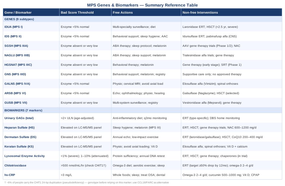
With both the biomarker and genetic landscapes mapped, the table below brings the key data together in a single reference view.
Summary Reference Table
10 Research Findings That Are Reshaping MPS Clinical Management
The clinical science of MPS has advanced rapidly since the first ERT approvals in the early 2000s, and several research findings have genuinely challenged standard assumptions about diagnosis timing, treatment goals, and what is achievable. Drawing on data from the MPS Registry, the Lysosomal Disease Network, and work from leading research groups including those of Chester Whitley at the University of Minnesota, Maurizio Scarpa at the Brains for Brain Foundation, and Patricia Dickson at UCLA, the following ten findings represent the most practice-shifting conclusions of recent years.
1. Pre-Symptomatic Treatment Window Is Much Narrower Than Previously Recognized
The original thinking about MPS I was that ERT could be started when symptoms appeared and still produce good outcomes. Registry data from thousands of MPS I patients have definitively shown that cognitive outcomes after HSCT are determined almost entirely by age at transplant and by cognitive status at the time of transplant — not by disease severity at birth. Children transplanted before 12 months of age with normal neurodevelopmental scores have outcomes indistinguishable from unaffected peers in the best-case scenarios. Every month of delay in severe MPS I narrows the outcome gap that treatment can close. This has driven the push for newborn screening inclusion in national programs globally.
2. Urinary GAGs Alone Are Not Sufficient for Monitoring — Plasma Biomarkers Are Essential
The historical reliance on total urinary GAGs as the primary monitoring biomarker has been progressively challenged by LC-MS/MS data showing that plasma heparan sulfate and dermatan sulfate provide superior sensitivity for detecting residual disease burden during ERT. Some patients on ERT normalize urine GAGs while plasma HS remains significantly elevated — and this residual plasma HS correlates with ongoing tissue storage and inferior functional outcomes. This finding argues for moving to LC-MS/MS plasma fractionation as the standard of care for treated patients.
3. CNS-Penetrating ERT Is Achievable — But Requires Novel Delivery Mechanisms
Standard IV ERT for MPS I, II, and III does not adequately penetrate the blood-brain barrier. This was once accepted as a fundamental limitation. The approval of pabinafusp alfa (intrathecal idursulfase for MPS II) in Japan and the positive Phase 2 data from intrathecal laronidase studies in MPS I have shown that CNS enzyme delivery is possible. These results have reframed the goal of MPS treatment from "somatic control" to "simultaneous somatic and neurological control," which is a fundamentally different and more ambitious therapeutic target.
4. Substrate Reduction Therapy May Be More Tractable for MPS III Than Direct Enzyme Replacement
Because HGSNAT encodes a transmembrane protein that cannot easily be delivered by the standard M6P receptor pathway, researchers pivoted to substrate reduction therapy — reducing the amount of HS synthesized in the first place, so less accumulates. Early clinical work with small-molecule heparan sulfate synthesis inhibitors has shown measurable HS reduction in CNS tissue in animal models, and Phase 1 studies in humans are underway. This approach is being actively studied for MPS IIIC in particular and could provide the first systemic-oral treatment option for Sanfilippo syndrome.
5. Gene Therapy Can Produce Durable Biochemical Correction — But Immune Responses Remain the Critical Challenge
AAV gene therapy for MPS I, IIIA, IIIB, and IVA has demonstrated the ability to produce sustained enzymatic correction lasting 12–36 months in early Phase 1/2 trials. The central challenge is not the science of gene insertion but the immune system's response to AAV capsids, which can neutralize the vector and prevent re-dosing. Multiple strategies are being evaluated: immunosuppression windows, capsid engineering, and non-AAV delivery platforms. The conclusion is that gene therapy for MPS is not a question of "if" but of managing the immune biology surrounding delivery.
6. Chitotriosidase Decline Predicts Long-Term ERT Outcomes Better Than Early GAG Reduction
While urinary GAGs respond quickly to ERT, it is the trajectory of chitotriosidase decline over the first 12–24 months that best predicts which patients will achieve durable somatic benefit. Patients whose chitotriosidase does not reach at least 50% reduction by month 12 on ERT tend to show inferior functional outcomes at 5 years. This finding supports using chitotriosidase not just as a monitoring tool but as an early surrogate endpoint for treatment adequacy, potentially informing dose adjustments or escalation to combination therapy.
7. Sleep Disorders in MPS III Are Neurologically Driven — Not Behavioral — and Require Neurological Management
The severe sleep inversion seen in MPS III was long attributed to behavioral factors and managed with sedating medications. Research documenting abnormal melatonin secretion patterns and hypothalamic neuronal loss in SGSH-deficient brains has reframed MPS III sleep disruption as a primary neurological symptom driven by hypothalamic storage damage. This changes the clinical approach: melatonin supplementation at physiological doses is supported by the mechanism, sedating antihistamines and benzodiazepines are discouraged (they impair cognition further), and the goal shifts to circadian rhythm support rather than sedation.
8. Cardiac Valve Disease in MPS Progresses Independently of ERT and Requires Dedicated Surveillance
A critical finding from long-term follow-up of ERT-treated MPS I, II, and VI patients is that valve disease — particularly aortic and mitral involvement — progresses on ERT at rates that do not substantially differ from natural history controls. This is attributed to the poor penetration of recombinant enzyme into avascular cardiac valve tissue. The clinical implication is that cardiologists managing MPS patients on ERT cannot assume that ERT protects the heart. Annual echocardiography and low thresholds for valve surgery are appropriate regardless of ERT duration.
9. Attenuated MPS Phenotypes Are Systematically Under-Diagnosed and Under-Treated
Large case series from adult MPS clinics have revealed that attenuated MPS I, II, and VI is consistently diagnosed 5–15 years later than severe forms, often after decades of misdiagnosis with conditions such as carpal tunnel syndrome, joint hypermobility syndrome, or early-onset cardiac valve disease. The enzymatic and genetic tools to diagnose MPS in adults are widely available, but clinical awareness is insufficient. The research finding that attenuated adult MPS patients started on ERT show meaningful functional improvements — even after years of untreated disease — has motivated advocacy for broader enzyme screening in adults with unexplained musculoskeletal and cardiac disease.
10. Biomarker-Guided Dosing May Replace Fixed-Weight ERT Protocols
The current ERT dosing paradigm is weight-based and fixed (e.g., 0.58 mg/kg weekly for MPS I). Emerging pharmacokinetic and pharmacodynamic data suggest that patients with different antibody levels, body compositions, and residual disease burdens have dramatically different enzyme exposure at the tissue level from the same weight-based dose. Research groups are developing individualized dosing algorithms based on plasma HS decline kinetics, chitotriosidase trajectories, and anti-drug antibody titers. This precision dosing approach has not yet reached routine clinical practice but represents where the field is heading and argues for comprehensive biomarker monitoring even in patients who appear clinically stable on standard ERT.
Complementary Approaches That May Support Quality of Life
Complementary modalities cannot treat the underlying enzyme deficiency or reverse GAG accumulation. But several have evidence supporting quality-of-life benefits in MPS — addressing the joint pain, respiratory compromise, behavioral distress, and fatigue that compound the disease burden. The following three approaches have the clearest human evidence relevant to MPS.
Breathing-Based Therapies
Respiratory involvement is present across most MPS types, driven by thoracic cage restriction, airway GAG deposition, obstructive apnea, and recurrent pulmonary infections. Breathing-based therapies — specifically techniques that train diaphragmatic function, improve chest wall compliance, and optimize airway clearance — address these mechanisms directly. In a connective tissue and thoracic environment compromised by GAG accumulation, respiratory muscle training can partially compensate for mechanical restriction.
A 2017 clinical review of respiratory management in lysosomal storage diseases, including MPS, supported the integration of airway clearance techniques and respiratory physiotherapy as adjuncts to ERT in patients with established restrictive lung disease. Inspiratory muscle training (IMT) using a threshold loading device has demonstrated measurable improvements in respiratory muscle strength in pediatric neuromuscular conditions with restrictive patterns analogous to MPS thoracic involvement. Protocols typically involve 30 breaths at 30–40% maximal inspiratory pressure, 5 days per week.
In practice, a respiratory physiotherapist experienced with pediatric neuromuscular or metabolic disease is the appropriate referral. Techniques include diaphragmatic breathing exercises (10 minutes twice daily), positive expiratory pressure (PEP) masks for airway clearance in patients with mucus retention, and positioning strategies to optimize respiratory mechanics during sleep. Evidence specific to MPS is limited and mostly observational; these techniques should be considered supportive rather than primary and integrated into the overall pulmonary management plan.
Massage Therapy
Joint stiffness, contractures, and musculoskeletal pain are among the most disabling somatic features of MPS, affecting daily function, sleep quality, and emotional wellbeing. Massage therapy — specifically techniques adapted for patients with connective tissue disease, skeletal dysplasia, and reduced cervical mobility — offers a non-pharmacological approach to managing the myofascial and joint components of this pain burden.
A 2019 study on massage therapy for joint stiffness in connective tissue disorders (including conditions with overlapping musculoskeletal pathology to MPS) demonstrated reductions in perceived stiffness and pain scores without adverse events when adapted protocols were used by experienced therapists familiar with structural limitations. For MPS specifically, gentle myofascial release and soft tissue techniques are preferred over deep tissue or high-pressure methods, given the fragility of GAG-compromised connective tissue and the risk of cervical injury in subtypes with instability.
Practically, massage should be conducted by therapists with experience in rare or metabolic conditions; a full musculoskeletal assessment including any cervical instability documentation must be reviewed before treatment. Sessions of 30–45 minutes weekly to biweekly are a reasonable starting point. Cervical manipulation is contraindicated in any MPS subtype with documented atlantoaxial instability (particularly MPS IVA and MPS I). Parents of affected children should work with pediatric physiotherapists rather than unspecialized massage practitioners.
Mindfulness Meditation and MBSR
Chronic disease burden, behavioral challenges, caregiver fatigue, and pain-related psychological distress are consistent features of life with MPS — for patients who are cognitively able to engage, and particularly for caregivers. Mindfulness-based stress reduction (MBSR) and adapted mindfulness programs have well-established effects on perceived pain intensity, anxiety, and quality of life in chronic disease populations with systemic, multi-organ involvement comparable to MPS.
A meta-analysis published in JAMA Internal Medicine covering 47 randomized trials of mindfulness meditation programs found moderate evidence for improvements in pain, anxiety, and depression in chronic illness populations. While no MPS-specific RCT of MBSR exists, the mechanism — downregulation of the sympathetic stress response, reduction of inflammatory cytokine levels including IL-6 and TNF-α, and improved sleep architecture — directly addresses several secondary burdens in MPS. Caregiver MBSR programs have specifically shown benefit in pediatric rare disease settings.
An 8-week MBSR program (2.5 hours per week plus daily home practice of 15–30 minutes) is the standard evidence-based format. App-based adaptations (Headspace, Calm, Insight Timer) provide lower-barrier entry points for patients or caregivers with limited time or mobility. Evidence is limited for cognitively impaired patients (such as advanced MPS III); benefits here are primarily for caregivers and mildly affected patients. Frequency: daily practice sustains the neurobiological effects; periodic refresher programs every 6–12 months help maintain consistency.
Conclusion
Mucopolysaccharidosis is a condition where the depth of your understanding directly influences the quality of decisions made on its behalf. The nine genes covered here define the molecular foundation of each subtype; the seven biomarkers translate that foundation into measurable signals of what is actually happening in the body week by week and year by year. Neither framework is a substitute for expert metabolic disease care, but both are tools for engaging with that care more effectively — asking sharper questions, interpreting results more fully, and recognizing earlier when something has changed.
The most useful next step depends on where you are. If you are newly diagnosed or supporting a newly diagnosed child, the priority is connecting with a center that has genuine MPS expertise and ensuring that biomarker baselines are established before any therapy begins. If you are already in care, the conversation worth having with your specialist centers on whether your current monitoring panel includes fractionated plasma GAGs by LC-MS/MS — not just total urinary GAGs — and whether gene therapy or substrate reduction trial eligibility has been reviewed recently. The science is moving faster than most clinical protocols have caught up with, and the gap between what is available in research settings and what is offered in routine care is narrowing but still real.
Musculoskeletal Eye Neurological Cardiovascular Respiratory Endocrine & Metabolic
Musculoskeletal: Bone Conditions Joint Conditions
Neurological: Brain Conditions Memory & Cognitive Conditions
Respiratory: Sleep & Breathing Disorders
Autoimmune: Connective Tissue Conditions
Ear, Nose & Throat: Hearing & Balance Conditions