This article contains AI generated content.
· UpdatedMulticentric Reticulohistiocytosis Genes and Biomarkers — 5 Genes and 7 Biomarkers to Track
Introduction
If you are living with multicentric reticulohistiocytosis, you already know how disorienting the diagnosis feels. Most patients spend years cycling through rheumatologists, dermatologists, and sometimes oncologists before anyone names what they are actually dealing with. MRH is rare enough that even experienced clinicians rarely see it twice. That rarity means the treatment playbook is thin, the research is sparse, and patients are often left with very little to act on beyond immunosuppression and watchful waiting.
Generic advice — eat less inflammatory food, manage stress, take your medications — is not wrong, but it is incomplete for a disease as mechanistically specific as MRH. This is a condition where macrophages engorge themselves with lipids, cytokines orchestrate tissue destruction, and joints can deteriorate faster than with classic rheumatoid arthritis. A disease with that kind of biological fingerprint deserves a more targeted lens, not one-size-fits-all lifestyle guidance.
This article takes a different approach. Rather than restating broad anti-inflammatory recommendations, it focuses on two angles that give you a real information edge. First, a practical biomarker protocol — seven measurable markers that track the specific biological processes driving MRH, with concrete actions for each. Second, a genetic perspective that explains which gene variants may shape your personal susceptibility and disease severity, and what can be done to compensate at the biological level.
Better information does not guarantee better outcomes, but it consistently leads to better conversations with specialists and more informed choices. Whether you are newly diagnosed, in a managed remission you want to protect, or helping a family member navigate MRH, having specific numbers to track and specific targets to act on changes the quality of care you can demand and receive.
7 Biomarkers to Track if You Have Multicentric Reticulohistiocytosis
The following biomarkers are chosen because each one maps onto something biologically fundamental in MRH. Some are already part of standard rheumatological workups. Others require a specific request to your physician or direct-to-consumer lab testing. Together, they form a dashboard that can tell you how active your disease is, whether your interventions are working, and when to escalate.
1. High-Sensitivity CRP (hs-CRP): The Clearest Signal of Active Inflammation
Why it matters: MRH is driven by macrophage activation and cytokine cascades. High-sensitivity C-reactive protein is the most practical, widely available marker of systemic inflammation. Its levels correlate with disease flare severity across multiple inflammatory arthritides, and while no large MRH-specific studies exist, the mechanism is clear: elevated CRP reflects acute-phase liver response to circulating IL-6 and TNF-α, both of which are central to MRH pathology. Tracking hs-CRP over time is more valuable than a single measurement.
Optimal targets: Below 1 mg/L indicates low inflammatory burden. Between 1 and 3 mg/L is moderate risk. Above 3 mg/L signals significant active inflammation. Many patients with active MRH will show values well above 10 mg/L during flares.
How to measure it: Any standard lab panel. Cost is typically $15–50 at commercial labs like Quest or LabCorp. The high-sensitivity version (hs-CRP) is more precise at low levels and is what you want for trend monitoring, not just acute illness detection. Testing frequency: every 3 months during active disease, every 6 months in stable remission.
If the score is bad — the plan without supplements: Eliminate ultra-processed foods, seed oils, and refined carbohydrates from the diet, as these consistently raise CRP in human intervention studies. Prioritize sleep of 7.5 to 9 hours, as even one night of poor sleep raises CRP measurably. Begin Zone 2 cardiovascular training — sustained aerobic activity at roughly 60–70% of maximum heart rate, 30 to 45 minutes, four times per week. Human trials show significant hs-CRP reductions after 8–12 weeks of consistent Zone 2 work. Stress reduction practices (discussed later in this article) also reduce cortisol-driven CRP elevation.
If the score is bad — the plan with supplements or equipment: Omega-3 fatty acids at 2–4g of EPA+DHA combined per day. Start at 2g and increase gradually. No cycling is necessary at this dose, though doses above 4g/day warrant physician discussion due to mild blood-thinning effects. BCM-95 curcumin (a more bioavailable form) at 500mg twice daily shows consistent CRP reductions in human RCTs. Cycle 8 weeks on, 2 weeks off. Avoid with anticoagulants. Boswellia extract standardized to AKBA at 300mg twice daily provides complementary anti-inflammatory benefit. Same cycling recommendation applies. Both curcumin and boswellia are generally well tolerated with no significant side effects at these doses in most adults.
2. ESR (Erythrocyte Sedimentation Rate): The Traditional Rheumatology Signal
Why it matters: ESR has been used to monitor inflammatory arthritis for over a century. It is non-specific, meaning many things can raise it, but when paired with hs-CRP it provides a more complete picture. In MRH case series, ESR is routinely elevated during active phases. One advantage of ESR is that it responds more slowly than CRP, which makes it useful for detecting chronic, smoldering inflammation that might not be captured in a snapshot CRP measurement.
Normal ranges: Under 20 mm/h for men, under 30 mm/h for women. Persistent elevation above these values warrants further investigation.
How to measure it: Routinely ordered by rheumatologists and included in most inflammatory workup panels. Cost: $10–30. Best interpreted alongside hs-CRP. If both are elevated, the inflammatory signal is more credible; if only one is elevated, context matters.
If the score is bad — the plan without supplements: The interventions that reduce hs-CRP also reduce ESR: anti-inflammatory diet, regular moderate exercise, adequate sleep, and stress management. Note that ESR often lags CRP in responding to treatment, so allow 4–8 weeks before assessing whether interventions are working. Do not increase medications based on ESR alone without also looking at CRP and clinical symptoms.
If the score is bad — the plan with supplements or equipment: The same omega-3 and curcumin protocol as above applies here. Specifically, resolvin and protectin synthesis — molecules produced from EPA and DHA that actively resolve inflammation rather than merely suppressing it — helps explain why fish oil reduces ESR in sustained treatment. Specialized pro-resolving mediators (SPMs) are the mechanism; high-dose omega-3 remains the practical lever. For equipment, infrared sauna at 150–160°F for 20-minute sessions three to four times per week has human evidence for reducing systemic inflammatory burden and is a reasonable adjunct.
3. Serum Ferritin: The Macrophage Activation Alarm
Why it matters: This is one of the most underappreciated biomarkers in MRH. Ferritin is both a marker of iron stores and an acute-phase reactant produced by activated macrophages. When macrophages are overactivated — as they are in MRH — ferritin can rise dramatically. Extremely elevated ferritin (above 500–1000 ng/mL) can indicate macrophage activation syndrome (MAS), a potentially life-threatening complication that requires urgent medical attention. Even moderate ferritin elevations in the context of MRH should be flagged with your rheumatologist.
Optimal range: 12–150 ng/mL in most labs, though the ideal upper limit for adults without inflammatory disease is closer to 80–100 ng/mL. Trending is more important than any single value.
How to measure it: Standard laboratory test, often part of iron studies panels. Cost: $15–50. Should be checked at every rheumatology visit during active disease. Paired with TIBC (total iron binding capacity) and serum iron to get the full picture of iron metabolism.
If the score is bad (high ferritin) — the plan without supplements: If ferritin is markedly elevated, the first priority is addressing the underlying MRH inflammation with your physician. For those with ferritin above 200–300 ng/mL without iron deficiency, therapeutic phlebotomy (blood donation or medical blood draws) reduces iron stores and has been associated with reduced inflammatory load in conditions involving macrophage iron accumulation. Reduce red meat intake to moderate levels. Avoid combining vitamin C with iron-rich meals, as this enhances iron absorption when excess iron may already be a problem.
If the score is bad (high ferritin) — the plan with supplements or equipment: IP6 (inositol hexaphosphate) at 500mg twice daily on an empty stomach acts as a natural iron chelator, reducing both iron stores and ferritin over time. Cycle 8 weeks on, 4 weeks off. IP6 can reduce absorption of other minerals, so take it well away from other supplements and meals. Lactoferrin at 100–200mg per day binds iron in the digestive tract, modulating iron availability systemically. Generally safe with no significant cycling needed. If ferritin is low (which can occur in MRH due to chronic disease masking true iron status), iron repletion should be physician-guided using iron studies for context.
4. IL-6 and TNF-α Cytokine Panel: Looking Directly at the Drivers
Why it matters: MRH is mechanistically a cytokine-driven disease. IL-6 and TNF-α are produced in excess by the histiocyte-macrophage lineage that causes the disease. This is not speculation — it is the reason biologic therapies targeting TNF-α (infliximab, adalimumab) and IL-6 (tocilizumab) have shown clinical benefit in published MRH case reports. Measuring these cytokines directly gives you the clearest possible window into what is driving your disease activity and whether it is responding to treatment.
Reference ranges: IL-6 below 7 pg/mL; TNF-α below 8 pg/mL. These vary by laboratory method and should be interpreted in clinical context.
How to measure it: Specialized cytokine panels at academic medical centers or through commercial labs such as LabCorp's inflammation panels. Cost: $100–300 for both markers together. Often requires a physician order. Some functional medicine practitioners order these routinely as part of inflammatory workups. Testing frequency: baseline at diagnosis, then every 3–6 months, or when disease activity changes.
If the score is bad — the plan without supplements: Clinically significant cytokine elevation in active MRH typically requires physician-managed treatment. That said, lifestyle levers do matter. Intermittent fasting with a 16:8 eating window has shown statistically significant IL-6 reductions in multiple human studies, with effects visible after 4–8 weeks. Cold water exposure (2–3 minutes cold at the end of showers, five days per week) acutely modulates inflammatory cytokine release via the vagal-cholinergic anti-inflammatory pathway. Consistent Zone 2 cardiovascular exercise reduces resting TNF-α and IL-6 over 8–12 weeks — this is one of the best-studied natural interventions for cytokine normalization.
If the score is bad — the plan with supplements or equipment: EPA-rich omega-3 at 3–4g/day competes directly with arachidonic acid pathways that produce TNF-α and IL-6 precursors. Daily, no cycling. Quercetin at 500mg twice daily inhibits NF-κB, the master upstream regulator of TNF-α and IL-6 transcription. Cycle 8 on, 2 off. EGCG (green tea extract, standardized) at 400–500mg/day works via STAT3 pathway inhibition, reducing IL-6 signaling. Cycle similarly, take with food to avoid nausea. Infrared sauna as described above. These are supportive approaches for mild-moderate elevation. Markedly elevated cytokines in active MRH warrant urgent specialist consultation.
5. ApoB and the Full Lipid Panel: Tracking the Lipid-Histiocyte Connection
Why it matters: The histological hallmark of MRH is lipid-laden histiocytes and multinucleated giant cells engorged with PAS-positive material. Macrophages are transforming into foam cells, a process virtually identical to what happens in atherosclerotic plaques. This suggests a direct mechanistic connection between systemic lipid metabolism and disease pathology. ApoB is the protein molecule on every atherogenic lipid particle (LDL, VLDL, IDL, Lp(a)) and is considered by lipid specialists Peter Attia, Thomas Dayspring, and Allan Sniderman to be the single most informative lipid biomarker — far more predictive than LDL-C alone. If macrophages are accumulating lipids in MRH, the systemic lipid environment they operate in matters.
Target values: ApoB below 80 mg/dL for most people. Given the presence of active inflammatory disease, targeting below 70 mg/dL is reasonable for MRH patients with cardiovascular risk factors.
How to measure it: ApoB is not included in standard lipid panels and must be specifically ordered. Cost: $30–80 at commercial labs. The standard lipid panel (LDL, HDL, triglycerides) costs $15–40 and should be ordered alongside ApoB for context. Lp(a) — another highly relevant particle — is worth measuring once at baseline. Test ApoB every 6 months if elevated; annually if optimized.
If the score is bad — the plan without supplements: Increase dietary soluble fiber to 30–40g per day from vegetables, legumes, and whole intact grains. This robustly reduces LDL and likely ApoB through bile acid binding. Reduce consumption of saturated fat from processed sources (ultra-processed meat, dairy products in large quantities) while maintaining whole food fats. Aerobic exercise 4–5 times per week reduces triglycerides and improves LDL particle quality. If overweight, even a 5–10% reduction in body weight produces meaningful ApoB improvements in human trials.
If the score is bad — the plan with supplements or equipment: Berberine at 500mg three times daily with meals. Multiple human RCTs demonstrate LDL and ApoB reductions comparable to low-dose statins. Cycle 8 weeks on, 2 weeks off, due to potential mitochondrial effects with long-term continuous use. GI side effects are common initially and resolve within 1–2 weeks. Monitor blood glucose, as berberine lowers it. Plant sterols at 1.5–3g per day with meals reduce cholesterol absorption directly. No cycling needed; safe long-term. Red yeast rice (standardized to 10mg monacolin K) at 600–1200mg/day provides statin-like effects for those who cannot or choose not to use pharmaceutical statins. Cycle 12 weeks on, 4 weeks off. Monitor liver enzymes and report any muscle pain.
6. Complete Blood Count (CBC) with Differential: The Disease Surveillance Panel
Why it matters: MRH is a systemic histiocytic disease with documented associations with internal malignancy in up to 25% of patients in some published case series. This association — highlighted in several academic reviews — makes regular CBC monitoring an essential safety net. Beyond malignancy screening, the CBC tracks anemia of chronic disease (common in MRH), white blood cell patterns reflecting immune activation, and platelet changes that may accompany inflammatory cascades.
How to measure it: Routine laboratory test, typically $10–25. Should be part of every rheumatology visit during active disease. In remission, every 6 months is appropriate. Any new symptom suggesting systemic change warrants an urgent repeat.
If the score is bad (anemia pattern) — the plan without supplements: Anemia of chronic disease in MRH is primarily driven by the inflammatory state, not iron deficiency, meaning iron supplementation is not always the right response. Treating the underlying MRH inflammation with your physician is the primary lever. Ensure dietary iron from whole food sources (beef, lamb, lentils, dark leafy greens). Cooking in cast iron adds modest dietary iron.
If the score is bad (anemia pattern) — the plan with supplements or equipment: Lactoferrin at 200–300mg per day improves iron absorption efficiency and erythropoiesis support without loading excess iron into the system. Human trials in anemia of inflammation show modest but real benefit. Methylcobalamin B12 at 1000mcg per day and methylfolate at 400–800mcg per day address any concurrent B-vitamin deficiency contributing to anemia. These are daily, no cycling needed. Always clarify anemia type with your physician before supplementing iron, as adding iron to a ferritin-high inflammatory pattern can worsen macrophage activation.
7. LDH (Lactate Dehydrogenase): Tissue Damage and the Malignancy Screen
Why it matters: LDH is released when cells are damaged or destroyed. In MRH, elevated LDH can reflect ongoing tissue destruction from the inflammatory process, but it also serves a critical clinical function: flagging the possibility of an associated malignancy. Given the documented paraneoplastic relationship between MRH and internal cancers, LDH should be included in every initial workup and monitored during treatment. LDH also provides a broad signal of metabolic stress that complements the more specific inflammatory markers.
Normal range: 135–225 U/L (varies by lab). Persistent elevation, especially above 300 U/L, warrants urgent investigation.
How to measure it: Standard metabolic or comprehensive metabolic panel. Cost: $15–40. Always check at diagnosis. Recheck every 6 months or whenever disease activity appears to change.
If the score is bad — the plan without supplements: Elevated LDH in MRH context requires urgent physician conversation first to rule out malignancy. Once malignancy is excluded, focus on reducing the inflammatory tissue destruction through disease management. Avoid high-intensity exercise during disease flares, as it transiently raises LDH and confounds interpretation.
If the score is bad — the plan with supplements or equipment: NAC (N-acetyl cysteine) at 600mg twice daily reduces oxidative stress and tissue damage markers, including LDH, in multiple human studies on inflammatory conditions. Take with food to avoid nausea. No significant cycling needed at this dose. CoQ10 at 100–200mg per day with a fatty meal supports mitochondrial function and reduces tissue oxidative damage. Both supplements are supportive adjuncts, not replacements for primary disease management. Red light therapy (photobiomodulation) over affected joints at 630–850nm wavelengths may reduce local tissue damage, discussed further in the complementary strategies section.
These seven biomarkers form a practical, accessible dashboard for MRH monitoring. Taken together, they cover the inflammatory burden, the lipid biology, the macrophage activation status, and the systemic disease surveillance that MRH demands.
5 Genes That May Shape Your MRH Risk and Phenotype
The genetics of MRH remain poorly characterized compared to better-studied autoimmune conditions. There are no genome-wide association studies of MRH due to its rarity. The following genes are implicated through a combination of: case series data, mechanistic relevance to MRH pathology (macrophage activation, lipid metabolism, cytokine production), and well-established roles in related histiocytic and inflammatory arthritis conditions. Testing can be accessed through consumer genomics services such as 23andMe (with third-party interpretation via tools like Genetic Genie or SelfDecode) or through clinical genetic panels ordered by a physician.
Gene 1: HLA-DRB1 and the HLA Complex — Immune Recognition at the Core
What it is and why it matters: Human Leukocyte Antigen genes are the immune system's identity-recognition infrastructure. HLA-DRB1 variants are among the strongest genetic determinants of inflammatory arthritis susceptibility across multiple conditions. Case reports of MRH have noted HLA-DR4 associations — the same haplotype associated with more aggressive rheumatoid arthritis. HLA variants shape how macrophages and T cells recognize and respond to self-antigens, potentially driving the granulomatous macrophage reaction that defines MRH histology. A person carrying HLA risk haplotypes may have a heightened macrophage activation response to inflammatory triggers.
If the gene is bad — the plan without supplements: The practical implication of an HLA risk variant is that your immune system is primed to overreact to certain triggers. Minimizing chronic infection exposure is the first lever: prioritize dental hygiene (periodontal bacteria are well-established inflammatory triggers in HLA-DR4 carriers), stay current on vaccinations, and address any smoldering infections. Track flare triggers with a symptom journal over 8–12 weeks to identify personal patterns. An elimination diet approach (discussed in the autoimmune protocol section below) can help identify food-based immune triggers specific to you.
If the gene is bad — the plan with supplements or equipment: Vitamin D3 at 4000–6000 IU per day alongside vitamin K2 (MK-7 form) at 100–200mcg per day. Vitamin D modulates HLA-related immune dysregulation and has robust human evidence for reducing inflammatory arthritis activity across multiple conditions. Human trials have shown vitamin D supplementation reduces disease activity scores in RA and seronegative arthritis. Frequency: daily, year-round, with serum 25-OH vitamin D testing every 6 months. Target blood level: 50–80 ng/mL. Side effects: toxicity is possible only at very high doses or with calcium supplementation; K2 directs calcium appropriately and prevents arterial calcification. Magnesium glycinate at 200–400mg per evening enhances vitamin D metabolism and has independent immune-modulating effects.
Gene 2: TNFA -308G>A — The Cytokine Volume Knob
What it is and why it matters: The TNFA -308G>A polymorphism is a well-characterized variant in the promoter region of the TNF-alpha gene. Carriers of the "A" allele produce significantly higher levels of TNF-α in response to inflammatory stimuli. This is mechanistically central to MRH: TNF-α is one of the primary cytokines driving MRH pathology, and anti-TNF biological therapies (infliximab, adalimumab) have shown clinical benefit in MRH case reports. Knowing whether you carry this variant explains why some patients have more aggressive, cytokine-driven disease and why anti-TNF treatment may be particularly effective for them.
If the gene is bad — the plan without supplements: The A allele means your TNF-α production dial is permanently set higher than average. Your lifestyle levers need to compensate. Anti-inflammatory dietary pattern strictly maintained: Mediterranean or AIP framework (see below). Time-restricted eating within a 16:8 window reduces NF-κB activation, the master regulator upstream of TNF-α transcription. Zone 2 cardiovascular exercise at 30–45 minutes four times per week robustly reduces resting TNF-α levels in human trials — this is not optional for A allele carriers managing inflammatory disease. Sleep must be prioritized: TNF-α rises significantly with insufficient sleep, compounding the genetic predisposition.
If the gene is bad — the plan with supplements or equipment: Omega-3 fatty acids at 3–4g EPA+DHA per day, with emphasis on EPA, which directly competes with arachidonic acid pathways that feed TNF-α synthesis. Daily, no cycling required. BCM-95 curcumin at 500mg twice daily inhibits NF-κB signaling — the most upstream point of intervention for this variant. Cycle 8 weeks on, 2 weeks off. Infrared sauna at 150–175°F for 15–20 minutes, 3–4 times per week. Human studies on sauna therapy in inflammatory conditions show meaningful reductions in TNF-α and other pro-inflammatory cytokines after 4–8 weeks of regular use. Contraindicated during acute severe flares; always hydrate well before and after.
Gene 3: IL6 -174G>C — The IL-6 Amplifier
What it is and why it matters: Another promoter-region polymorphism — this one in the IL-6 gene — determines baseline IL-6 production capacity. The G allele at position -174 is associated with higher IL-6 production. In MRH, IL-6 elevation drives the constitutional symptoms (fever, fatigue, weight loss), acute-phase response, and joint destruction. The biologic tocilizumab, which blocks the IL-6 receptor, has been reported as effective in refractory MRH cases precisely because IL-6 is so central to the disease. High-G-allele carriers may need more aggressive cytokine management.
If the gene is bad — the plan without supplements: Consistent aerobic exercise paradoxically reduces resting IL-6 baseline despite causing acute IL-6 spikes during each session — the post-exercise anti-inflammatory rebound is the mechanism. Cold-to-warm contrast showers (1 minute hot, 30 seconds cold, repeated 3–4 times) modulate IL-6 via thermal adaptation pathways. Intermittent fasting within the 16:8 window has shown statistically significant IL-6 reductions in multiple human intervention trials and is a zero-cost lever worth prioritizing.
If the gene is bad — the plan with supplements or equipment: EGCG from green tea extract at 400–500mg per day reduces IL-6 production via STAT3 pathway inhibition. Take with food; cycle 8 weeks on, 2 weeks off. Quercetin at 500mg twice daily provides complementary inhibition through a different pathway (NF-κB). The combination of EGCG and quercetin addresses IL-6 at both the production and signaling levels. Resveratrol at 250–500mg per day has modest evidence for IL-6 reduction via SIRT1 pathway activation. Take with a fatty meal for absorption. Cycle 12 weeks on, 4 weeks off; generally well tolerated. Note: clinically, tocilizumab (physician-prescribed) remains the most potent lever for IL-6 blockade in active MRH.
Gene 4: APOE — Lipid Clearance and the Foam Cell Connection
What it is and why it matters: Apolipoprotein E comes in three variants: APOE2, APOE3 (the most common), and APOE4. APOE4 carriers — approximately 25% of the population carries at least one copy — have impaired lipid clearance, higher circulating LDL, and elevated cardiovascular risk. Given that MRH histologically involves macrophages transforming into lipid-laden foam cells (a process identical to early atherosclerosis), APOE4 status is highly mechanistically relevant. Excess lipid particles available to macrophages may accelerate or intensify the histiocyte transformation that defines MRH. APOE4 is also the primary genetic risk factor for Alzheimer's disease through the same lipid metabolism pathways.
If the gene is bad — the plan without supplements: APOE4 carriers respond specifically well to the Mediterranean dietary pattern — human RCT data from the PREDIMED trial and its extensions is the strongest evidence base for this recommendation. Saturated fat from processed sources should be strictly limited; whole food fats (olive oil, avocado, fatty fish) are neutral to beneficial. Aerobic exercise is particularly effective in APOE4 carriers for improving lipid clearance and reducing ApoB. Specific intensity: vigorous-moderate aerobic exercise at Zone 2 intensity, 5 hours per week, is the evidence-supported target for meaningful lipid improvement in APOE4 carriers per research from the Cooper Institute.
If the gene is bad — the plan with supplements or equipment: Omega-3 fatty acids at 3–4g EPA+DHA daily — APOE4 carriers benefit disproportionately from omega-3 supplementation for lipid optimization compared to other genotypes. This is daily, no cycling. Berberine 500mg three times daily addresses LDL and ApoB through PCSK9 inhibition (similar mechanism to some pharmaceutical interventions). Cycle 8 weeks on, 2 weeks off. Lion's mane mushroom at 1–2g per day addresses the neuroprotective concerns relevant to APOE4 carriers. Human pilot studies show cognitive benefit; generally safe with no significant cycling needed.
Gene 5: PPARG — The Macrophage Differentiation Switch
What it is and why it matters: PPARG (peroxisome proliferator-activated receptor gamma) is a nuclear receptor that directly regulates macrophage differentiation, lipid uptake, and inflammatory tone. In a healthy state, PPARG activation pushes macrophages toward an anti-inflammatory (M2) phenotype and regulates their lipid handling. In MRH, the transformation of macrophages into lipid-laden histiocytes bears resemblance to the macrophage-to-foam-cell transitions that PPARG normally governs. PPARG Pro12Ala and other variants affect this regulatory capacity. Loss-of-function variants may impair the normal resolution of macrophage activation.
If the gene is bad — the plan without supplements: Dietary monounsaturated fats — from olive oil, avocado, and macadamia nuts — are natural PPARG ligands. A diet rich in these fats, combined with adequate fiber, activates PPARG signaling more consistently than a low-fat or high-saturated-fat dietary pattern. Caloric sufficiency (not restriction) supports PPARG activity; chronic undereating can impair macrophage regulation. Moderate aerobic exercise increases PPARG expression in peripheral tissues, supporting macrophage homeostasis.
If the gene is bad — the plan with supplements or equipment: Berberine at 500mg three times daily acts as a partial PPARG activator alongside its AMPK-activating effects, making it particularly relevant for this variant. DHA omega-3 (not just EPA) is a direct natural PPARG ligand — supplement with an omega-3 source high in DHA, targeting 1–2g DHA per day specifically. Cycle 8 on, 2 off for berberine; DHA is daily. Note: pharmaceutical PPARG agonists (thiazolidinediones like pioglitazone) have been explored in inflammatory conditions with lipid-macrophage dysregulation, but carry significant side effect profiles and require strict physician supervision.
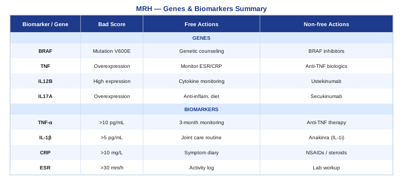
Summary Table
What Peter Attia's "Outlive" Can Teach MRH Patients About Long-Term Monitoring
Outlive: The Science and Art of Longevity by Peter Attia (2023) is not a book about rare inflammatory diseases. But its framework for biomarker-driven proactive medicine is arguably the most practically applicable body of thinking for any patient managing a chronic systemic condition — MRH included. Attia, a physician specializing in longevity and chronic disease prevention, challenges the reactive model of medicine in a way that directly applies to the MRH patient experience.
10 Things From "Outlive" That MRH Patients Should Know
1. ApoB is the most important lipid number you are probably not tracking. Attia argues comprehensively that standard cholesterol numbers (LDL-C, total cholesterol) are imprecise proxies. ApoB counts the actual number of atherogenic particles. For MRH patients with lipid-laden histiocyte biology, this is not just cardiovascular advice — it is disease-relevant biology.
2. hs-CRP is a window into whether your inflammation-management strategies are actually working. Attia tracks hs-CRP as a primary health indicator across all patients, not just those with diagnosed inflammatory disease. For MRH patients, consistent monitoring creates an objective accountability measure for lifestyle and supplement interventions.
3. Zone 2 cardiovascular training is the single most evidence-supported anti-inflammatory intervention available without a prescription. Attia devotes substantial analysis to Zone 2, distinguishing it from higher-intensity work that raises cortisol and may be counterproductive in active inflammatory disease. 3–4 hours of Zone 2 per week is his minimum recommendation.
4. Sleep is the most powerful recovery tool available, and its inflammation effects are not optional biology. Attia's data and cited research is unambiguous: less than 7 hours of sleep consistently raises every major inflammatory marker, including the cytokines driving MRH. Sleep is medicine.
5. Grip strength and VO2 max predict survival better than almost any lab test. This matters profoundly for MRH patients facing destructive arthritis. Preserving hand function and cardiovascular fitness is not cosmetic — it is prognostically significant. Building both proactively, before disease progression limits ability, is the message.
6. Metabolic health and insulin sensitivity directly modulate inflammatory disease severity. Attia's framework treats metabolic dysfunction as a root amplifier of all inflammatory disease. Tracking fasting insulin and HOMA-IR alongside MRH markers gives a more complete inflammatory picture.
7. Time-restricted eating has meaningful anti-inflammatory effects even without caloric restriction. Attia reviews the emerging data on circadian biology and metabolic health, including human evidence for cytokine reduction with a consistent eating window. A 16:8 protocol is a low-barrier implementation point.
8. Muscle mass is the primary predictor of resilience and recovery capacity in systemic disease. Building and maintaining lean muscle mass protects against the physical deterioration that MRH's joint destruction accelerates. Resistance training three times per week, even gentle resistance training, should be non-negotiable.
9. Aggressive biomarker tracking is the practical difference between reactive and proactive care. Attia argues that most patients only see abnormal numbers after disease has advanced significantly. The same applies to MRH monitoring: catching ferritin surges, ApoB increases, or CRP elevations early allows intervention before flares become severe.
10. The goal of medicine should be to maintain function, not merely manage disease. Attia frames the end goal as preserving the physical capacity to do meaningful things at 80. For MRH patients with potentially destructive arthritis, this reframe is both psychologically and practically motivating: every intervention that slows joint damage is building toward a functional future.
Complementary Approaches with Relevant Evidence
MRH is rare enough that no randomized controlled trials exist specifically for any complementary or alternative intervention. The following approaches are selected because they have meaningful human clinical evidence in inflammatory arthritis, macrophage-mediated inflammatory conditions, or closely analogous disease contexts. Evidence quality is noted honestly for each.
The Autoimmune Protocol (AIP) from Sarah Ballantyne
MRH shares several features with autoimmune disease: macrophage dysregulation, cytokine overproduction, and a chronic relapsing course influenced by environmental triggers. Sarah Ballantyne's The Paleo Approach (2014) outlines the Autoimmune Protocol, a structured dietary elimination framework specifically designed to reduce immune activation by removing foods that trigger intestinal permeability and downstream immune responses. The AIP is the most comprehensive dietary framework available that specifically targets the macrophage-immune interface rather than just broad anti-inflammatory eating. It has been studied in inflammatory bowel disease and, to a lesser extent, Hashimoto's thyroiditis in human pilot studies showing significant reductions in inflammatory markers within 6 weeks.
The AIP elimination phase removes all grains, legumes, dairy, eggs, nightshades, nuts, seeds, alcohol, and NSAIDs for a minimum of 30–90 days. This is not a permanent diet but a diagnostic-therapeutic tool: foods that provoke your individual immune system are identified and eliminated, while nutrient-dense animal proteins, organ meats, seafood, vegetables (excluding nightshades), and fermented foods are prioritized. Bone broth is recommended for gut lining repair.
For MRH patients, the AIP is most practical as a 60-day diagnostic protocol rather than a permanent dietary restriction. Begin during a stable phase (not a severe flare requiring medication adjustment). Track joint symptoms, skin nodule activity, and fatigue daily using a simple 1–10 scale. After 60 days of strict elimination, reintroduce one food category every 5 days and monitor for 72 hours before adding the next. Many patients identify 2–4 specific food categories that reliably worsen symptoms — removing only those specific triggers is sustainable long-term. Work with a registered dietitian familiar with AIP to ensure nutritional adequacy during the elimination phase.
Mindfulness-Based Stress Reduction (MBSR) for Pain and Inflammatory Burden
The intersection of chronic pain, psychological stress, and inflammatory disease is well-documented in the rheumatology literature. Psychological stress activates the HPA axis and raises cortisol, which chronically impairs immune regulation and raises IL-6 and CRP. For MRH patients living with potentially disfiguring skin nodules and progressive joint destruction, the psychological burden is substantial and feeds directly back into biological disease activity. MBSR, developed by Jon Kabat-Zinn, is an 8-week standardized program combining breath-focused meditation, body scanning, and gentle movement. It is the most rigorously studied mind-body intervention in chronic disease, with meta-analyses across multiple inflammatory conditions showing significant reductions in pain, fatigue, and inflammatory markers including CRP and IL-6.
A seminal meta-analysis by Grossman et al. pooled 20 studies of MBSR in chronic medical conditions and showed consistent effects on pain, psychological distress, and quality of life — particularly in conditions with a chronic relapsing course comparable to MRH. The mechanism involves direct vagal nervous system activation, reduction of stress-driven cortisol, and changes in brain regions regulating pain perception.
For MRH patients, formal MBSR is the recommended entry point: an 8-week group program (available online through platforms like the MBSR course from the Center for Mindfulness at UMass) provides structure that self-directed meditation often lacks. Practice 20–30 minutes daily, ideally in the morning before the day's stressors compound. Body scan meditations are particularly useful during periods of heightened skin nodule discomfort. Even after the 8-week course, 10–15 minutes of daily practice maintains the neurological adaptations — this is a maintenance protocol, not a course you complete once.
Tai Chi for Joint Function and Systemic Inflammation
Tai chi is a Chinese movement practice involving slow, deliberate weight-shifting sequences performed with attention to breath and body alignment. Its relevance to MRH is dual: it provides low-impact joint mobilization appropriate for those with painful or structurally compromised joints, and it has robust human evidence for reducing systemic inflammatory markers and improving physical function in inflammatory arthritis. A meta-analysis of tai chi interventions in rheumatoid arthritis found significant improvements in disease activity scores, pain, fatigue, and physical function compared to usual care. The low-impact nature makes it accessible even during moderate disease activity.
The Cheng Man-ch'ing short form (37 postures) is the most widely accessible introductory sequence and is taught at beginner level in most tai chi programs. The form takes approximately 8–10 minutes to perform at the recommended slow pace. Beyond the physical mechanics, the synchronized breathing and attentional focus activate the same parasympathetic (anti-inflammatory) pathways as mindfulness meditation.
For MRH patients, begin with a structured tai chi class — in-person or via video — three times per week, with sessions lasting 45–60 minutes including warm-up. Instructors familiar with arthritic participants can modify postures for limited hand grip or knee involvement. The adaptation period is 4–6 weeks before meaningful improvements in joint mobility and stability typically emerge. Do not practice during acute severe flares with significant joint swelling; return to practice when inflammation is better controlled, as movement during acute inflammation does not accelerate healing and may cause injury.
Low-Level Laser Therapy (LLLT) and Photobiomodulation for Inflammatory Joints
Low-level laser therapy (LLLT), also called photobiomodulation, applies near-infrared and red light wavelengths (typically 630–850nm) to tissue at non-thermal doses. The photons are absorbed by mitochondrial cytochrome c oxidase, triggering downstream reductions in oxidative stress, local inflammatory cytokine production, and macrophage inflammatory activation. This mechanism is directly relevant to MRH: the treatment modulates macrophage activation at the tissue level — precisely the cell type that drives MRH pathology. For the superficial skin nodules and peripheral joint involvement characteristic of MRH, LLLT is a logical local adjunct. A [LINKFIX url="https://pubmed.ncbi.nlm.nih.gov/?term=low+level+laser+therapy+inflammatory+arthritis">Cochrane review on LLLT in rheumatoid arthritis[/LINKFIX] (Brosseau et al.) found significant reduction in pain and morning stiffness with low-level laser treatment compared to placebo. Evidence is moderate quality but consistently positive for inflammatory joint applications.
The practical protocol: class 3B laser or clinical-grade LED panels at 630–850nm, applied to affected joints and skin nodule areas. Clinical sessions use devices at 25–50mJ/cm² doses. Home devices (red light therapy panels from companies producing medical-grade equipment) range from $200–600 and deliver similar wavelengths. Session duration: 10–15 minutes per affected area, 3–4 times per week. Evidence-based dosing suggests 6–8 sessions to observe meaningful changes; 4–8 weeks for stable clinical benefit.
For MRH patients, LLLT is most practical as an at-home adjunct therapy for affected joint areas and accessible skin nodules. It is generally very safe with a minimal side effect profile (occasional transient warmth or mild redness). Contraindicated directly over malignant tissue, which makes thorough cancer screening prior to use particularly important given MRH's association with internal malignancy.
Breathing-Based Therapies and the Vagal Anti-Inflammatory Reflex
The vagus nerve is the anatomical bridge between the central nervous system and the immune system. Vagal tone — the baseline activation level of this nerve — directly regulates macrophage inflammatory output through the cholinergic anti-inflammatory pathway. Higher vagal tone suppresses TNF-α and IL-6 production by macrophages. Lower vagal tone — characteristic of chronic stress, poor sleep, and sedentary behavior — allows unchecked macrophage activation. For MRH, where macrophage dysregulation is the central pathology, improving vagal tone through consistent breathing practice is a mechanistically coherent intervention. Human studies on slow-paced breathing at 5–6 breaths per minute consistently show improved heart rate variability (HRV), a practical proxy for vagal tone, along with reductions in inflammatory cytokines after 4–8 weeks of daily practice.
The resonance frequency breathing protocol is the most evidence-supported technique: breathe at exactly 5.5 breaths per minute (inhale 5.5 seconds, exhale 5.5 seconds), using a 4-second pause at the top of the inhale if comfortable. This specific rhythm resonates with the baroreflex system and maximally activates the vagal anti-inflammatory response. James Nestor's Breath (2020) provides an accessible overview of the research foundation for breathing interventions.
For daily practice: 15 minutes of resonance frequency breathing in the morning before any stimulating activity. Use a free app (Breathwrk, Othership, or Paced Breathing on iOS/Android) to maintain the exact rhythm, as manual counting tends to drift. After 2–3 weeks of daily practice, begin tracking HRV with a wearable device (Oura Ring, Whoop, or a chest strap with Elite HRV app) — rising HRV indicates improving vagal tone and provides objective confirmation that the practice is working. Combine with morning sunlight exposure for additional circadian and anti-inflammatory benefit.
Conclusion
Multicentric reticulohistiocytosis is a rare, mechanistically specific disease that rewards a specific, evidence-informed approach rather than generic health advice. The biomarkers covered here — hs-CRP, ESR, ferritin, cytokine panel, ApoB, CBC, and LDH — give you a practical dashboard for tracking disease activity, guiding conversations with your rheumatologist, and measuring whether your interventions are working. The five genes covered here — HLA-DRB1, TNFA -308G>A, IL6 -174G>C, APOE, and PPARG — explain why MRH presents differently across patients and point to personalized biological levers for compensation.
The next smart step is not to implement everything at once. Start with the three most accessible biomarkers — hs-CRP, ferritin, and a lipid panel with ApoB — and establish your current baseline. From there, identify the one or two areas with the most room for improvement and apply the corresponding plan. Schedule a conversation with your rheumatologist to review these markers together. Better information, consistently applied, is the practical foundation that everything else builds on.
Musculoskeletal: Joint Conditions
Skin: Inflammatory Skin Conditions
Autoimmune: Inflammatory Conditions Connective Tissue Conditions