This article contains AI generated content.
· UpdatedMultiligamentous Knee Injury Genes And Biomarkers - 6 Genes And 6 Biomarkers To Track
Introduction
A multiligamentous knee injury is not a pulled muscle or a minor sprain. It is one of the most complex orthopedic events the lower extremity can sustain — two or more major knee ligaments torn simultaneously, often alongside damage to cartilage, menisci, and sometimes neurovascular structures. Recovery is long, unpredictable, and deeply individual. If you have been through it, you already know that generic advice about rest, ice, and physiotherapy barely scratches the surface of what the body actually needs to rebuild.
What standard rehabilitation protocols almost never address is why two people with near-identical injuries can have radically different outcomes. One person regains full stability and returns to sport within a year. Another develops chronic instability, persistent swelling, or accelerated cartilage breakdown. The difference is not always effort or technique. It often comes down to biology — specifically, to differences in the genes governing connective tissue quality and inflammatory signaling, and to the state of the measurable biological markers that reflect healing at any given moment.
Most sports medicine content sits somewhere between oversimplified and inaccessibly technical. What is actually useful is a middle path: knowing which specific biological variables are worth measuring, understanding what they mean when they are out of range, and having a practical, evidence-grounded plan for correcting them. Generic anti-inflammatory advice is too broad. Specific, tracked, actionable data is something entirely different.
This article takes two complementary approaches. The first covers six measurable biomarkers that track inflammation, cartilage stress, anabolic drive, and tissue remodeling throughout recovery — with concrete plans for each when results are poor. The second examines six genes with meaningful evidence in ligament injury susceptibility and healing capacity, along with strategies to compensate for unfavorable variants. Between the two, there is far more useful information here than a single clinical visit is likely to provide — and with it, a more grounded basis for making better decisions every step of the way.
6 Biomarkers Worth Tracking During Recovery
Biomarkers do something that symptoms alone cannot: they make invisible processes visible. Swelling tells you something is inflamed, but not whether it is resolving, stalling, or driving secondary joint damage. Fatigue during rehabilitation does not tell you whether your anabolic hormones are sufficient to rebuild connective tissue. The right panel turns guesswork into data, and data into better decisions.
The six markers below were selected for their direct relevance to the specific biology of multiligamentous knee injury — ligament healing, cartilage stress, inflammation resolution, vascular support, and anabolic recovery capacity. They range from inexpensive and widely available to more specialized but increasingly accessible. For each, a realistic cost range is included alongside both supplement-free and supplement-assisted correction strategies.
1. hs-CRP — The Inflammation Traffic Light
Why it matters: High-sensitivity C-reactive protein is produced by the liver in response to systemic inflammation. After a multiligamentous knee injury, CRP rises sharply and should gradually decline as healing progresses. If it remains elevated weeks or months into recovery, it signals chronic, unresolved inflammation that actively interferes with tissue repair. Persistent elevated hs-CRP degrades collagen synthesis, impairs growth factor signaling, and increases the risk of secondary cartilage damage over time. Peter Attia has described chronically elevated hs-CRP as one of the most underrated signals in musculoskeletal recovery — not just a cardiovascular risk marker.
Target range: Below 1.0 mg/L is ideal during active healing. Between 1.0–3.0 mg/L suggests moderate chronic inflammation worth addressing. Above 3.0 mg/L during mid-to-late recovery is a clear signal to intervene.
How to measure it
hs-CRP is a standard blood test available from any major laboratory. In the United States, it costs $15–40 through services like Labcorp or Quest Diagnostics, and is frequently covered by insurance when ordered by a physician. At-home finger-prick tests cost $50–75 and are less precise but useful for tracking trends. Measure at baseline two to four weeks post-injury or post-surgery, then every four to eight weeks during active rehabilitation to assess whether inflammation is resolving on schedule.
If the score is bad, the plan without supplements
Dietary changes have the single largest impact on hs-CRP outside of resolving the injury itself. A Mediterranean-style diet — emphasizing oily fish, olive oil, vegetables, legumes, and nuts while reducing refined carbohydrates and industrial seed oils — has been shown to reduce hs-CRP by 20–30% within six to twelve weeks in multiple clinical studies. Sleep is the second major lever: consistently sleeping fewer than six hours per night raises CRP significantly. Achieving 7.5–9 hours of quality sleep during recovery is not optional comfort — it is a biological requirement. Beginning low-load, low-impact movement (pool walking, stationary cycling with no resistance) within the first weeks post-injury also prevents the inflammatory stagnation that comes from complete immobility.
If the score is bad, the plan with supplements or equipment
Omega-3 fatty acids (EPA+DHA): 2–4 grams per day of combined EPA and DHA has robust evidence for reducing CRP in musculoskeletal and cardiovascular contexts. Multiple meta-analyses on PubMed confirm anti-inflammatory effects at these doses. Cycle: daily throughout active recovery, then reduce to 1–2g for maintenance. Side effects: mild GI discomfort if taken on an empty stomach; mild blood-thinning effect (relevant if surgery is planned — discuss with your surgeon).
Curcumin with piperine: 500–1000 mg curcuminoids with black pepper extract, taken twice daily with meals. Evidence for hs-CRP reduction in inflammatory conditions is consistent across human trials. Cycle: 8–12 weeks on, then reassess with a follow-up blood test. Side effects: GI sensitivity at higher doses; avoid if on anticoagulant medications.
Whole-body cryotherapy or cold water immersion: 10–15 minutes at 10–15°C, three to four times per week during peak inflammatory phases. While evidence for systemic CRP reduction via cold immersion alone remains debated, local anti-inflammatory effects and recovery quality improvements are well-documented in sports science. Use in the first four to six weeks post-injury, then transition to heat-dominant protocols.
2. Vitamin D (25-OH) — The Healing Enabler
Why it matters: Vitamin D is far more than a bone mineral. Receptors for it are found throughout musculoskeletal tissue, including ligament fibroblasts, myocytes, and synovial cells. Deficiency impairs satellite cell function needed for muscle regeneration, reduces collagen synthesis rates, increases systemic inflammation, and has been consistently associated with both higher ligament injury risk and slower post-surgical healing. Multiple studies have found that athletes presenting with significant knee injuries have meaningfully lower 25-OH vitamin D levels than uninjured controls. Thomas Dayspring and others in functional medicine have argued that the conventionally acceptable minimum of 20 ng/mL is insufficient for optimal musculoskeletal function, with recovery-optimized levels likely requiring 40–60 ng/mL.
Target range: 40–60 ng/mL (100–150 nmol/L) during active recovery. Below 30 ng/mL represents frank deficiency that should be corrected before expecting optimal rehabilitation progress.
How to measure it
A standard 25-OH vitamin D blood test costs $30–60 without insurance and is commonly included in annual comprehensive panels or ordered by primary care physicians. At-home finger-prick testing is available through services such as Everlywell or Imaware for $60–80. Measure at the start of recovery, then retest after eight to twelve weeks of correction to verify adequate absorption and dose response.
If the score is bad, the plan without supplements
Midday sun exposure between 10am and 2pm — with large body surface areas (arms, legs, torso) exposed for 15–30 minutes — can raise vitamin D by the equivalent of 1000–3000 IU per session in fair-skinned individuals. This is strongly latitude- and season-dependent. At northern latitudes above 40°N during winter months, sun-based correction is effectively impossible. Dietary sources provide limited correction: oily fish (salmon, mackerel, sardines) and egg yolks are the most concentrated food sources, but achieving 40–60 ng/mL through food alone is not realistic for most people.
If the score is bad, the plan with supplements or equipment
Vitamin D3 with K2: For levels below 30 ng/mL, 5,000–10,000 IU of D3 daily for eight to twelve weeks, always co-administered with 100–200 mcg MK-7 (vitamin K2 menaquinone) to direct calcium appropriately toward bone rather than soft tissue. After confirmed correction, reduce to 2,000–4,000 IU daily for maintenance. Side effects: toxicity is rare below 10,000 IU daily, but monitoring calcium levels is advisable with long-term high-dose use. K2 co-supplementation significantly reduces the theoretical hypercalcemia risk.
UVB light therapy devices: Sperti Vitamin D lamps and similar medical-grade UVB devices deliver calibrated ultraviolet-B doses for those who cannot access adequate outdoor sun exposure. Cost: $200–350 for a home device. Protocol: two to five minute sessions, three to four days per week, following manufacturer's skin-type guidelines. Useful particularly during autumn and winter months in northern regions.
3. COMP — The Cartilage Stress Gauge
Why it matters: Cartilage Oligomeric Matrix Protein is released from articular cartilage and synovial tissue when they experience mechanical stress or structural damage. In a multiligamentous knee injury, the loss of stabilizing ligaments subjects cartilage to abnormal load distribution — a primary driver of post-traumatic osteoarthritis, which affects a significant proportion of MLI patients within 10–15 years of injury. Serum COMP rises with joint loading and injury severity, providing an early warning signal for cartilage stress before structural changes become apparent on MRI. Research published in rheumatology journals has established COMP as a prognostic marker for radiographic osteoarthritis progression in post-traumatic joint conditions.
Target range: Normal reference ranges vary by laboratory but most place the upper limit at 12–15 U/L. In the acute injury phase, elevations are expected and appropriate. During recovery at three to six or more months post-injury or surgery, persistent elevation above 15 U/L warrants attention and load management review.
How to measure it
COMP is not typically included in standard panels. It requires a specialty lab order and costs $80–150 through commercial labs (Labcorp, Quest) or through sports medicine and rheumatology practices that routinely include it in joint disease workups. It is not routinely covered by insurance but is within reach for motivated patients. The recommended schedule is testing at three to six months post-reconstruction, then every six months through the first two years of recovery to track the cartilage stress trajectory.
If the score is bad, the plan without supplements
Load management is the primary intervention. COMP rises in response to abnormal or excessive joint loading. Ensuring that rehabilitation progression does not outpace ligament graft maturation, incorporating pool-based or unweighted exercise (underwater treadmill, antigravity treadmill) during high-stress phases, and addressing movement quality deficits — quadriceps inhibition, valgus knee collapse, poor hip control — all reduce cartilage overload significantly. Body weight is also directly relevant: each excess kilogram adds approximately three to four kilograms of additional compressive load through the medial knee compartment during walking. A 5–10% body weight reduction in overweight individuals has measurable downstream impact on COMP levels.
If the score is bad, the plan with supplements or equipment
Undenatured Type II Collagen (UC-II): 40 mg daily — not grams; this is an oral immune tolerance protocol, not a structural supplement. A randomized controlled trial found UC-II superior to glucosamine plus chondroitin for knee function scores. The mechanism involves inducing immune tolerance to cartilage collagen antigens, reducing the low-grade immune-mediated component of cartilage degradation. Cycle: daily for three to six months, then reassess. Side effects: minimal; occasional mild GI sensitivity.
Glucosamine sulfate + chondroitin sulfate: 1,500 mg glucosamine and 1,200 mg chondroitin daily. Evidence is mixed for established osteoarthritis, but more consistently positive for early-stage cartilage stress in younger individuals in the post-traumatic window. Cycle: three months on, then evaluate response. Side effects: occasional GI discomfort; theoretical blood sugar interactions with glucosamine sulfate at higher doses.
Knee unloading or functional braces: Offloading braces that shift compressive force away from a damaged compartment reduce cartilage stress measurably during weight-bearing rehabilitation phases. Cost: $300–800 for quality functional bracing. This is a particularly useful non-pharmacological intervention during the high-load phases of physical therapy.
4. MMP-3 — The Tissue Remodeling Signal
Why it matters: Matrix metalloproteinase-3 (stromelysin-1) is an enzyme that degrades extracellular matrix components, including the collagen networks that form ligament and cartilage structure. Elevated serum MMP-3 reflects ongoing tissue breakdown — appropriate and expected acutely, but pathological when it persists. Sustained MMP-3 elevation indicates that degradation is outpacing repair and that synovial inflammation has not resolved. MMP-3 has been studied as a serum and synovial fluid marker in post-traumatic knee conditions, where it correlates with joint destruction rate and inflammatory synovitis severity.
Target range: Serum MMP-3 reference ranges are approximately 3–13 ng/mL for adult females and 4.5–24 ng/mL for adult males, though this varies between laboratories. Persistent elevation above the upper normal limit beyond three to four months post-injury is a signal to investigate and address inflammatory drivers.
How to measure it
MMP-3 is available through specialty panels at commercial labs. Cost: $70–130. Some functional medicine and sports medicine physicians routinely include it alongside other joint health markers. Measure at six to eight weeks post-injury or surgery, and again at three and six months to track whether the tissue remodeling trajectory is moving in the right direction.
If the score is bad, the plan without supplements
MMP-3 is upregulated by IL-6 and TNF-alpha — inflammatory cytokines driven by poor sleep, dietary inflammatory load, and metabolic stress. The most effective free interventions focus directly on these upstream drivers: improving sleep depth and duration (prioritizing slow-wave sleep, where nocturnal growth hormone peaks), eliminating processed foods, refined carbohydrates, and industrial vegetable oils, and maintaining regular moderate movement that promotes the anti-inflammatory myokines released by working muscle. Time-restricted eating protocols (16:8 or similar) have preliminary evidence for reducing inflammatory cytokine levels without compromising caloric adequacy during recovery.
If the score is bad, the plan with supplements or equipment
Boswellia serrata extract (AKBA fraction): 100–250 mg of standardized AKBA (acetyl-11-keto-beta-boswellic acid) daily. Human trials show meaningful MMP and leukotriene inhibition relevant to joint inflammation. Clinical trials on Boswellia for joint inflammation have demonstrated significant improvements in knee pain and functional scores with standardized extracts. Cycle: 8–12 weeks; side effects are minor and primarily GI. Combining with curcumin produces additive effects on NF-kB signaling.
Astaxanthin: 12 mg daily. A potent marine carotenoid antioxidant with human evidence for reducing oxidative drivers of MMP expression. Cycle: continuous during recovery; no significant adverse effects at standard doses. Very high doses over prolonged periods may cause mild skin discoloration.
Green tea extract (EGCG): 400–800 mg EGCG daily. Epigallocatechin gallate inhibits the NF-kB and AP-1 transcription factors that upregulate MMP-3. Cycle: 8–12 weeks; take with food to support liver tolerance at higher doses. Avoid use in pregnancy or alongside hepatotoxic medications.
5. IGF-1 — The Anabolic Healing Signal
Why it matters: Insulin-like Growth Factor 1 is the primary downstream mediator of growth hormone's tissue-building effects. It promotes fibroblast proliferation and collagen synthesis in ligament tissue, drives satellite cell differentiation in muscle, and supports bone remodeling throughout recovery. Low IGF-1 during recovery means the anabolic machinery for tissue repair is running at reduced capacity — a finding common in sedentary recovery periods, excessive caloric restriction, or disrupted sleep. Peter Attia has described IGF-1 optimization as one of the most underutilized levers in surgical and post-trauma recovery.
Target range: Reference values are age-dependent but generally fall between 100–400 ng/mL. For adults in active recovery from significant injury, targeting the upper third of the age-appropriate range is preferred to maximize anabolic drive for tissue synthesis.
How to measure it
IGF-1 is a standard blood test available from any major laboratory. Cost: $30–70, and it is frequently included in endocrine or sports medicine panels. Diurnal variation is minimal compared to growth hormone itself, making single morning measurements reliable and repeatable. Measure at baseline early in recovery, then at three-month intervals to track anabolic status throughout rehabilitation.
If the score is bad, the plan without supplements
Sleep optimization: Approximately 70–80% of daily growth hormone — and therefore the IGF-1 it stimulates — is secreted during slow-wave sleep. Consistently achieving 7.5–9 hours with high sleep quality (cool room at 67–68°F, dark environment, fixed sleep-wake schedule) is the single most impactful free intervention available. Even one week of restricting sleep to five hours measurably reduces circulating IGF-1 levels.
Resistance training: Compound resistance exercises (squats, deadlifts, presses) produce the strongest acute GH and IGF-1 release. Even partial range-of-motion work on uninjured limbs and upper body during knee recovery maintains systemic anabolic drive that benefits ligament healing. Frequency: three to four sessions per week.
Avoid caloric restriction: Extended caloric deficit during active tissue repair suppresses IGF-1 meaningfully. Maintaining adequate protein (1.6–2.2g per kg body weight) and total caloric intake — even during periods of reduced physical activity — is essential for sustaining anabolic signaling.
If the score is bad, the plan with supplements or equipment
Zinc bisglycinate: 25–40 mg elemental zinc daily. Zinc deficiency directly impairs growth hormone receptor sensitivity and IGF-1 production. Deficiency is common in athletes and in anyone eating a restricted diet during injury recovery. Cycle: daily; take with food to reduce nausea. At doses above 25 mg, supplement 1–2 mg copper daily to prevent zinc-induced copper depletion.
Bovine colostrum: 10–20g daily, taken 30 minutes before exercise or on an empty stomach in the morning. Colostrum contains IGF-1 directly as well as IGF-binding proteins. Studies in athletes have demonstrated improved IGF-1 levels and lean mass outcomes with colostrum supplementation. Cycle: twelve weeks on, four weeks off. Side effects: minimal; avoid in dairy allergy.
Pre-sleep amino acid stack (arginine + lysine): 3–5g arginine plus 3g lysine taken 30–60 minutes before sleep on an empty stomach. Arginine at this dose stimulates nocturnal growth hormone secretion. Evidence is modest but consistent in individuals with low baseline GH/IGF-1. Avoid doses above 10g arginine due to GI side effects. Do not take alongside phosphodiesterase inhibitors.
6. Fibrinogen — The Coagulation and Chronic Inflammation Marker
Why it matters: Fibrinogen is both a clotting factor and an acute-phase inflammatory protein synthesized by the liver. In the acute phase of injury, it rises sharply and plays a critical scaffolding role in the early healing response. However, chronically elevated fibrinogen beyond the acute window signals sustained systemic inflammation and is a meaningful predictor of poor tissue healing trajectories. Thomas Dayspring has identified fibrinogen as one of the most underappreciated inflammatory biomarkers in routine clinical practice, noting that many patients with normal CRP still carry significantly elevated fibrinogen as a marker of smoldering systemic inflammation.
Target range: 200–400 mg/dL during non-acute recovery phases. Persistent elevation above 450 mg/dL more than six to eight weeks post-injury warrants dietary, lifestyle, and if needed, targeted supplementation intervention.
How to measure it
Fibrinogen is part of many standard coagulation panels and comprehensive metabolic workups. Cost: $15–40 as a standalone test, and it is frequently included in sports medicine or functional medicine comprehensive panels. Measure at six to eight weeks post-injury, then quarterly throughout the recovery period.
If the score is bad, the plan without supplements
The most effective dietary intervention for reducing fibrinogen is lowering refined carbohydrate and sugar intake, which drives hepatic fibrinogen synthesis through insulin resistance and elevated insulin pathways. A low-glycemic diet rich in fiber, leafy vegetables, and omega-3-containing foods reliably reduces fibrinogen over eight to twelve weeks in the majority of individuals. Regular moderate aerobic activity — even pool-based during the early knee recovery period — is also independently associated with fibrinogen reduction independent of weight loss.
If the score is bad, the plan with supplements or equipment
Omega-3 EPA+DHA at higher dose: 3–4 grams daily exerts a moderate fibrinogen-lowering effect in addition to its hs-CRP benefits, making omega-3s the most justified first supplement in the recovery stack from a dual-target perspective.
Nattokinase: 2,000–4,000 FU (fibrinolytic units) per day, taken on an empty stomach between meals. Nattokinase is a serine protease derived from fermented soybeans with documented fibrinolytic activity in human studies. Important: do not use nattokinase within two to three weeks of surgery or alongside anticoagulant medications such as warfarin or heparin. Cycle: four to eight weeks with fibrinogen follow-up testing. Side effects: increased bleeding time; take seriously in the pre- and post-surgical context.
Lumbrokinase: An alternative fibrinolytic enzyme with potentially stronger systemic activity than nattokinase. 20–40 mg twice daily on an empty stomach. Increasingly used in integrative medicine for chronic inflammation and hypercoagulability. The same anticoagulant precautions apply. Discuss with your physician before using either fibrinolytic enzyme if you have any cardiovascular or coagulation history.
With a clear picture of the key measurable markers and how to move them, the next layer of personalization involves the genes that set the structural and inflammatory stage for the injury in the first place.
6 Genes That Shape Ligament Injury Risk and Healing
Genetic testing for musculoskeletal injury susceptibility has moved from academic research toward practical accessibility. Companies like 23andMe provide raw genotype data that can be analyzed through tools such as SelfDecode, Rhonda Patrick's SNPedia-based resources, or the analytical frameworks developed by researchers like Ali Torkamani at the Scripps Research Translational Institute. Gary Brecka's clinical approach to functional genomics — particularly around methylation and nutrient-gene interactions — provides a useful framework for understanding how unfavorable genetic variants can be partially compensated through targeted nutritional support.
For multiligamentous knee injury specifically, six gene variants stand out in the peer-reviewed literature for their relevance to ligament structure, inflammatory signaling, matrix remodeling, and healing capacity. For most of these, the evidence is meaningful but not yet at the level of clinical genetic screening — think of them as useful context rather than definitive predictors.
COL5A1 — The Ligament Collagen Blueprint
What it does: COL5A1 encodes the alpha-1 chain of type V collagen, a critical regulator of collagen fibril diameter and three-dimensional organization in ligaments and tendons. The rs12722 (C/T) polymorphism is among the most studied genetic variants in sports injury research. The TT genotype is associated with reduced COL5A1 expression, producing thinner and less organized collagen fibrils with lower tensile strength. Multiple studies have linked this variant to significantly elevated susceptibility to ACL rupture and multiligamentous injury. Research by Posthumus and colleagues confirmed this association in cohorts of ACL-ruptured athletes. The TT genotype does not guarantee injury, but it represents a structural vulnerability that becomes especially clinically significant under repeated high-load, high-deceleration sports demands.
If the gene variant is unfavorable, the plan without supplements
The primary compensation is mechanical: optimizing how loads are transmitted through the knee to reduce peak stress on structurally compromised ligament tissue. This means prioritizing neuromuscular training — particularly valgus-control work (especially relevant in female athletes, where this variant amplifies existing biomechanical risk), single-leg stability progressions, eccentric hamstring loading protocols, and landing mechanics training. COL5A1-compromised ligaments respond to gradual mechanical stimulation with improved fibril organization over time. Long progressive loading phases matter more than rapid return-to-load protocols. Avoid high-volume plyometric work early in sport reintroduction phases, and invest in frequent single-leg proprioceptive work throughout the maintenance phase even years post-recovery.
If the gene variant is unfavorable, the plan with supplements or equipment
Hydrolyzed collagen (15g) + vitamin C (50mg), taken 60 minutes before exercise: A well-cited study by Shaw et al. (American Journal of Clinical Nutrition, 2017) demonstrated that this specific combination and timing tripled collagen synthesis markers in ligament and tendon tissue. This is the most targeted and evidence-supported supplement strategy for structurally compromised collagen, and it is directly applicable to COL5A1-related weakness. Cycle: daily during active rehabilitation, for at least 12 continuous weeks. Side effects: caloric (collagen contains approximately 60 kcal per 15g); low risk overall.
Copper bisglycinate: 2–4 mg daily. Copper is a required cofactor for lysyl oxidase, the enzyme responsible for crosslinking collagen fibrils after they are synthesized — a post-translational step that COL5A1-affected tissue is already less efficient at performing. Without adequate copper, crosslink formation is impaired regardless of how much collagen precursor is available. Cycle: daily alongside zinc supplementation. Do not supplement high-dose zinc without concurrent copper.
COL1A1 — The Structural Collagen Anchor
What it does: COL1A1 encodes the most abundant collagen in the human body — type I collagen — which serves as the primary structural protein in ligaments, tendons, and bone. The Sp1 binding site polymorphism (rs1800012, G/T) has been associated across multiple studies with reduced collagen quality and quantity, and with elevated ligament and tendon injury risk. The TT genotype carries the most significant structural impairment. This gene overlaps in clinical significance with COL5A1 and the two unfavorable variants together create a particularly high-risk structural profile. Evidence is primarily from observational sports injury research; direct causation is well-supported but specific healing trajectory studies are still limited.
If the gene variant is unfavorable, the plan without supplements
The same progressive load-conditioning approach applies as with COL5A1, with additional emphasis on long-duration isometric contractions. Sustained isometric holds at moderate intensities (50–70% of maximum voluntary contraction, held 30–45 seconds) have been shown to stimulate collagen gene expression in tendon fibroblasts without the high peak-stress loading that risks structural failure in compromised tissue. Frequency: daily isometric protocol sessions during early recovery, transitioning to eccentric-isotonic loading by weeks eight to twelve. The key principle is that collagen remodeling requires mechanical stress — the goal is calibrated, consistent stress, not avoidance.
If the gene variant is unfavorable, the plan with supplements or equipment
The identical collagen plus vitamin C protocol described for COL5A1 applies with equal rationale here. In addition: orthosilicic acid (as BioSil or similar): 10–25 mg daily. Silicon in bioavailable form participates in the hydroxylation of proline and lysine residues during collagen triple-helix formation — a step critical to the mechanical strength of the resulting fibril. Small human trials have shown improvements in collagen density-related markers with orthosilicic acid supplementation over 20 weeks. Cycle: 20 weeks on; reassess. Side effects: minimal at recommended doses.
MMP3 — The Degradation Accelerator
What it does: The MMP3 gene has two relevant variants: the rs679620 SNP and the 5A/6A promoter polymorphism. The 5A allele in the promoter region is associated with significantly higher baseline MMP-3 expression compared to the 6A allele. Individuals carrying the 5A/5A genotype may experience faster matrix degradation in response to joint injury and inflammation — meaning the race between tissue breakdown and repair is harder to win. This variant has been associated in observational data with more rapid cartilage degradation following joint injury and may explain why some patients develop post-traumatic OA features faster than anatomically predicted. This is largely based on observational and in-vitro data; direct human ligament healing trials are still limited.
If the gene variant is unfavorable, the plan without supplements
Reducing all upstream drivers of MMP-3 expression becomes essential. This includes eliminating dietary sources of advanced glycation end-products (AGEs from high-temperature processed foods), minimizing exposure to smoking (a major MMP upregulator), aggressively managing sleep quality and cortisol burden (elevated cortisol drives MMP expression through glucocorticoid-responsive elements), and maintaining a rehabilitation approach that minimizes inflammatory spikes. The priority is keeping the inflammatory environment consistently low rather than occasionally good.
If the gene variant is unfavorable, the plan with supplements or equipment
Boswellia AKBA directly inhibits MMP-3 and leukotriene B4 at the gene expression level, making it particularly well-matched to this variant. Combine with green tea extract (EGCG): 400–800 mg daily. EGCG inhibits the NF-kB and AP-1 pathways that drive MMP-3 transcription from the 5A promoter. This combination targets MMP-3 overexpression at two complementary points. Cycle: 8–12 weeks; take with food; monitor liver enzymes if using at higher doses for extended periods.
TGFB1 — The Healing Orchestrator
What it does: Transforming Growth Factor Beta 1 is a master regulator of fibrosis, scar formation, and connective tissue remodeling. The rs1800469 (C/T) polymorphism influences TGF-β1 circulating levels. The CC genotype is associated with higher TGF-β1 expression, which can lead paradoxically to excessive fibrosis and thick, disorganized scar tissue rather than well-organized ligament healing — producing a stiff, hypomobile joint outcome. The TT genotype, by contrast, may heal more slowly due to reduced growth factor signaling driving repair. Both extremes are clinically relevant to MLI recovery quality. Evidence for these variants in ligament healing specifically is still largely inferred from connective tissue and OA research rather than direct MLI studies.
If the gene variant is unfavorable (CC — high TGF-β1), the plan without supplements
Emphasis on early mobilization and controlled progressive range-of-motion loading is critical. Scar tissue is organized — or disorganized — by the mechanical stresses applied to it. Movement within the pain-free range, beginning as early as the surgical and rehabilitation protocol permits, prevents the dense adhesive fibrosis that high-TGF-β1 genotypes are prone to. Instrument-assisted soft tissue mobilization (IASTM), used by physical therapists to address early scar tissue formation, is particularly useful here. Stretching and joint mobilization should be a core component of every physiotherapy session.
If the gene variant is unfavorable, the plan with supplements or equipment
For CC genotype (high TGF-β1): Trans-resveratrol at 500 mg–1g daily has documented TGF-β1-suppressing activity in human tissue studies, relevant for reducing pathological fibrosis risk. Cycle: 8–12 weeks; take with meals. Potential hormonal interactions exist at very high doses; avoid in pregnancy.
For TT genotype (low TGF-β1): The collagen plus vitamin C protocol supports healing in low-TGF-β1 contexts by supplying structural precursors independent of growth factor signaling. Ensuring adequate IGF-1 status (see biomarker section) becomes especially important since IGF-1 can partially compensate for insufficient TGF-β1 in driving fibroblast activity.
IL6 — The Inflammatory Amplifier
What it does: The IL6 gene carries a well-studied promoter polymorphism at position -174 (rs1800795, G/C). The GG genotype is associated with higher basal and injury-stimulated IL-6 production. Elevated IL-6 in the acute injury phase is protective and necessary; persistent IL-6 elevation drives CRP, upregulates MMP-3, suppresses anabolic IGF-1 signaling, and accelerates cartilage catabolism. The GG variant may contribute to the chronic low-grade inflammation that some MLI patients experience well into their recovery without a clear structural explanation.
If the gene variant is unfavorable, the plan without supplements
IL-6 upregulation is directly tied to metabolic health status. Insulin resistance and excess visceral adipose tissue are among the strongest drivers of chronic IL-6 elevation beyond the tissue-repair context. Body composition management, carbohydrate quality, and regular moderate exercise are the foundational free interventions. An important nuance: working muscle releases IL-6 as a myokine with anti-inflammatory systemic effects — this is distinct from the pathological IL-6 released by adipose tissue and injured joints. Active, progressive rehabilitation suppresses the damaging form while stimulating the protective form.
If the gene variant is unfavorable, the plan with supplements or equipment
The omega-3 and curcumin/Boswellia stack described in the biomarker sections directly targets IL-6 and applies with full rationale here. Add quercetin: 500–1,000 mg daily. Quercetin has human evidence as an IL-6 inhibitor through NF-kB modulation, and its bioavailability is significantly improved when combined with vitamin C. Human trials examining quercetin and inflammatory cytokines support its role in reducing systemic inflammatory markers at these doses. Cycle: 8 weeks, then reassess with hs-CRP. Side effects: minimal; occasional mild headache at high doses.
VEGF — The Vascularization Variable
What it does: Vascular Endothelial Growth Factor drives angiogenesis — the formation of new capillary networks into healing tissue. Ligaments already have notoriously poor vascular supply, which is a primary reason they heal so much more slowly than muscle. VEGF polymorphisms (notably rs2010963 and rs3025039) affect the amount of VEGF produced in response to hypoxic or mechanical stimuli. Low-VEGF genotypes may experience particularly delayed ligament vascularization post-reconstruction, extending the critical "ligamentization" window — the phase during which a graft transforms into functional ligament tissue — which typically spans 12–24 months and may be extended further in these individuals. Much of the evidence for these specific variants in ligament healing comes from animal models and small observational studies; human-specific ligament healing data remains limited.
If the gene variant is unfavorable, the plan without supplements
Heat exposure is a potent physiological stimulus for VEGF production. Regular sauna use — 15–20 minutes at 80–90°C, four to five times per week — significantly upregulates VEGF and heat shock proteins that protect and accelerate healing tissue. Rhonda Patrick's research summaries on sauna physiology extensively document this connection. This is accessible, low-cost after initial investment, and has multiple documented recovery benefits beyond VEGF including growth hormone release and cardiovascular adaptation. Begin sauna use after the acute inflammatory phase has passed (typically weeks 2–4 post-injury or post-surgery).
If the gene variant is unfavorable, the plan with supplements or equipment
Hyperbaric oxygen therapy (HBOT): Intermittent hyperoxia from HBOT stimulates VEGF and tissue angiogenesis in healing structures. Cost: $100–250 per session at clinics; home units are available at $5,000–15,000. Evidence in post-surgical soft tissue healing is promising but still largely case-series and small controlled trials rather than large RCTs. Cycle: 20–40 sessions of 60 minutes at 1.5–2.4 ATA. Requires medical supervision and is contraindicated in certain pulmonary and ENT conditions.
Pycnogenol (pine bark extract): 100–200 mg daily. Pycnogenol has documented effects on VEGF-mediated microcirculation improvement in human trials, particularly for conditions involving impaired peripheral perfusion. Cycle: daily throughout recovery; side effects minimal at recommended doses.
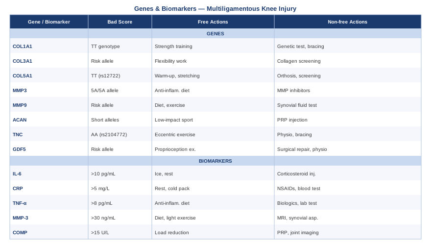
The genetic and biomarker frameworks give you a structural and biochemical map of your recovery. What follows is a resource that pulls many of these threads together in a practical, accessible format — and challenges some assumptions that still dominate mainstream rehabilitation thinking.
The Huberman Lab Podcast on Injury Recovery That Changes the Framework
Andrew Huberman's episode "Science of Healing Wounds, Tendons, Muscle & Bones" (Huberman Lab, 2021) is one of the most science-dense, practically useful resources on connective tissue recovery available outside of academic journals. It synthesizes research from exercise physiology, endocrinology, and molecular cell biology into actionable protocols — and challenges several dominant assumptions in standard sports rehabilitation.
1. Collagen synthesis has a narrow feeding window before exercise
The body is most responsive to dietary collagen precursors approximately 60 minutes before loading exercise. Consuming 15g hydrolyzed collagen with 50mg vitamin C before physical therapy exploits a biologically active window when fibroblasts are primed for protein uptake. This reframes supplementation timing — it is not about daily intake averages, but about biochemical synchronization with tissue-building cues.
2. Complete rest actively delays ligament healing
Tendons and ligaments receive nutrients primarily through mechanical diffusion rather than vascular supply. Movement is not merely safe during recovery — it is required for nutrient delivery to healing tissue. Controlled loading within pain-free ranges accelerates matrix organization and reduces the window during which graft and healing tissue are most vulnerable to re-injury.
3. Deep sleep is where connective tissue is actually rebuilt
Growth hormone secretion during slow-wave sleep is the dominant anabolic driver of collagen synthesis. Even modest improvements in deep sleep duration — through temperature regulation (67–68°F bedroom), consistent sleep timing, and eliminating late alcohol — produce outsized effects on healing speed. This makes sleep architecture a legitimate clinical target, not just a wellness recommendation.
4. Inflammation is necessary but must be time-limited
Inflammation is not the enemy of healing — it is the first stage of it. However, if inflammatory signaling fails to resolve by weeks three to five, it transitions into a chronic state that actively degrades the repair process rather than supporting it. This is the mechanistic rationale for tracking hs-CRP and intervening when elevation persists — not for suppressing the initial acute response.
5. The autonomic nervous system modulates healing rate
Autonomic balance between sympathetic and parasympathetic tone directly modulates healing rate through cytokine signaling networks. Chronic psychological stress measurably slows tendon and ligament healing. Huberman outlines practical breathing interventions — physiological sighs and cyclic hyperventilation followed by extended exhale holds — that rapidly shift autonomic state and reduce cortisol burden.
6. Heat and cold have opposite and complementary roles across recovery phases
Cold reduces acute inflammation in the first 24–72 hours. Heat exposure — sauna or far-infrared — upregulates heat shock proteins and VEGF, both critical for ligament vascularization and repair. The most common mistake is applying cold throughout recovery when heat should dominate from week two or three onward. Protocol misalignment with recovery phase is common and consequential.
7. Blood flow restriction training protects muscle mass when the knee cannot load
BFR training uses partial arterial occlusion to produce a metabolic stimulus equivalent to heavy loading at very low absolute weights. For MLI patients who cannot safely load the knee, BFR allows maintenance of quadriceps and hamstring cross-sectional area — preventing the substantial muscle atrophy that creates joint instability and impairs ligament graft protection during the long recovery window.
8. Hormonal context has measurable effects on healing speed
Higher testosterone levels are associated with faster ligament repair and stronger collagen synthesis. Estrogen at specific phases of the menstrual cycle reduces collagen synthesis rates and increases ligamentous laxity — explaining the well-documented sex-specific injury rate differences at particular cycle phases in competitive athletes. Understanding hormonal context is clinically useful for planning the highest-intensity rehabilitation sessions.
9. PRP has a real but limited and context-dependent role
Huberman reviews the platelet-rich plasma literature honestly, noting that while early enthusiasm was high, evidence for PRP in ligament healing specifically is mixed. Where outcomes have been more positive is in the combination of PRP administration with early controlled mobilization rather than PRP followed by immobilization. Standalone PRP without concurrent movement protocol does not significantly improve outcomes in most trials.
10. Biological readiness matters more than time elapsed for return to sport
The "return to sport at nine months" guideline for ACL reconstruction is a population-average timestamp, not a biological readiness indicator. Tissue maturation markers — quadriceps symmetry indices, movement quality assessments, and increasingly biomarkers like COMP and collagen turnover — are more meaningful guides for return-to-load decisions than the calendar alone. This is the scientific basis for the biomarker-guided approach to recovery described throughout this article.
Complementary Approaches with Real Evidence for Knee Ligament Recovery
Standard surgical and physiotherapy pathways remain the core of multiligamentous knee injury management. But several evidence-supported complementary modalities can meaningfully reduce pain, accelerate healing biology, and improve functional outcomes alongside primary treatment. The three below were selected for the strength and relevance of their clinical evidence for this specific type of injury.
Low-Level Laser Therapy (Photobiomodulation)
Low-level laser therapy — also called photobiomodulation — uses specific wavelengths of red and near-infrared light (typically 630–1000 nm) to stimulate mitochondrial cytochrome c oxidase activity, increase cellular ATP production, reduce local inflammatory mediators, and accelerate tissue repair processes. For multiligamentous knee injury, the cellular mechanism is directly relevant: photobiomodulation has documented effects on collagen fibroblast proliferation, macrophage polarization toward the healing phenotype, and post-surgical edema reduction. These effects are applicable whether recovery is non-surgical or post-reconstruction.
A systematic review of LLLT in musculoskeletal post-surgical conditions found significant reductions in post-operative pain and accelerated functional recovery in treated patients. For joint and soft tissue applications, wavelengths of 820–850 nm at doses of 4–8 J/cm² per session appear most effective based on dose-response data in the literature.
For practical use in MLI recovery, clinical LLLT is available at physiotherapy clinics and sports medicine centers at $30–80 per session. Home devices — from brands such as Joovv, Mito Red, or PlatinumLED Biomax — offer therapeutic near-infrared panels for $400–1,200 one-time. Suggested protocol: 10–20 minutes daily over the knee, five to seven days per week during the first three to six months of recovery. Evidence is strongest for pain and inflammation reduction; evidence for structural graft healing specifically is promising but still developing. The risk-to-benefit ratio is very favorable.
Massage Therapy
Therapeutic massage in the context of multiligamentous knee injury addresses something distinct from relaxation: it manages secondary muscle guarding, connective tissue adhesion, and lymphatic drainage in a joint that is simultaneously immobilized and undergoing aggressive mechanical rehabilitation. The quadriceps, hamstrings, iliotibial band, and calf musculature become significantly dysfunctional following both the injury and surgical reconstruction, and this muscle inhibition impairs the neuromuscular stability that protects healing ligament structures from additional stress.
A randomized controlled trial published in the Journal of Athletic Training found that therapeutic massage significantly reduced quadriceps inhibition and improved functional strength outcomes in post-ACL reconstruction patients compared to standard physiotherapy alone. The mechanisms include pain gating through large-diameter mechanoreceptor stimulation, reduction in protective co-contraction patterns, and improvement in lymphatic drainage around the surgical site.
Practically, regular sports massage once or twice per week — focusing on the quadriceps, iliotibial band, calf complex, and hip external rotators — provides measurable benefit during the rehabilitation phase. Massage directly over any surgical incision is contraindicated until confirmed tissue healing, typically eight to twelve weeks post-surgery. Cost: $60–120 per session; a foam roller and trigger point tools ($20–60 one-time) provide partial benefit between clinical sessions. Always communicate the surgery timeline clearly to the massage therapist to ensure appropriate technique selection.
Mindfulness-Based Stress Reduction (MBSR)
Pain following multiligamentous knee injury has a significant central sensitization component. After weeks or months of persistent nociceptive input, the central nervous system's pain processing can become dysregulated, amplifying pain signals beyond what the current tissue state justifies. Mindfulness-Based Stress Reduction — an eight-week structured program developed by Jon Kabat-Zinn at the University of Massachusetts — addresses this pathway directly through attentional retraining and interoceptive awareness. It also reduces cortisol and sympathetic tone, which — as discussed in the genetics section — directly affects ligament healing rate through cytokine signaling.
A 2016 meta-analysis in JAMA Internal Medicine demonstrated that MBSR significantly reduced chronic pain intensity and pain-related disability across musculoskeletal conditions. Effect sizes were modest to moderate but consistent across studies with minimal harm risk — a favorable profile for a population already managing a significant medical recovery.
For MLI recovery, an eight-week MBSR program requires 30–45 minutes per day of formal practice. Programs are available online through the Brown University Mindfulness Center and through well-structured apps such as Waking Up or Insight Timer. An important practical insight: combining formal mindfulness practice with physical therapy sessions — attending to body sensations during exercises rather than distracting from them — has been shown to improve proprioceptive calibration, which is directly relevant to the neuromuscular retraining demands of knee ligament rehabilitation. Start with 10-minute body scan practices and build gradually over the first two weeks.
Conclusion
A multiligamentous knee injury is one of the body's most demanding recovery challenges. The biology behind it — connective tissue structural quality, inflammatory regulation, anabolic capacity, cartilage stress response — varies significantly between individuals, and that variation matters more than most rehabilitation protocols acknowledge. The six biomarkers covered here (hs-CRP, vitamin D, COMP, MMP-3, IGF-1, and fibrinogen) offer a concrete, largely affordable window into that biology in real time. The six genes (COL5A1, COL1A1, MMP3, TGFB1, IL6, and VEGF) add a deeper layer of structural and functional context that makes recovery strategy genuinely personal rather than generic.
None of this replaces the orthopedic surgeon, the physiotherapist, or the imaging that guides structural decisions. But it gives you better questions to ask, better data to bring to appointments, and better tools for optimizing the biological environment in which repair is happening. The most practical next step is straightforward: request a blood panel that includes hs-CRP, 25-OH vitamin D, fibrinogen, and IGF-1 — all four are affordable, widely available, and immediately actionable. If genetic testing is accessible, raw data from consumer platforms can be analyzed for the variants discussed here at no additional cost. From that foundation, decisions about supplementation, training load progression, and complementary therapies move from guesswork to evidence. Recovery from an MLI is measured in months and years. Better, more specific information makes every one of those months count more.
Musculoskeletal: Joint Conditions Tendon & Ligament Conditions Sports Injuries
Autoimmune: Inflammatory Conditions Connective Tissue Conditions