This article contains AI generated content.
· UpdatedMultiple Epiphyseal Dysplasia — 6 Genes And 7 Biomarkers To Track
Introduction
Living with multiple epiphyseal dysplasia means navigating a condition that most clinicians encounter only a handful of times in a career. You may have spent years with unexplained joint pain, a diagnosis that arrived late, or a medical team that defaults to general osteoarthritis management without fully engaging with the underlying mechanics. That gap between the complexity of your condition and the breadth of standard advice is real, and it matters.
MED is not one disease — it is a family of related but distinct skeletal disorders, each rooted in a different gene, each affecting cartilage and bone development through a slightly different mechanism. Generic joint-health advice built for common wear-and-tear arthritis will often miss the mark for someone with a COMP variant or an SLC26A2 mutation. The biological pathways are different, and so are the most meaningful intervention targets.
This article approaches MED from two directions that complement each other. The first is genetic: understanding which gene is involved, what it disrupts, and what the current science suggests for supporting the affected pathway — from exercise and diet adjustments to targeted supplementation. The second is biomarker-based: identifying which blood and tissue markers are most informative for tracking bone turnover, joint stress, inflammation, and cartilage breakdown, along with what to do when those numbers drift in the wrong direction.
Neither direction promises a reversal of a genetic condition. What both offer is a clearer map — one that helps you make better-informed decisions about movement, nutrition, supplements, and monitoring, in conversation with a specialist who knows your case. Clearer information, applied carefully, does lead to better outcomes. That is the grounded hope this article is built on.
The Genetic Architecture of MED — What Your Variant May Mean and What You Can Do
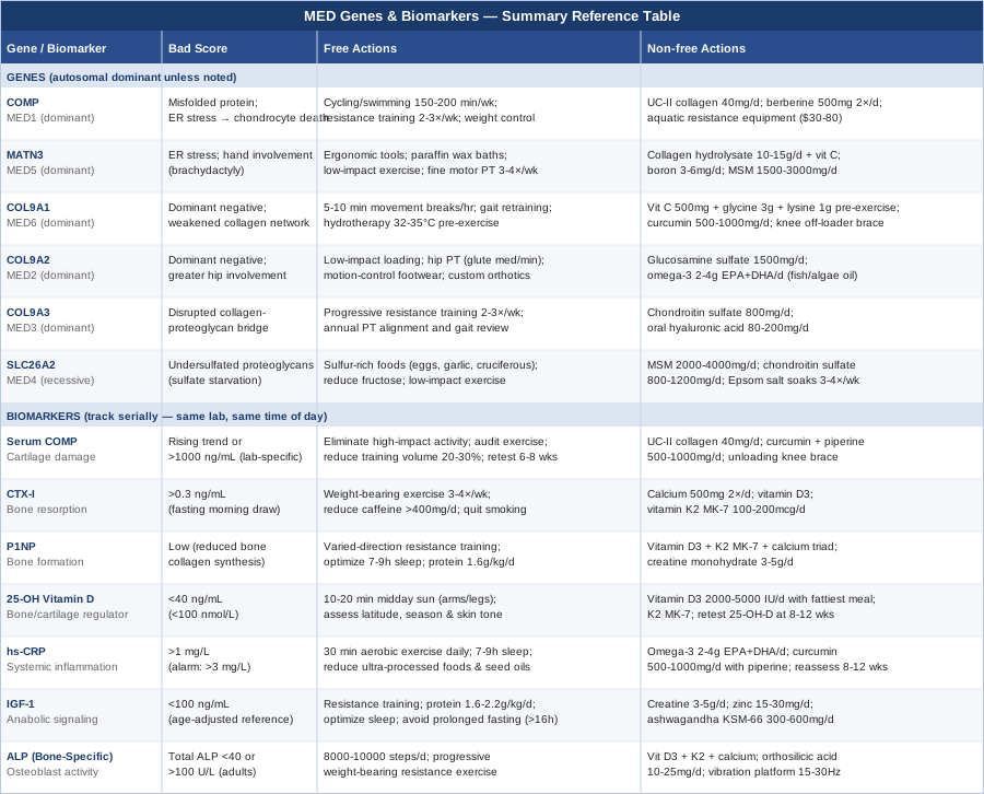
Multiple epiphyseal dysplasia is caused by variants in at least six distinct genes, with most cases following autosomal dominant inheritance (one faulty copy is enough) and a smaller subset following autosomal recessive inheritance. Knowing your specific gene matters enormously because each one disrupts cartilage biology through a different molecular mechanism — and that determines which compensatory strategies are most biologically rational.
The genetic landscape of MED is catalogued in depth at the NCBI GeneReviews entry for Multiple Epiphyseal Dysplasia, which serves as the primary clinical reference for genotype-phenotype correlations.
Gene 1: COMP (Cartilage Oligomeric Matrix Protein)
What this gene does: COMP encodes a large pentameric glycoprotein that functions as a structural organizer of the extracellular matrix in cartilage and tendon. It acts like molecular scaffolding — helping collagen fibrils assemble correctly and anchoring them in place. When COMP carries a pathogenic variant, the misfolded protein accumulates inside chondrocytes (cartilage cells), triggering endoplasmic reticulum (ER) stress. The cell tries to process a protein it cannot fold properly, and this ER stress cascade leads to premature chondrocyte death, disorganized matrix, and abnormal epiphyseal ossification.
COMP variants account for roughly 30–40% of autosomal dominant MED cases and produce MED1 (also called pseudoachondroplasia when severe). Joint pain typically begins in childhood or adolescence, with knees, hips, and ankles most affected.
If the gene variant is present — the plan without supplements: The primary non-pharmacological target is reducing mechanical overload on affected joints while preserving muscle mass and cardiovascular health. High-impact loading on compromised cartilage accelerates matrix breakdown faster than the tissue can repair.
Practical framework: - Exercise mode: Replace running and jumping with cycling, swimming, or elliptical training. Aquatic exercise in particular offloads 60–90% of body weight while maintaining joint mobility and muscle activity. Aim for 150–200 minutes per week of low-impact aerobic activity. - Resistance training: Preserve muscle mass around vulnerable joints, especially quadriceps, gluteus medius, and hip external rotators. These muscles act as force absorbers. Use controlled, slow tempo resistance training (3–4 seconds eccentric phase) to minimize joint peak forces. Frequency: 2–3 sessions per week. - Weight management: Each kilogram of body mass reduction translates to approximately 3–4 kg reduction in knee joint force during walking. If BMI is above 25, this becomes a high-priority lever. - Sleep position: Use a pillow between the knees when sleeping on your side to reduce hip adduction loading at night. - Mobility work: Daily low-load stretching of hip flexors, hamstrings, and thoracic spine reduces compensatory loading patterns. 10–15 minutes daily.
If the gene variant is present — the plan with supplements or equipment: The ER stress mechanism of COMP mutations opens a specific supplementation rationale: support protein quality control and reduce cellular stress.
- Undenatured type II collagen (UC-II): 40 mg/day. Works via oral tolerance, modulating cartilage-specific immune responses. Evidence includes a 2016 randomized trial showing UC-II superior to glucosamine/chondroitin for joint comfort in osteoarthritis subjects. Cycling: continuous use is acceptable; reassess every 6 months. Side effects: minimal, occasional mild GI discomfort. - Vitamin C (ascorbic acid): 500–1000 mg/day with meals. Required as a cofactor for prolyl and lysyl hydroxylase enzymes that stabilize collagen triple-helix structure. Note that megadoses (above 2000 mg/day) may increase kidney stone risk in susceptible individuals. Side effects at this dose: rare loose stools. - Berberine: 500 mg twice daily with meals (cycle 8 weeks on, 4 weeks off). Has demonstrated AMPK activation and ER stress reduction in cell studies. Evidence remains largely preclinical for cartilage-specific applications, but the ER stress pathway is plausible. Interaction warning: may potentiate hypoglycemic medications. - Aquatic resistance equipment: Pool resistance gloves and ankle floats allow progressive overload in zero-impact conditions. One-time cost of $30–80; highly practical for joint-protective strength work.
Gene 2: MATN3 (Matrilin-3)
What this gene does: Matrilin-3 is an extracellular matrix adaptor protein expressed almost exclusively in cartilage. It links collagen networks to proteoglycans (aggrecan, versican) and helps coordinate matrix assembly in the growth plate. Like COMP, pathogenic MATN3 variants cause protein misfolding and ER stress — but through a structurally distinct mechanism involving a von Willebrand factor A-like domain. The resulting phenotype (MED5) often presents with hand involvement (brachydactyly), which is a distinguishing clinical feature compared to other MED subtypes.
If the gene variant is present — the plan without supplements: - Fine motor joint protection: Avoid grip tools that place sustained compressive load on interphalangeal joints. Use ergonomic handles, jar openers, and typing wrist supports. - Physical therapy focus: Emphasize proprioceptive training for hands and wrists (putty exercises, fine motor coordination tasks), not just large joint work. 20 minutes, 3–4 days per week. - Heat therapy: Paraffin wax baths for hands (10–15 minutes before exercise) reduce stiffness and improve tissue extensibility. Cost: $40–60 for a home paraffin unit. - Activity modification for knees and hips: Same low-impact framework as COMP, with additional attention to hand-grip activities during resistance training (avoid barbell exercises that load finger joints; use straps or machines).
If the gene variant is present — the plan with supplements or equipment: - Collagen hydrolysate (bovine or marine, 10–15 g/day): Provides a concentrated source of hydroxyproline, proline, and glycine — the amino acid building blocks of collagen and matrilin proteins. A 2019 trial published in the British Journal of Sports Medicine demonstrated increased collagen synthesis markers after hydrolyzed collagen supplementation combined with vitamin C. Take 30 minutes before exercise or physiotherapy. - Boron: 3–6 mg/day (from food or supplement). Boron influences bone and cartilage metabolism via effects on vitamin D activation and collagen cross-linking. Dietary sources include raisins, almonds, avocado. Supplement form: sodium tetraborate or calcium fructoborate. - MSM (methylsulfonylmethane): 1500–3000 mg/day in divided doses. Provides bioavailable sulfur for glycosaminoglycan synthesis. Evidence for joint pain reduction is moderate-quality; more relevant for MED subtypes with sulfate pathway involvement (see SLC26A2 below). Side effects: generally well tolerated; occasional GI upset at higher doses. Cycling: continuous acceptable; 12-week reassessment recommended.
Gene 3: COL9A1 — Type IX Collagen Alpha-1
What this gene does: Type IX collagen is a fibril-associated collagen that wraps around type II collagen fibrils in cartilage, acting as a molecular bridge between the collagen network and the surrounding proteoglycan matrix. COL9A1 mutations cause MED6 through a dominant negative mechanism — the mutant chain is incorporated into collagen trimers but disrupts their function, weakening the entire network even when only one copy is affected.
If the gene variant is present — the plan without supplements: - Movement consistency over intensity: Regular, low-load movement maintains cartilage nutrition (cartilage is avascular and depends on cyclic compression/relaxation for nutrient diffusion). Short, frequent movement breaks (5–10 minutes every 45–60 minutes of sitting) are more protective than single long exercise sessions. - Posture and alignment work: Gait retraining with a physiotherapist to distribute joint loads more evenly. Lateral wedge insoles can reduce medial compartment knee load by 5–10% in valgus-aligned knees. - Hydrotherapy: Warm water immersion (32–35°C) for 20 minutes before resistance training reduces muscle guarding and allows fuller joint range of motion under safe loading conditions.
If the gene variant is present — the plan with supplements or equipment: - Vitamin C, glycine, and lysine combination: These three cofactors support the hydroxylation and cross-linking steps in collagen biosynthesis. A practical approach: 500 mg vitamin C + 3 g glycine + 1 g lysine, taken together before exercise or physiotherapy. No significant cycling needed; adjust based on tolerance. - Curcumin (with piperine for bioavailability): 500–1000 mg curcuminoids/day. Inhibits NF-κB-driven collagenase (MMP-13) expression in chondrocytes. A 2014 RCT showed significant reduction in knee osteoarthritis scores with curcumin versus placebo. Take with fat-containing meal for absorption. Cycling: 12 weeks on, 4 weeks off. Caution with anticoagulant medications. - Knee unloading brace: For medial or lateral compartment load concentration, an off-loader knee brace ($150–400) distributes force away from the most damaged zone. Evidence-based for OA; reasonable application in MED-related knee joint degradation.
Gene 4: COL9A2 — Type IX Collagen Alpha-2
What this gene does: COL9A2 encodes a second structural chain of the type IX collagen heterotrimer. Variants here (MED2) produce a phenotype largely overlapping with COL9A1-MED but with some differences in severity and joint distribution. The molecular disruption is similar — dominant negative integration into the collagen scaffold — but the exact structural domain affected differs, which can influence phenotypic expression.
If the gene variant is present — the plan without supplements: - Same foundational joint-protection principles as COL9A1: low-impact loading, regular movement cycling, physical therapy, weight management. - Particular attention to hip joint: COL9A2 variants may have slightly greater hip involvement. Hip-targeted physiotherapy (strengthening of gluteus medius and minimus, piriformis stretching) reduces the hip joint reaction force by improving muscle-force vectors during walking. - Footwear: Cushioned, motion-control footwear reduces peak joint impact transients by 20–30%. Custom orthotics may provide additional benefit if gait analysis reveals significant pronation or supination.
If the gene variant is present — the plan with supplements or equipment: - Glucosamine sulfate: 1500 mg/day (sulfate form preferred over hydrochloride). Provides substrate for glycosaminoglycan chains that surround collagen fibrils. The GAIT trial (NIH-funded) showed mixed results for pain, but glucosamine sulfate specifically showed benefit in moderate-to-severe knee OA in subgroup analyses. Cycling: 3 months on, 1 month off to assess response. Side effects: mild GI symptoms; caution in shellfish allergy. - Omega-3 fatty acids (EPA+DHA): 2–4 g/day combined EPA+DHA from fish oil or algae. Reduces prostaglandin E2 and leukotriene B4 synthesis, attenuating synovial inflammation around affected joints. Take with the largest meal of the day. Cycling: continuous; reassess at 3 months. Mildly blood-thinning at higher doses; discuss with physician if on anticoagulants.
Gene 5: COL9A3 — Type IX Collagen Alpha-3
What this gene does: The third chain of the type IX collagen heterotrimer. COL9A3 variants (MED3) tend to produce one of the milder forms of autosomal dominant MED, with joint pain and stiffness becoming symptomatic more often in adulthood. The α3 chain carries the glycosaminoglycan side chain attachment site — a chondroitin sulfate chain that anchors type IX collagen to aggrecan networks. Disruptions here specifically impair the collagen-proteoglycan bridge in the cartilage matrix.
If the gene variant is present — the plan without supplements: - Progressive resistance training starting early in adulthood: Because symptom onset is often delayed, there is a meaningful window to build protective muscle mass and joint stability before secondary OA develops. Two to three days per week of progressive resistance training targeting lower limb and core musculature. - Periodic physiotherapy review: Even during asymptomatic phases, annual physiotherapy assessment of gait, joint alignment, and muscle balance can catch loading asymmetries before they cause accelerated cartilage breakdown.
If the gene variant is present — the plan with supplements or equipment: - Chondroitin sulfate: 800 mg/day. Particularly relevant here given the α3 chain's role in chondroitin sulfate attachment. Chondroitin sulfate provides the exact molecular substrate that the COL9A3 disruption affects downstream. Evidence quality is moderate; a 2015 meta-analysis in Annals of the Rheumatic Diseases supported a small-to-moderate effect on pain and function. Cycling: 3 months on, 1 month off. Side effects: minimal, occasional GI discomfort. - Aggrecan-supporting compounds: Hyaluronic acid (oral, 80–200 mg/day from high-molecular-weight source) supports the hyaluronan component of the proteoglycan network that aggrecan integrates with. Evidence for oral HA in joint health is emerging; a 2016 Japanese RCT showed reduced knee pain scores. No significant side effects or cycling requirements.
Gene 6: SLC26A2 (DTDST — Diastrophic Dysplasia Sulfate Transporter)
What this gene does: SLC26A2 encodes a sulfate transporter in the cell membrane. Chondrocytes (and other cells) import sulfate from the extracellular environment to synthesize glycosaminoglycans — the long, sulfated sugar chains that give cartilage its compressive resilience. When both copies of SLC26A2 are mutated (autosomal recessive MED, also called MED4 or recessively inherited MED), chondrocytes are starved of sulfate. The result is undersulfated aggrecan and other proteoglycans — structurally weakened cartilage that cannot resist compressive loads normally.
This is mechanistically very different from the ER stress-dominant pathway of COMP and MATN3. The target here is sulfate availability, not protein folding quality control.
If the gene variant is present — the plan without supplements: - Dietary sulfur-rich foods: Increase intake of foods naturally high in organosulfur compounds and inorganic sulfate: eggs, garlic, onions, leeks, cruciferous vegetables (broccoli, cabbage, Brussels sprouts), meat. These provide precursors that can partly compensate for reduced transporter efficiency. - Avoid sulfate depletion: High intake of fructose may reduce hepatic sulfate availability (fructose is sulfated during metabolism); consider reducing added sugars. This is a relatively low-certainty recommendation but carries no downside. - Load management: As above — low-impact exercise, weight maintenance, regular movement. Undersulfated proteoglycans are particularly vulnerable to compressive overload.
If the gene variant is present — the plan with supplements or equipment: - MSM (methylsulfonylmethane): 2000–4000 mg/day in divided doses (with meals). MSM is an organic sulfur compound that crosses cell membranes and contributes to the intracellular sulfur pool. Most directly relevant for SLC26A2-MED among all MED subtypes. The transporter deficiency means cells rely more heavily on alternative sulfur sources. A 2006 RCT showed MSM reduced knee OA pain by 25% versus placebo over 12 weeks. Cycling: 12 weeks on, 4 weeks off. Side effects: GI discomfort if started at full dose; titrate up over 2 weeks. - Chondroitin sulfate: 800–1200 mg/day. Provides an exogenous source of sulfated glycosaminoglycans. Particularly biologically relevant here because it bypasses the transporter step — the sulfated sugar chains arrive already assembled. Cycling: continuous or 3 months on / 1 month off cycles. - Sodium sulfate (inorganic, as Epsom salt footsoaks or bath): Transdermal sulfate absorption is modest but measurable. 20-minute Epsom salt soaks (1–2 cups in warm bath) 3–4 times per week. Cost: low. Evidence is preliminary but mechanistically plausible for this specific genetic subtype where intracellular sulfate supply is the rate-limiting factor.
With the genetic picture mapped, tracking a set of quantifiable blood and tissue markers allows you to monitor how the condition is actually progressing in your body — and whether the interventions above are making a measurable difference.
7 Biomarkers Worth Tracking in Multiple Epiphyseal Dysplasia
These seven markers were selected for their direct relevance to the biological processes most disrupted in MED: cartilage turnover, bone remodeling, inflammation, and growth signaling. Experts like Peter Attia (structural health monitoring, bone density biomarkers), Thomas Dayspring (lipid-adjacent metabolic markers), and orthopedic researchers tracking serum cartilage proteins collectively inform this selection.
Biomarker 1: Serum COMP (Cartilage Oligomeric Matrix Protein)
Why it matters: COMP is released from cartilage into synovial fluid and bloodstream when chondrocytes are stressed or cartilage matrix is degraded. Elevated serum COMP is a marker of active cartilage breakdown and is used as a research biomarker for early OA progression. In MED caused by COMP gene mutations, the protein itself is structurally abnormal, which may affect interpretation — but rising levels over time still signal accelerating cartilage damage. In non-COMP MED subtypes, serum COMP functions as a straightforward joint stress indicator.
How to measure it: Specialized laboratory blood test (ELISA method). Not part of standard panels — requires a specialty lab order. Cost range: $80–200. Ask a rheumatologist or sports medicine physician to order it. Reference ranges are laboratory-specific; serial measurements (same lab, same time of day) are more informative than single values.
If the score is elevated — the plan without supplements: Elevated COMP in a known MED patient usually signals excessive mechanical joint loading. Audit current physical activity for high-impact elements (running, jumping, heavy squats) and replace with equivalent low-impact alternatives. Reduce training volume temporarily by 20–30% and retest in 6–8 weeks.
If the score is elevated — the plan with supplements or equipment: UC-II undenatured collagen (40 mg/day) and curcumin with piperine (500–1000 mg/day) have the most direct evidence for reducing cartilage breakdown markers. An unloading knee brace during high-activity periods provides equipment-level joint stress reduction.
Biomarker 2: CTX-I (C-Terminal Telopeptide of Type I Collagen)
Why it matters: CTX-I is a fragment of type I collagen released into the bloodstream when osteoclasts resorb bone. It is the gold-standard bone resorption marker. Elevated CTX-I in MED indicates accelerated bone turnover, which often presages trabecular bone quality deterioration and increased fracture risk in already-compromised epiphyseal structures. Peter Attia tracks CTX-I as part of his longevity bone health protocol; it is actionable, affordable, and reproducible.
How to measure it: Fasting morning blood test (bone resorption markers are diurnally variable; morning fasting is essential for consistency). Cost: $30–80 on most standard panels or as an add-on to a metabolic panel. Optimal range: below 0.3 ng/mL (age-dependent; postmenopausal women have higher physiological norms).
If CTX-I is elevated — the plan without supplements: Impact exercise (walking, resistance training) suppresses bone resorption by increasing mechanical load signaling. Even 20 minutes of weight-bearing exercise 3–4 days per week measurably lowers CTX-I within weeks. Eliminate calcium depleting habits: reduce caffeine above 400 mg/day, avoid very high sodium intake, quit smoking if applicable.
If CTX-I is elevated — the plan with supplements or equipment: Calcium (500 mg twice daily with meals, calcium carbonate with food or calcium citrate independent of meals) and vitamin D3 (see below) are first-line. Vitamin K2 (MK-7 form, 100–200 mcg/day) activates osteocalcin, directing calcium into bone rather than soft tissue. Cycling: continuous, reassess CTX-I at 3 months.
Biomarker 3: P1NP (Procollagen Type I N-Terminal Propeptide)
Why it matters: Where CTX-I measures breakdown, P1NP measures formation — it is the most sensitive marker of new bone collagen synthesis. The CTX-I/P1NP ratio captures the net bone remodeling balance: a high CTX-I with low P1NP means net loss; a rising P1NP with stable CTX-I means the interventions are working. In MED, monitoring this ratio over time provides an early signal of whether secondary osteoporosis is developing in the compromised epiphyseal regions.
How to measure it: Fasting morning blood test (same as CTX-I; the two are usually ordered together). Cost: $40–100 when combined with CTX-I. Labs may report it as "total P1NP" or "intact P1NP" — total P1NP is the standard clinical measure.
If P1NP is low — the plan without supplements: Progressive resistance training and impact loading are the strongest stimuli for bone formation. Specifically, varied loading directions (not just axial compression) stimulate osteoblast activity most effectively. Incorporate lateral movements, rotational movements, and varied-angle resistance exercises. Sleep optimization (7–9 hours) supports the growth hormone pulse during deep sleep that drives P1NP.
If P1NP is low — the plan with supplements or equipment: Vitamin D3 (2000–4000 IU/day; adjust based on serum 25-OH vitamin D levels) + K2 MK-7 (100–200 mcg/day) + calcium are the triad. Creatine monohydrate (3–5 g/day, no loading phase needed) has emerging evidence for bone formation support — a 2022 meta-analysis found modest positive effects on bone mineral density with resistance training. No cycling needed for creatine; it is one of the most safety-studied supplements available.
Biomarker 4: 25-OH Vitamin D
Why it matters: Vitamin D is not just a bone mineral regulator — it modulates chondrocyte differentiation, collagen synthesis, and inflammatory signaling in cartilage. Low 25-OH vitamin D is associated with accelerated cartilage loss in OA studies, and given that MED patients face a higher baseline risk of secondary osteoarthritis, maintaining optimal vitamin D levels is a genuinely actionable lever. Deficiency is common, particularly in northern climates and in individuals who spend most of the day indoors.
How to measure it: Standard blood test. Cost: $20–50 on most panels; often covered by insurance with a clinical indication. Optimal functional range (not just sufficiency): 40–60 ng/mL (100–150 nmol/L) per Peter Attia's longevity protocol. Standard clinical "sufficient" threshold of 20 ng/mL is considered too low by many functional medicine and sports medicine clinicians.
If 25-OH vitamin D is low — the plan without supplements: Daily sun exposure (10–20 minutes of midday sunlight on arms and legs, without sunscreen for this duration) produces 1000–5000 IU of vitamin D3 depending on skin tone, latitude, and season. This is the most physiological approach but insufficient at northern latitudes in winter.
If 25-OH vitamin D is low — the plan with supplements or equipment: Vitamin D3 (cholecalciferol, not D2) at 2000–5000 IU/day with the fattiest meal of the day for absorption. Always co-supplement with vitamin K2 MK-7 to prevent inappropriate calcium deposition. Retest 25-OH vitamin D after 8–12 weeks to calibrate dose. Side effects: essentially absent at these doses; toxicity requires sustained intake above 10,000 IU/day for months.
Biomarker 5: hs-CRP (High-Sensitivity C-Reactive Protein)
Why it matters: Systemic low-grade inflammation accelerates cartilage matrix degradation by upregulating matrix metalloproteinases (MMP-3, MMP-13) and inflammatory cytokines (IL-1β, TNF-α) in joint tissue. In MED, secondary inflammatory arthritis developing in young adulthood is a significant source of functional decline. hs-CRP is a sensitive, inexpensive, and reproducible systemic inflammation index. Thomas Dayspring includes it in cardiovascular risk assessment; in the MED context, it also serves as a joint-degradation risk signal.
How to measure it: Standard blood test (fasting not required). Cost: $15–40. Optimal: below 0.5 mg/L. Moderate risk: 1–3 mg/L. High risk for tissue-level inflammatory damage: above 3 mg/L (after ruling out acute infection).
If hs-CRP is elevated — the plan without supplements: Anti-inflammatory lifestyle interventions with the strongest evidence: aerobic exercise (even moderate-intensity, 30 minutes most days), 7–9 hours of sleep, reduced ultra-processed food intake, stress reduction. Eliminating trans fats and reducing omega-6/omega-3 ratio through dietary changes (more fatty fish, less seed oil) typically lowers hs-CRP within 4–8 weeks.
If hs-CRP is elevated — the plan with supplements or equipment: Omega-3 fatty acids (2–4 g EPA+DHA/day) are the most evidence-backed anti-inflammatory supplement — a 2013 meta-analysis confirmed their hs-CRP-lowering effect. Curcumin (500–1000 mg/day with piperine) independently reduces NF-κB activity and hs-CRP. Cycling for both: continuous use acceptable; reassess hs-CRP at 8–12 weeks. Combination is reasonable and synergistic; reduce total EPA+DHA to 2 g/day if also on any antiplatelet medications.
Biomarker 6: IGF-1 (Insulin-Like Growth Factor 1)
Why it matters: IGF-1 is the primary anabolic mediator for cartilage and bone. Chondrocytes express IGF-1 receptors, and IGF-1 stimulates proteoglycan synthesis, collagen production, and chondrocyte survival. Low IGF-1 is associated with reduced cartilage repair capacity and accelerated OA. In MED patients — particularly those who received growth hormone-related interventions in childhood, or who have adult growth hormone deficiency — tracking IGF-1 provides a meaningful window into anabolic capacity for cartilage maintenance.
How to measure it: Blood test (fasting preferred). Cost: $40–80. Optimal adult range varies by age but typically 100–250 ng/mL for adults aged 30–60 (exact optimal ranges are debated; consult an endocrinologist for interpretation in the context of individual growth history and MED).
If IGF-1 is suboptimal — the plan without supplements: IGF-1 is highly responsive to lifestyle: resistance training (especially compound movements like squats and deadlifts or their low-impact equivalents — leg press, Romanian deadlift) acutely and chronically raises IGF-1. Sleep quality is critical — growth hormone (which drives IGF-1 synthesis) is secreted primarily in slow-wave sleep. Maintaining protein intake at 1.6–2.2 g/kg body weight supports IGF-1 production. Intermittent fasting beyond 16–18 hours can suppress IGF-1; avoid prolonged fasting if IGF-1 is already suboptimal.
If IGF-1 is suboptimal — the plan with supplements or equipment: Creatine monohydrate (3–5 g/day) supports IGF-1 signaling at the muscular level. Zinc (15–30 mg/day with food) is a cofactor in IGF-1 receptor signaling; zinc deficiency is independently associated with reduced IGF-1. Ashwagandha (KSM-66 extract, 300–600 mg/day) has shown IGF-1 increases in some resistance training trials. Cycling for ashwagandha: 8 weeks on, 2 weeks off. Side effects: occasional mild sedation; avoid during pregnancy.
Biomarker 7: ALP (Alkaline Phosphatase, Bone-Specific)
Why it matters: Bone-specific alkaline phosphatase (BSALP) is an enzyme produced by osteoblasts and is a direct marker of bone formation activity — particularly in growth plate and newly forming bone. It provides a more bone-specific signal than total ALP (which can also reflect liver and intestinal activity). In MED, where epiphyseal ossification is already disrupted, tracking BSALP helps distinguish between normal bone remodeling, accelerated pathological turnover, and the suppressed formation seen in advancing secondary OA.
How to measure it: Bone-specific ALP can be ordered as a standalone test or is sometimes included in comprehensive bone panels. Total ALP is on most metabolic panels and gives a rough indication. Bone-specific: $60–120 at specialty labs. Total ALP: $10–30 on standard metabolic panel. Optimal total ALP for adults: 40–100 U/L; very low suggests reduced bone formation activity; very high may indicate accelerated turnover or Paget-like activity.
If BSALP is low — the plan without supplements: Increase weight-bearing mechanical load through progressive resistance training and impact-appropriate activities. Low BSALP in an MED patient often signals physical inactivity, not just disease severity. Even walking 8,000–10,000 steps daily raises bone formation markers measurably within weeks.
If BSALP is low — the plan with supplements or equipment: Vitamin D3 + K2 + calcium triad (as above). Strontium ranelate (historically used for osteoporosis) stimulates BSALP directly but has cardiovascular risk concerns and is restricted in many countries — discuss risk-benefit with an endocrinologist or rheumatologist. Silicon (as orthosilicic acid, 10–25 mg/day) supports bone matrix formation and has a reasonable safety profile; evidence quality is preliminary. A whole-body vibration platform (15–30 Hz, 10–15 minutes/day, $200–600 for home units) stimulates osteoblast activity with minimal joint impact load.
What "Outlive" by Peter Attia Reveals About Musculoskeletal Longevity — 10 Things That Should Change How You Think
Peter Attia's 2023 book Outlive: The Science and Art of Longevity is one of the most rigorously evidence-referenced books on long-term health optimization available to general readers. While not written specifically for MED, its framework for musculoskeletal resilience, bone health, and cartilage preservation maps directly onto the functional challenges of living with this condition — and challenges several assumptions that conventional orthopedic care tends to reinforce.
1. Muscle mass is a longevity organ, not an aesthetic preference
Attia makes the case that skeletal muscle is the most important organ for long-term functional survival. For MED patients, this reframes the conversation: the goal is not simply pain management but building the largest possible "structural reserve" — muscle mass that absorbs force, protects joints, and maintains independence as the disease progresses. He recommends targeting a minimum of 1.6 g protein per kg/day and progressive resistance training starting no later than the 30s, with the recognition that it becomes much harder to build muscle in later decades.2. VO2 max is the single most predictive longevity biomarker
Attia cites data showing that individuals in the top quartile of VO2 max for their age have a 45% lower all-cause mortality risk compared to those in the bottom quartile. For MED patients who may be limited in high-impact aerobic activity, this is an urgent argument for finding a mode that preserves cardiovascular capacity — cycling, rowing, swimming, or elliptical — rather than accepting deconditioning as inevitable.3. Zone 2 training preserves mitochondrial health in chondrocytes too
Attia's discussion of Zone 2 aerobic training (conversational pace, primarily fat-burning) emphasizes mitochondrial efficiency. What is less discussed in orthopedics is that chondrocytes — usually described as metabolically quiet cells — are significantly influenced by mitochondrial health. Dysregulated mitochondrial function accelerates chondrocyte apoptosis in OA tissue. Zone 2 cardiovascular training protects mitochondrial integrity systemically.4. Bone density peaks by the mid-30s — the window is not infinite
Attia emphasizes the lifetime arc of bone density: the time to build skeletal capital is childhood through the mid-30s. MED disrupts normal epiphyseal ossification from the start, which means the baseline bone mineral density trajectory is already compromised. This makes early intervention with weight-bearing exercise, calcium, and vitamin D not just beneficial but critical — you cannot fully compensate for the lost window, but you can significantly mitigate the consequence.5. Inflammation is the force multiplier for chronic disease
Outlive frames chronic low-grade inflammation as the common upstream driver of most organ-system decline. In MED, joint inflammation accelerates cartilage matrix degradation through the same inflammatory cascades. Attia's practical anti-inflammatory toolkit — consistent sleep, time-restricted eating, omega-3 supplementation, stress reduction — maps directly onto what will slow secondary arthritis progression in MED joints.6. Insulin sensitivity directly affects joint tissue health
Attia presents data linking poor insulin sensitivity to accelerated OA progression through advanced glycation end-products (AGEs) that crosslink and stiffen collagen fibers. Disordered collagen — already structurally weakened in MED — is particularly vulnerable to AGE cross-linking. Maintaining insulin sensitivity (through low-glycemic diet, resistance training, sleep) is a meaningful cartilage-protective strategy, not just a metabolic one.7. Sleep is non-negotiable for tissue repair
Deep sleep is when growth hormone secretion peaks, driving the IGF-1-mediated anabolic window for cartilage and bone repair. Attia cites data showing that even modest sleep restriction (6 hours versus 8 hours) reduces anabolic hormone output by 20–30%. For MED patients relying on whatever residual repair capacity their chondrocytes retain, protecting sleep quality is a direct investment in joint longevity.8. The stability-mobility-strength framework for joint health
Attia's discussion of physical training distinguishes between stability (proprioceptive control), mobility (range of motion under load), and strength (force production). Most exercise programs address strength while neglecting stability and mobility — the two capacities that, when lost, create the aberrant loading patterns that accelerate cartilage breakdown. For MED, stability training (single-leg balance, perturbation training, foot-ground awareness work) is arguably more protective than strength training alone.9. Tracking matters more than guessing
A recurring theme in Outlive is that subjective assessment is a poor substitute for objective measurement. Attia tracks bone density (DEXA), inflammatory markers, hormone levels, and functional capacity metrics serially over time. This is exactly the argument for tracking the biomarker panel above: pain as a feedback signal is delayed and imprecise. Biomarkers give you a 6–12 month early warning system.10. The biggest risk is doing too little, not too much
Perhaps the most counterintuitive point in Outlive for patients with painful skeletal conditions: the evidence strongly favors more structured physical activity, not less. Deconditioning accelerates every downstream complication — muscle loss, bone loss, inflammation, insulin resistance, cardiovascular deterioration. The prescription is not rest; it is smarter loading, with low-impact modalities substituted for high-impact ones as needed.Beyond genetics and biomarkers, there are a handful of evidence-supported physical modalities that address MED's most pressing functional challenges: joint pain, mobility restriction, muscle guarding, and the psychological toll of living with a chronic progressive condition.
Complementary Approaches With Meaningful Clinical Evidence
Low-Level Laser Therapy (Photobiomodulation)
Low-level laser therapy (LLLT) uses near-infrared light (typically 780–1100 nm wavelength) to penetrate tissue and stimulate mitochondrial cytochrome c oxidase, increasing local ATP production, reducing oxidative stress, and modulating inflammatory cytokine expression. For MED joints experiencing secondary inflammatory arthritis, LLLT addresses the inflammatory and mitochondrial components simultaneously — two biologically relevant targets given what is known about chondrocyte energy metabolism and matrix preservation.
A 2009 Cochrane systematic review on LLLT for osteoarthritis concluded that photobiomodulation produces clinically meaningful reductions in pain and improvements in function compared to placebo laser. The analysis included multiple randomized controlled trials with 2–8 week treatment courses. The effect was most consistent for knee osteoarthritis, the joint most commonly and severely affected in MED. Wavelengths of 810–830 nm and doses of 3–8 J/cm² per session showed the strongest signal.
For practical application in MED, LLLT can be applied at home using a consumer-grade red/near-infrared light therapy device ($150–500, with 660 nm and 850 nm wavelengths being the most versatile). Target the most symptomatic joints for 10–15 minutes per joint, 3–5 times per week. Begin at the lower end of treatment time and assess tolerance. Evidence is not strong enough to recommend LLLT as a standalone treatment, but as an adjunct to exercise and joint protection strategies, the risk-benefit profile is favorable given MED's chronic, low-grade inflammatory joint environment.
Tai Chi
Tai chi is a Chinese movement practice combining slow, flowing sequences with weight transfer, balance challenge, and intentional breath control. It trains exactly the stability and proprioceptive capacities that Peter Attia identifies as most protective for joint longevity, and it does so at a mechanical load low enough to be appropriate even for significantly compromised joints. Balance and proprioception deficits in MED develop partly because abnormal joint surfaces send unreliable sensory signals to the nervous system — tai chi directly targets this sensorimotor gap.
A landmark 2016 randomized controlled trial in the Annals of Internal Medicine compared tai chi to physical therapy for knee osteoarthritis and found that 12 weeks of tai chi produced equivalent improvements in pain and function, with superior mental health and depression outcomes. The 108-movement Yang style and the shorter 24-movement Sun style are both appropriate; the Sun style is particularly well-suited for individuals with limited hip and knee range of motion because it uses a higher, narrower stance.
For MED application: begin with a beginner class (community centers, online programs) under the supervision of a qualified instructor who understands your joint limitations. Inform the instructor about which joints are most affected. Two to three sessions per week of 30–45 minutes each is the evidence-based dosage. Most forms can be adapted for individuals who cannot stand for extended periods — many movements can be modified to a chair-based version during flares.
Massage Therapy
Therapeutic massage addresses one of the most under-acknowledged consequences of chronic joint pain: the secondary muscle guarding, compensatory movement patterns, and myofascial trigger points that develop around affected joints over years of protective loading. In MED, the hips, knees, and lumbar paraspinal muscles are particularly prone to chronic tension from altered gait mechanics. This secondary myofascial component often contributes as much to functional limitation as the primary cartilage pathology.
A 2015 RCT in the Journal of General Internal Medicine found that a full-body massage protocol over 8 weeks significantly reduced pain and improved function in knee OA patients compared to sham or no-treatment controls, with effects persisting at 24-week follow-up. The effect was attributed to both mechanical (myofascial release, improved circulation) and neurological (descending pain inhibition modulation) mechanisms. Evidence specifically for MED is absent, but the muscle guarding and myofascial tension patterns are shared.
Practical application: seek a massage therapist with experience in musculoskeletal or orthopedic massage. Initial series of 4–8 weekly sessions focused on hip flexors, iliotibial band, quadriceps, and lumbar paraspinal muscles — all regions that commonly become secondary pain generators in MED-related altered gait. Monthly maintenance sessions thereafter. Communicate clearly about joint fragility and the need to avoid direct compression on epiphyseal regions. Deep transverse friction over the joint itself is contraindicated; work should focus on the surrounding musculature.
Biofeedback
Biofeedback uses real-time physiological monitoring (surface EMG, heart rate variability, skin conductance) to train voluntary regulation of normally automatic body processes — muscle tension, pain amplification, and autonomic nervous system arousal. In a chronic pain condition like MED-related arthritis, central sensitization (the nervous system's tendency to amplify pain signals over time) becomes a significant contributor to the total pain experience that is distinct from peripheral joint pathology. Biofeedback addresses this central component.
A review published by the Association for Applied Psychophysiology and Biofeedback found that EMG biofeedback training reduces chronic musculoskeletal pain with moderate-to-strong effect sizes, and HRV biofeedback reduces the autonomic hyperarousal that amplifies pain perception. These are conditions-agnostic findings with mechanistic plausibility for any chronic joint pain syndrome, including MED. For gait-specific applications, force plate biofeedback has been studied in knee OA to retrain loading symmetry — directly relevant for MED patients with asymmetric joint involvement.
Practically: clinical biofeedback is offered by neuropsychologists, physiotherapists with advanced training, and pain psychologists. 8–12 sessions is a typical introductory course; effects are durable because the patient learns a skill, not just receives a treatment. Home HRV biofeedback devices ($100–200, paired with apps like HeartMath Inner Balance) provide a cost-effective way to practice daily between sessions.
Conclusion
Multiple epiphyseal dysplasia is a complex genetic condition, but complexity does not mean passivity. Understanding which gene is involved opens targeted interventions at the biological pathway level. Tracking the right biomarkers transforms vague symptom monitoring into an early-warning system with actionable thresholds. The complementary physical modalities covered here add meaningful, evidence-grounded tools for managing the secondary joint damage and central pain sensitization that accumulate over years.
None of this replaces the care of a medical team — ideally one that includes an orthopedic specialist with rare skeletal disease experience, an endocrinologist for bone health monitoring, and a physiotherapist who understands joint protection programming. But the information in this article gives you a more complete map to bring to those conversations.
A practical next step: if you do not yet know your specific gene variant, genetic testing through a clinical genetics service is the most important first action. From there, ordering a baseline panel of the biomarkers above — particularly 25-OH vitamin D, CTX-I, P1NP, hs-CRP, and IGF-1 — gives you a data foundation to build from. Then choose one lifestyle intervention from the exercise section and one supplement from the relevant gene section and commit to 12 weeks before reassessing. Small, measurable steps consistently applied outperform complex multi-supplement protocols that never become habits.
Musculoskeletal: Bone Conditions Joint Conditions
Autoimmune: Inflammatory Conditions Connective Tissue Conditions