This article contains AI generated content.
· UpdatedMultiple Pterygium Syndrome – 9 Genes and 6 Biomarkers to Track
Introduction
Multiple Pterygium Syndrome (MPS) is one of those conditions where the name barely hints at what daily life actually looks like for those navigating it. The characteristic skin webs (pterygia) that form across joints — most often the neck, knees, elbows, and fingers — are striking, but they represent only the visible surface of a condition that runs far deeper. Beneath those webs are joint contractures, growth delays, scoliosis, and in more severe forms, life-threatening complications during fetal development. If you or someone you love has received this diagnosis, the first thing to understand is that MPS is not a single disease — it is a family of related presentations, each driven by a specific disruption in one highly specific molecular pathway.
That pathway is the neuromuscular junction (NMJ) — the interface where motor neurons hand off electrical signals to muscle fibers. During fetal development, movement is not incidental; it actively shapes how joints, tendons, and skin form. When genes controlling NMJ signaling are disrupted, fetal movement is reduced, and the connective tissue forming around immobile limbs does so abnormally — producing the webs, contractures, and skeletal anomalies that define MPS. Understanding this mechanism is the starting point for making sense of everything else.
Generic health advice rarely meets the complexity of a rare genetic condition. Recommendations built for common musculoskeletal or inflammatory diseases do not automatically translate here. The molecular specifics matter enormously: which gene is affected determines which biochemical pathway is compromised, which treatments help or harm, and what the realistic trajectory looks like. This is precisely why this article takes a deeper approach, anchored to those specifics rather than broad generalities.
None of what follows replaces specialist care. But better information leads to better conversations with medical teams, better questions about genetic testing, and more informed decisions about which supportive strategies are worth pursuing. This article covers nine genes linked to MPS, six biomarkers worth tracking over time, flexibility science directly applicable to joint contractures, and complementary approaches with genuine clinical evidence — a layered toolkit for navigating this condition more actively and intelligently.
Summary
Multiple Pterygium Syndrome traces back to mutations in genes that control how nerve signals reach fetal muscles. Nine key genes are involved — from CHRNG, the most common driver of Escobar syndrome, to DOK7 and MUSK, where a surprising finding about beta-agonists has changed clinical management, to MYBPC1, which underlies the most severe lethal form. Each gene disrupts a different step in the same critical pathway, and each opens a specific set of interventions worth understanding.
Beyond the genetic picture, six biomarkers — creatine kinase, aldolase, inflammatory markers, IGF-1, acetylcholine receptor antibodies, and comprehensive genetic sequencing — offer an objective window into what is actually happening in the body over time, how to measure them, and what to do when results fall outside the optimal range.
This article also draws on the neuroscience of flexibility and joint mobility, distilling ten practically relevant insights about how to get more from stretching and rehabilitation. Rounding it out are four complementary approaches — yoga, massage therapy, breathing-based training, and biofeedback — selected specifically because they have real clinical evidence for the physical challenges MPS creates.
If you have been looking for something more useful than generic reassurance, this is a good place to start.
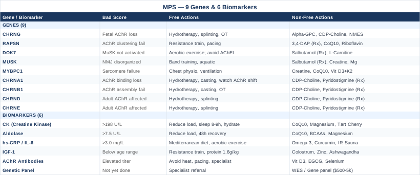
What the Genetics of Multiple Pterygium Syndrome Actually Tell You
MPS is almost entirely explained by disruptions in the genes governing how the neuromuscular junction forms and functions during fetal life. When those genes fail, fetal movement is impaired — and since normal movement is required for normal joint and connective tissue development, the result is the characteristic pattern of pterygia and contractures. The nine genes below each disrupt a different step in this chain. For each one, the practical question is not "can I change the mutation?" (you cannot), but rather: which downstream pathway is disrupted, and what can be done to support it?
CHRNG: The Most Common Driver of Escobar Syndrome
What the gene does: CHRNG encodes the gamma subunit of the fetal nicotinic acetylcholine receptor (AChR) — the receptor on fetal muscle fibers that responds to acetylcholine released by motor neurons. In adults, this gamma subunit is eventually replaced by an epsilon subunit, but during fetal development, the gamma-containing receptor is essential for muscle contraction and movement.
What happens when it is mutated: Loss-of-function mutations in CHRNG impair or eliminate fetal AChR function. Without effective signaling at the NMJ, fetal muscles cannot contract, fetal movement is dramatically reduced (fetal akinesia), and the cascade of contractures and pterygia follows. CHRNG mutations are the most frequently identified cause of Escobar syndrome — the milder, non-lethal form of MPS. A landmark study by Morgan et al. (2006), published in the American Journal of Human Genetics, confirmed CHRNG as the primary gene following linkage analysis and sequencing in affected families.
If the gene is mutated — the plan without supplements:
The non-pharmacological priority is maximizing neuromuscular engagement through structured physical rehabilitation:
- Aquatic physical therapy (hydrotherapy): Three to four sessions per week, 30–45 minutes each. Buoyancy reduces load on contracted joints while allowing greater range of motion. Evidence for hydrotherapy in joint contracture management is strong across analogous conditions. - Static progressive splinting: Custom splints applied at near-maximum tolerable stretch for 30–60 minutes daily. Most effective when started early and maintained consistently. - Serial casting: For severe contractures, applied by an orthopaedic specialist in progressive increments over several weeks. - Occupational therapy: Targeted at grip, fine motor skills, and adaptive strategies for daily tasks.
If the gene is mutated — the plan with supplements and equipment:
Supporting the cholinergic pathway nutritionally may offer marginal benefit in terms of neuromuscular signaling efficiency. Evidence is extrapolated from related conditions rather than CHRNG-MPS specifically:
- Alpha-GPC (alpha-glycerylphosphorylcholine): 300–600 mg daily, taken in the morning. A direct acetylcholine precursor that supports neuromuscular transmission where some receptor function remains. Side effects: mild gastrointestinal discomfort at higher doses; rare reports of headache. - CDP-Choline (Citicoline): 500 mg twice daily. Provides both choline and cytidine for acetylcholine and phospholipid synthesis. Well-tolerated. No cycling required at standard doses. - Pantothenic Acid (Vitamin B5): 500 mg daily. Required for acetyl-CoA synthesis, which is a building block for acetylcholine production. Minimal side effects. - Neuromuscular electrical stimulation (NMES): Applied to affected muscle groups, 20-minute sessions, three to five times per week. NMES drives muscle contractions in the presence of impaired voluntary signaling. Consult a physiotherapist for appropriate frequency and intensity parameters. - Huperzine A: 50–100 mcg, used carefully. An acetylcholinesterase inhibitor that increases synaptic acetylcholine availability. Cycle strictly: 2 weeks on, 2 weeks off to prevent receptor desensitization. Side effects: nausea and bradycardia at higher doses; avoid in those on other cholinergic medications.
RAPSN: The Scaffolding Gene
What the gene does: RAPSN encodes rapsyn, a protein that clusters and anchors nicotinic AChRs at the neuromuscular junction. Without it, AChRs cannot be concentrated at the synaptic membrane efficiently, and NMJ transmission becomes unreliable even when the receptors themselves are structurally normal.
What happens when it is mutated: RAPSN mutations are classically associated with congenital myasthenic syndrome, but they also appear in the MPS spectrum — particularly in presentations combining pterygia with myasthenic weakness. Even a modest reduction in rapsyn function significantly reduces the density of functional AChRs at the synapse.
If the gene is mutated — the plan without supplements:
- Resistance training at submaximal intensities: Low-load, high-repetition protocols three times per week to build neuromuscular efficiency without pushing NMJ circuits to fatigue. - Pacing strategies: Work-to-rest ratios of approximately 1:2 during physical tasks to avoid cumulative NMJ fatigue. - Ergonomic and positioning support: Adaptive seating, joint braces, and environmental modifications to reduce unnecessary muscular load throughout the day.
If the gene is mutated — the plan with supplements and equipment:
- 3,4-Diaminopyridine (3,4-DAP): A prescription medication used in congenital myasthenic syndromes including RAPSN-related cases. It prolongs the nerve terminal action potential, increasing acetylcholine release per signal. Requires medical supervision. Typical dosing: 10–80 mg/day divided into three to four doses. Side effects: perioral and digital tingling, gastrointestinal upset; cardiac monitoring recommended. - Riboflavin (Vitamin B2): 400 mg daily. Supports mitochondrial function in muscle cells and has shown modest benefit in some congenital myasthenic contexts. - CoQ10: 200–400 mg daily with a fat-containing meal. Supports mitochondrial energy production in muscle cells. Generally well-tolerated; fat-soluble formulations improve absorption significantly.
DOK7: The Activator Gene
What the gene does: DOK7 encodes an adapter protein that activates MuSK (Muscle Specific Kinase) — a receptor on the muscle fiber surface that is essential for NMJ formation and maintenance. DOK7 is the upstream signal that tells MuSK to begin organizing the synapse; without it, AChRs cannot cluster properly regardless of receptor availability.
What happens when it is mutated: DOK7 mutations cause a form of congenital myasthenic syndrome with limb-girdle pattern weakness — and in the fetal period, the kind of movement impairment that generates MPS features. One important clinical distinction: patients with DOK7-related disease often respond poorly or even worsen with acetylcholinesterase inhibitors (like pyridostigmine), which are commonly used in other myasthenic conditions. Beeson et al. identified in a landmark Science paper that beta-agonists like salbutamol produce significant clinical improvement in DOK7 patients — a finding that substantially changed management for this genotype.
If the gene is mutated — the plan without supplements:
- Avoid cholinesterase inhibitors unless a neuromuscular specialist with DOK7 experience specifically directs otherwise — they can worsen function in this genotype. - Consistent aerobic conditioning: Low to moderate intensity walking, cycling, or swimming to maintain cardiovascular and muscular baseline without triggering NMJ fatigue. - Targeted mobility work: DOK7 disease commonly weakens hip flexors and shoulder girdle; specific physiotherapy protocols for these regions prevent secondary contracture.
If the gene is mutated — the plan with supplements and equipment:
- Salbutamol (Albuterol): 4–16 mg/day orally (prescription required). Clinical trials and case series have consistently demonstrated significant strength and endurance improvements in DOK7-CMS with oral salbutamol. This is a prescription beta-2 agonist and must be used under medical supervision. Side effects: tremor, tachycardia, hypokalemia; monitor potassium levels during use. - Ephedrine: 25–75 mg/day (where legally available and under medical oversight). An alternative sympathomimetic shown to benefit some congenital myasthenic subtypes. Side effects: hypertension, tachycardia, anxiety; use with caution. - L-Carnitine: 1–2 g/day with meals. Supports fatty acid utilization in muscle mitochondria. Generally well-tolerated; mild gastrointestinal effects possible at higher doses.
MUSK: The Organizing Kinase
What the gene does: MuSK receives the DOK7 signal and orchestrates the clustering and stabilization of AChRs at the NMJ. It is also directly activated by agrin, a nerve-derived signal. Its activity is essential throughout both fetal NMJ formation and ongoing NMJ maintenance in adults.
What happens when it is mutated: MUSK mutations produce a CMS/MPS phenotype closely paralleling DOK7 disease — significant NMJ dysfunction, limb-girdle weakness, and fetal akinesia in utero. Management closely follows the DOK7 model: beta-agonists tend to outperform cholinesterase inhibitors, which may worsen symptoms.
If the gene is mutated — the plan without supplements:
- Resistance band training for proximal muscle groups: Three times weekly, 20–30 minutes per session, focusing on shoulder and hip girdle. - Aquatic exercise: Full-body, joint-friendly conditioning that maintains muscle engagement without high-impact loading. - Sleep positioning: Positioning pillows and orthotics during rest to prevent overnight contracture progression at affected joints.
If the gene is mutated — the plan with supplements and equipment:
- Salbutamol or Ephedrine: Same protocol as DOK7, under medical supervision. - Creatine monohydrate: 3–5 g/day. Supports ATP resynthesis in muscles experiencing metabolic stress from impaired NMJ efficiency. No loading phase required; ensure adequate hydration. No cycling required for long-term maintenance dosing. - Magnesium glycinate: 300–400 mg/day at night. Magnesium modulates NMJ function and muscle membrane excitability. Well-tolerated; mild laxative effect at higher doses.
MYBPC1: The Gene Behind the Lethal Form
What the gene does: MYBPC1 encodes the slow-twitch myosin binding protein C — a structural protein within the sarcomere (the contractile unit of muscle fiber). It regulates the interaction between myosin and actin, affecting the force and speed of muscle contraction at the most fundamental level.
What happens when it is mutated: MYBPC1 mutations underlie the lethal form of multiple pterygium syndrome, characterized by hydrops fetalis, severe fetal akinesia, and typically perinatal death. In rare survivors, profound hypotonia and weakness dominate the clinical picture. Unlike the AChR-related mutations, this is a sarcomeric failure rather than a signaling failure — the contractile machinery itself is structurally compromised.
If the gene is mutated — the plan without supplements:
- Multidisciplinary rehabilitation from infancy: Speech therapy, physical therapy, and respiratory support are the primary pillars in those who survive. - Chest physiotherapy: Manual techniques to assist with secretion clearance and prevent respiratory complications, performed daily or as directed by a respiratory therapist. - Mechanical ventilation and respiratory support: Many severe MYBPC1 survivors require at least intermittent ventilatory assistance, particularly during sleep.
If the gene is mutated — the plan with supplements and equipment:
- Creatine monohydrate: 3–5 g/day. Evidence from sarcomeric myopathy and Duchenne muscular dystrophy research suggests creatine helps maintain ATP availability in mechanically impaired muscle. Begin at 2 g/day and titrate up over two weeks. - CoQ10: 200–400 mg/day with meals. Supports mitochondrial energy production; beneficial across conditions of impaired muscle metabolism. - Vitamin D3 with K2: 2,000–5,000 IU D3 daily paired with 100–200 mcg K2 (MK-7 form). Vitamin D plays a direct role in muscle fiber development, immune regulation, and calcium handling in muscle. Test serum 25(OH)D first; target 40–70 ng/mL.
CHRNA1, CHRNB1, CHRND, and CHRNE: The Other Receptor Subunit Genes
These four genes encode the alpha, beta, delta, and epsilon subunits of the nicotinic acetylcholine receptor, respectively. While CHRNG provides the fetal-specific gamma subunit, mutations in these other subunits affect the structural and functional integrity of the AChR complex across development.
CHRNA1 mutations disrupt the ligand-binding domain of the AChR — where acetylcholine physically attaches. Even subtle changes here significantly alter ion channel kinetics. CHRNB1 mutations affect the structural assembly of the AChR pentamer; without the correct beta subunit, functional receptor complexes cannot form efficiently. CHRND (delta) and CHRNE (epsilon) mutations affect the adult AChR isoform more strongly than the fetal one — meaning that symptoms may actually shift as the child grows and the developmental gamma-to-epsilon subunit transition occurs. In some cases, this transition brings improvement; in others, it introduces a new symptom phase that needs distinct management.
If any of these genes are mutated — the plan without supplements:
The same core approach applies as with CHRNG: hydrotherapy, serial casting, adaptive splinting, and occupational therapy. A critical additional step is monitoring for changes in neuromuscular function specifically during the fetal-to-adult receptor subunit transition in early childhood, since this is when the clinical picture may shift and rehabilitation needs should be reassessed.
If any of these genes are mutated — the plan with supplements and equipment:
- CDP-Choline: 500 mg twice daily. Same rationale as CHRNG. - Pyridostigmine (for CHRNA1, CHRNB1, CHRND, CHRNE cases without DOK7/MUSK involvement): This acetylcholinesterase inhibitor can improve NMJ transmission when the receptor is structurally present but underperforming. Prescription required; medical supervision essential. Typical starting dose: 30–60 mg three to four times daily. Side effects: cholinergic excess effects including bradycardia, excessive secretions, and gastrointestinal cramping; dose-dependent and manageable with titration. - Omega-3 fatty acids (EPA and DHA): 2–3 g combined daily. Supports neuronal membrane fluidity and may enhance receptor function in the phospholipid-rich synaptic membrane environment.
Epigenetic Considerations Across All MPS Genes
The mutations driving MPS are heritable and structural — you cannot undo them. But epigenetic factors can influence how severely those mutations express, and this is an area where intervention has real leverage.
DNA methylation of gene promoter regions in CHRN genes can reduce expression even in the absence of coding mutations; adequate methyl donor intake (folate, methylcobalamin, betaine) supports healthy methylation patterns that keep gene expression from being further suppressed. Exercise-induced epigenetic changes in muscle tissue are well-documented: regular physical activity increases histone acetylation around genes involved in NMJ maintenance and muscle protein synthesis, effectively boosting compensatory expression from the unaffected allele or supporting pathway genes. For the fetal development period specifically, maternal nutrition — adequate choline, folate, and vitamin D during pregnancy — appears to influence how severely affected genes express in the developing fetus.
For those already living with MPS: consistent physical activity, optimizing B12, folate, and choline intake, and avoiding methylation-disrupting factors like chronic alcohol use or severe nutritional deficiency represent tangible, evidence-based epigenetic levers — even if their effect sizes are modest compared to the primary genetic disruption.
6 Biomarkers Worth Tracking Over Time
Biomarkers do not diagnose MPS — genetic testing does that. But once a diagnosis is established, biomarkers become an objective monitoring layer: a way to track what is happening physiologically, catch developing complications early, and gauge how the body is responding to rehabilitation and supplementation. The six below are the most clinically meaningful for someone navigating MPS on an ongoing basis.
1. Creatine Kinase (CK): The Muscle Stress Indicator
Why it matters: CK is the primary blood marker of muscle cell damage and turnover. In neuromuscular conditions underlying MPS — particularly those with sarcomeric involvement like MYBPC1 — CK can be elevated when muscle fibers are under excessive stress, inadequately innervated, or undergoing subclinical degeneration. Tracking CK over time reveals whether muscle tissue is stable, deteriorating, or responding to rehabilitative work.
How to measure it: Standard serum CK panel, available through any primary care physician. Cost range: $20–$60, often covered by insurance when neuromuscular disease is documented. Normal range is approximately 22–198 U/L in adults (varies by sex and lab). CK-MM (the muscle-specific isoform) can be separately quantified if total CK is abnormal, providing more precise insight.
If the score is elevated — the plan without supplements:
- Reduce training intensity if exercise load has recently increased. - Increase sleep duration to prioritize muscle recovery — 8–9 hours minimum. - Assess hydration status: dehydration independently elevates CK by concentrating the enzyme in serum. - Review any recent medication changes — statins, corticosteroids, and several other commonly used drugs elevate CK as a direct side effect.
If the score is elevated — the plan with supplements and equipment:
- CoQ10: 200–400 mg daily with a fatty meal. Supports mitochondrial function and has been shown in multiple studies to reduce exercise-induced CK elevation. Take in the ubiquinol form for better absorption over 50 years of age. - Magnesium glycinate: 300–400 mg at night. Magnesium deficiency compromises muscle cell membrane integrity and can independently elevate CK. - Tart cherry extract: 480 mg standardized extract or 30 ml concentrate twice daily. Well-studied for reducing muscle damage markers including CK, particularly in the recovery window after physical activity. Cycling: Can be used continuously, though most evidence comes from 7–14 day periods around intensive physical work.
2. Aldolase: The Companion to CK
Why it matters: Aldolase is a glycolytic enzyme found in muscle and liver that rises when muscle tissue is stressed or damaged. It often moves in parallel with CK and serves as a useful confirmatory marker, particularly when CK results are ambiguous or disproportionate to the clinical picture. In conditions where muscle cell integrity is chronically challenged — as in NMJ-related MPS — aldolase provides an additional metabolic window.
How to measure it: Serum aldolase test, typically ordered alongside a CK panel in neuromuscular workup. Cost: $30–$80. Normal adult range: 1.0–7.5 U/L. Note that liver disease also elevates aldolase, so hepatic causes should be ruled out when both aldolase and liver enzymes are abnormal.
If the score is elevated — the plan without supplements:
- Evaluate total physical load relative to recovery capacity and adjust to sustainable levels. - Enforce 48-hour minimum recovery between sessions targeting the same muscle groups. - Rule out hepatic involvement with a basic metabolic panel.
If the score is elevated — the plan with supplements:
- Same core protocol as for elevated CK (CoQ10, magnesium, tart cherry). - Branched-chain amino acids (BCAAs): 5–10 g before and after exercise. BCAAs reduce muscle protein catabolism during training and have been shown to lower muscle damage markers in the post-exercise recovery window. Side effects: minimal; mild gastrointestinal discomfort at doses above 15 g.
3. High-Sensitivity CRP and Interleukin-6: The Inflammation Panel
Why it matters: Chronic low-grade inflammation is an increasingly recognized driver of symptom burden in neuromuscular and connective tissue conditions. High-sensitivity CRP (hs-CRP) is the most sensitive routine marker of systemic inflammation; IL-6 is one of the primary cytokines that drives CRP production and is a direct mediator of pain signaling and fatigue. In MPS, elevated inflammatory markers may reflect ongoing tissue remodeling around contracted joints, secondary inflammatory processes, or intercurrent infections that disproportionately affect those with reduced mobility and pulmonary reserve.
How to measure it: hs-CRP is a standard lab test costing $20–$50. IL-6 requires a separate order and is slightly less common but available at most reference laboratories ($50–$150). For hs-CRP, optimal values are below 1.0 mg/L; values above 3.0 mg/L indicate high systemic inflammatory risk. Peter Attia recommends tracking hs-CRP as a key longevity biomarker precisely because it captures inflammatory burden that standard metabolic panels miss.
If the score is elevated — the plan without supplements:
- Anti-inflammatory dietary pattern: Mediterranean-style eating emphasizing olive oil, oily fish, leafy greens, and legumes while reducing refined carbohydrates and industrially processed seed oils. - Sleep optimization: Even one night of poor sleep meaningfully elevates IL-6. Consistent sleep-wake timing matters as much as total duration. - Moderate aerobic exercise: 30 minutes most days at moderate intensity reduces hs-CRP by a clinically meaningful amount over 8–12 weeks in most populations — more consistently than most pharmacological anti-inflammatory agents at this level.
If the score is elevated — the plan with supplements and equipment:
- Omega-3 fatty acids (EPA + DHA): 2–4 g daily total. Consistently shown across multiple systematic reviews and meta-analyses to produce dose-dependent reductions in both hs-CRP and IL-6. Take with meals containing fat; at higher doses, mild blood-thinning effects are possible — relevant for those on anticoagulants. - Curcumin (BCM-95 or Meriva bioavailable formulation): 500–1,000 mg daily. Inhibits NF-κB — a primary driver of inflammatory cytokine production including IL-6. Standard curcumin powder has very poor bioavailability; formulation matters. Side effects: minimal at these doses; high-dose curcumin may interact with blood thinners. - Infrared sauna: 20–30 minutes, three to four times weekly. Emerging evidence supports infrared sauna use for reducing systemic inflammatory markers; additionally, the heat exposure may provide therapeutic benefit for joint stiffness and tissue pliability directly relevant to MPS.
4. IGF-1: The Anabolic Signal Marker
Why it matters: IGF-1 (Insulin-like Growth Factor 1) is the primary mediator of muscle protein synthesis and tissue growth, and it serves as an indirect indicator of growth hormone status. In children and adults with MPS — particularly those with short stature and muscle hypotrophy — IGF-1 levels reveal whether anabolic signaling is sufficient to support the muscle growth and repair that rehabilitation is working to stimulate. Low IGF-1 in the context of MPS means the body is not effectively driving the healing and adaptation that physical therapy attempts to generate.
How to measure it: Standard serum IGF-1, available through any endocrine-aware primary care physician or specialist. Cost: $50–$150. Interpretation requires age- and sex-matched reference ranges, as IGF-1 peaks in adolescence and declines significantly with age. Thomas Dayspring and Peter Attia both include IGF-1 in routine longevity panels for its role in muscle maintenance and metabolic signaling.
If the score is low — the plan without supplements:
- Progressive resistance training: The most potent natural IGF-1 stimulant available. Even moderate-intensity resistance protocols reliably increase both systemic and local muscle IGF-1 (mechano-growth factor isoform). - Adequate protein intake: 1.6–2.2 g/kg body weight daily, prioritizing leucine-rich sources (eggs, fish, legumes) which drive mTOR signaling and downstream IGF-1 production. - Sleep optimization: Growth hormone — which drives IGF-1 — is released primarily during slow-wave sleep. A consistent sleep window aligned with circadian rhythm (ideally 10 pm–6 am) maximizes pulsatile growth hormone secretion.
If the score is low — the plan with supplements and equipment:
- Colostrum: 10–20 g/day. Contains IGF-1, IGF-2, and multiple growth factors that may modestly support IGF-1 signaling. Evidence is still early-stage but the biological rationale is sound for muscle-support conditions. - Zinc bisglycinate: 15–30 mg daily with food. Zinc is required for growth hormone receptor function and downstream IGF-1 production. Cycling note: take 1–2 mg of copper alongside any long-term zinc supplementation to prevent copper depletion. - Ashwagandha (KSM-66 extract): 600 mg daily. Multiple RCTs in adults have demonstrated significant improvements in muscle recovery, strength, and modest IGF-1 increases. Side effects: sedation at high doses; avoid during pregnancy.
5. Acetylcholine Receptor Antibodies: Ruling Out an Autoimmune Layer
Why it matters: In some MPS presentations — particularly those with a myasthenic clinical picture — autoimmune antibodies targeting AChRs may coexist with the genetic disruption. This effectively adds an acquired autoimmune component on top of the underlying gene mutation, changing the management picture significantly. AChR antibodies open additional therapeutic options (immunosuppression, intravenous immunoglobulin) that would not otherwise be considered.
How to measure it: Serum AChR antibody panel covering binding, blocking, and modulating antibodies. Available at specialty reference laboratories; cost: $100–$400. Positive results warrant urgent referral to a neuromuscular specialist with autoimmune experience.
If the score is elevated — the plan without supplements:
- Referral to a neuromuscular neurologist for evaluation and management of the myasthenic component. - Avoid high ambient temperatures — heat worsens AChR antibody-mediated NMJ dysfunction and can precipitate myasthenic crisis. - Implement pacing strategies and structured rest periods to manage fatigable weakness between medical treatment decisions.
If the score is elevated — the plan with supplements and equipment:
- Vitamin D3: 4,000–5,000 IU daily, paired with K2 (100–200 mcg MK-7 form). Low vitamin D is consistently associated with worse autoimmune outcomes across multiple conditions. Target serum 25(OH)D of 50–70 ng/mL; monitor every three to six months. - EGCG (green tea extract): 400–800 mg standardized EGCG daily. Demonstrates immunomodulatory properties relevant to autoimmune conditions in emerging research. Caution: hepatic stress reported at doses above 1,200 mg EGCG/day; stay within the recommended range. - Selenium as selenomethionine: 100–200 mcg daily. Supports regulatory T-cell function, which is relevant in autoimmune presentations. Do not exceed 400 mcg/day — selenium toxicity (selenosis) is real at high doses.
6. Comprehensive Genetic Panel: The Foundation Layer
Why it matters: Genetic testing is not a biomarker in the traditional sense, but it functions as the foundational diagnostic layer on which all other monitoring and intervention decisions rest. Knowing exactly which gene is mutated — and whether the mutation is homozygous or heterozygous — determines which biochemical pathways are most compromised, which medications are likely to help or harm, what the likely trajectory looks like over time, and which family members face elevated risk.
How to measure it: Whole exome sequencing (WES) or a targeted neuromuscular gene panel through a clinical genetics service. Cost: $500–$5,000 depending on panel scope, insurance coverage, and country. Major accredited genetics laboratories include GeneDx, Ambry Genetics, and Blueprint Genetics. Many insurance plans cover WES when appropriate clinical criteria are documented.
Once the gene is identified — the plan:
Genetic results do not alter the underlying mutation but guide every subsequent decision. Share results with a neuromuscular specialist familiar with congenital myasthenic syndromes and MPS. Determine whether the mutation affects fetal-only AChR (CHRNG) or adult AChR subunits, as this shapes expected symptom trajectory over childhood. Use the specific gene finding to select supplement and medication approaches from the preceding sections. Consider genetic counseling for family planning, as most forms of MPS follow autosomal recessive inheritance patterns.
Stretching, Flexibility, and Neuromuscular Adaptation: What the Science Actually Says
Andrew Huberman, a neuroscientist at Stanford University, has devoted substantial podcast content to the science of flexibility, joint mobility, and neuromuscular adaptation — topics that sit at the center of what people with MPS contend with every day. His approach is useful here not because it was designed for MPS specifically, but because it explains flexibility at the neural level rather than the purely mechanical one — which aligns precisely with the NMJ-centered biology underlying this condition. The following ten insights are drawn from his episodes and the research they reference, selected for direct relevance to the joint contracture management challenges of MPS.
1. Flexibility Is Primarily a Nervous System Phenomenon
The principal limiter of range of motion is not muscle fiber length — it is neural inhibition. The nervous system maintains a protective "safety margin" that prevents muscles from stretching to their mechanical limit. Static stretching over time works by gradually convincing the nervous system to tolerate a greater range, not by physically lengthening the muscle tissue. This reframing matters for MPS because contracture management is, at its core, a process of reprogramming neural thresholds — not just pulling at tissues.
2. Thirty to Sixty Seconds Per Hold Is the Minimum Effective Duration
Research reviewed by Huberman consistently shows that 30–60 second holds are the minimum duration needed to induce lasting changes in stretch tolerance. Shorter holds provide temporary mobility but do not drive meaningful neural adaptation. For MPS contractures: 60–90 second holds at the point of comfortable discomfort, repeated three to four times per affected joint, represent the evidence-based floor.
3. Daily Brief Sessions Beat Weekly Long Sessions
Five minutes of daily stretching produces greater cumulative flexibility gains than 35 minutes once per week. The nervous system adapts through repetition frequency, not volume spikes. For MPS management: 5–10 minutes of targeted stretch work for each major affected joint, performed every day without exception, outperforms intensive but infrequent sessions.
4. Tissue Temperature Significantly Affects Stretch Effectiveness
Connective tissue and muscle respond measurably better to stretching when pre-warmed — either through light exercise, a hot shower, or external heat packs applied for five to ten minutes before stretching. Warm collagen is significantly more extensible than cold collagen. For MPS: pre-stretch hydrotherapy or a brief warm bath before contracture work can meaningfully amplify each session's effectiveness at zero additional cost.
5. PNF Stretching Outperforms Passive Static Methods
Proprioceptive Neuromuscular Facilitation (PNF) — where a muscle is contracted against resistance for 5–10 seconds, then immediately relaxed and passively stretched — exploits autogenic inhibition to achieve greater range than static stretching alone. Studies referenced by Huberman show PNF produces up to two to three times greater acute flexibility gains. For those with sufficient muscle function in MPS, PNF protocols guided by a trained physiotherapist can substantially accelerate contracture management progress.
6. Slow Exhales Reduce Neural Resistance to Stretch
Deliberate slow exhalation during a stretch reduces sympathetic nervous system tone and activates the parasympathetic system, lowering the neural "resistance" signal the body sends against being stretched. A 4-second inhale followed by a slow 6–8 second exhale at peak stretch is the recommended protocol — immediately applicable and free to implement in any stretching session.
7. Cold Exposure After Stretching Can Blunt Adaptation
Counterintuitively, cold water immersion immediately after a flexibility session may slow the nervous system's incorporation of the new range of motion. Huberman recommends avoiding cold exposure for at least 4–6 hours after stretch-focused training when the goal is long-term range improvement. For MPS rehabilitation programs that combine contrast therapy and stretching, the sequence matters.
8. Sleep Is When Neuromuscular Adaptations Are Consolidated
Motor adaptations acquired during waking hours are consolidated during slow-wave and REM sleep. Sleep restriction meaningfully blunts how much of the day's neuromuscular work is retained. For MPS: protecting sleep duration and quality is not a soft wellness recommendation — it is mechanistically central to whether rehabilitation gains are retained or lost.
9. Active Use of Gained Range Locks It In
Gaining passive range of motion through stretching is only part of the adaptation; the nervous system needs to actively use the new range through strengthening exercises. Research cited by Huberman shows that training strength through a newly expanded range of motion anchors the neural patterns far more durably than stretching alone. Physical therapy for MPS should combine passive contracture stretching with active strengthening exercises through the expanded range immediately afterward.
10. Consistency Sustains Gains; Stopping Reverses Them Rapidly
Flexibility gains are cumulative but not permanent. Research shows that without maintenance, most gains are lost within four to eight weeks of stopping a stretching routine. For MPS, contracture management must be structured as a permanent daily practice rather than a finite course of rehabilitation. Even five minutes daily is sufficient to sustain previously earned range; stopping is not a plateau — it is a reversal.
Complementary Approaches with Clinical Evidence
The following approaches were selected for two reasons: genuine relevance to the physical challenges MPS creates, and meaningful human clinical evidence behind them. This is not an exhaustive list of everything that might help — it is a curated selection of what has enough evidence to be worth seriously considering.
Yoga for Joint Mobility and Contracture Management
Yoga offers a structured, adaptable framework for improving range of motion, joint proprioception, and movement patterning — all directly relevant to the contractures and pterygia that define MPS. Unlike generic passive stretching, yoga incorporates intentional breath control alongside progressive joint loading in supported positions, engaging the neural flexibility mechanisms discussed in the Huberman section above in a systematic way.
Multiple systematic reviews and randomized trials in arthritis, fibromyalgia, and neuromuscular populations have demonstrated that yoga-based programs improve range of motion and reduce pain-related disability in joint contracture conditions. While MPS-specific yoga trials do not exist (the condition is too rare to generate condition-specific RCTs), the underlying mechanisms — neuromotor adaptation and connective tissue response to gentle progressive load — are well-established and directly applicable.
For realistic application in MPS: Begin with chair yoga or supine yoga styles, which provide full joint support and avoid loading contracted joints in ways that could strain them. Work with a certified adaptive yoga instructor who has experience modifying for physical limitations. Three to four sessions per week of 20–40 minutes each is a reasonable starting target. Focus on slow, breath-accompanied transitions rather than achieving specific posture endpoints. Progress gradually under guidance and in consultation with the treating physiotherapist.
Massage Therapy for Soft Tissue and Contracture Support
Manual massage addresses the soft tissue components of MPS — contracted muscles, shortened fasciae, and bound connective tissue around affected joints. Regular therapeutic massage does not reverse the gene mutation, but it maintains tissue pliability, reduces secondary pain, supports local circulation, and may slow progressive soft tissue binding by reducing adhesion formation between tissue layers.
Clinical evidence supports massage therapy for contracture prevention and management in analogous conditions. A randomized controlled trial in scar and burn contractures — one of the closest mechanistic models for pterygia soft tissue management — showed that twice-weekly massage for 12 weeks significantly reduced tissue tightness and improved joint range of motion compared to standard care alone. While MPS differs in etiology, the connective tissue dynamics that massage addresses are directly comparable.
For realistic application: Weekly or twice-weekly sessions with a licensed massage therapist experienced in contracture management are the starting framework. Avoid aggressive deep tissue techniques directly over active pterygia or around recent surgical sites. Myofascial release techniques are particularly relevant and can be taught for between-session self-application using a foam roller or therapy ball. Sessions of 30–60 minutes; communicate clearly with the therapist about which joints are most restricted and whether any areas are postsurgical.
Breathing-Based Therapies for Respiratory and Autonomic Support
In moderate to severe MPS presentations, respiratory muscles may be involved, and chest wall compliance can be reduced by thoracic pterygia or coexisting scoliosis. Breathing-based therapies — including respiratory physiotherapy, diaphragmatic retraining, and inspiratory muscle training (IMT) — address both the mechanical and autonomic dimensions of respiratory compromise.
IMT using resistive breathing devices has been studied in neuromuscular conditions including congenital myopathies, with systematic reviews finding significant improvements in respiratory muscle strength and quality of life measures. While direct MPS evidence is absent, the mechanism is applicable wherever respiratory muscle weakness or reduced chest wall compliance is present — both features that can appear in the MPS spectrum.
For realistic application: Begin with assessment by a respiratory physiotherapist to establish baseline maximal inspiratory pressure (MIP). IMT devices such as Threshold IMT are trained at 30–50% of MIP, 30 breaths per session, once or twice daily, for a minimum of 8 weeks before reassessing. Diaphragmatic breathing exercises — 10 slow breaths with conscious belly expansion, performed five minutes daily — support autonomic regulation and can be practiced independently at no cost. This is a foundational tool for those with any respiratory involvement in MPS.
Biofeedback for Neuromuscular Reeducation
Electromyography (EMG) biofeedback provides real-time visual or auditory feedback of muscle activation, allowing patients to consciously modulate muscle tension and motor recruitment patterns. In MPS — where both insufficient voluntary activation and abnormal compensatory co-contraction patterns can coexist — neuromuscular biofeedback enables the kind of targeted motor relearning that conventional exercise programs cannot easily achieve.
Reviews of EMG biofeedback in neuromuscular rehabilitation have found consistent evidence for effectiveness in improving voluntary motor control in patients with upper and lower motor neuron disorders. Applications in children with cerebral palsy — another congenital condition affecting voluntary motor control — have been particularly well-studied, offering a clinically relevant model for the MPS rehabilitation context.
For realistic application: Sessions are conducted by a physiotherapist trained in biofeedback, with surface EMG sensors placed over target muscles. Sessions run 30–45 minutes, one to two times weekly, over an 8–12 week initial course. Increasingly, home biofeedback devices with wearable sensors and companion apps allow between-session practice to reinforce gains. For MPS, the most useful targets are typically muscles around the knees and in the cervical spine region — both common contracture sites — as well as any muscle groups that have been surgically released and require retraining.
Conclusion
Multiple Pterygium Syndrome is rare and genetically complex, but it is far from opaque. The biological pathways involved — neuromuscular junction signaling, acetylcholine receptor function, sarcomeric structure — are increasingly well understood, and that understanding translates into specific, actionable guidance. Knowing which of the nine implicated genes is involved shapes which supports make sense. Tracking the six biomarkers outlined here creates an objective baseline that makes medical conversations more productive and monitoring more meaningful.
No supplement protocol, yoga practice, or biofeedback course changes the underlying mutation. But they can meaningfully support the affected pathways, improve functional capacity, reduce secondary complications, and slow the accumulation of additional impairment over time. Pursued carefully, with appropriate medical oversight and realistic expectations, these strategies compound in ways that matter.
The next smart step is to confirm that genetic testing is complete if it has not already been done, to request the relevant biomarker panel from a physician who understands neuromuscular conditions, and to build a rehabilitation team that includes a physiotherapist with neuromuscular disease experience. Daily stretching, adequate protein, targeted supplementation, and regular lab monitoring may seem like small steps — but they are the kind that accumulate into meaningful gains over months and years of consistent effort.
Musculoskeletal: Joint Conditions Muscle Conditions Spine Conditions
Neurological: Nerve Conditions
Autoimmune: Connective Tissue Conditions