This article contains AI generated content.
· UpdatedMuscular Dystrophy Genes and Biomarkers: 8 Genes and 6 Biomarkers to Track
Introduction
Living with muscular dystrophy — or caring for someone who does — means navigating a condition where the details matter enormously. The word "muscular dystrophy" covers dozens of genetically distinct diseases with different speeds of progression, different organ involvements, and different responses to the same interventions. Yet the advice most people receive tends to stay at the surface: manage symptoms, avoid overexertion, see a physiotherapist, take corticosteroids if indicated. That is not bad advice, but it is incomplete in ways that increasingly do not have to be accepted.
What genetics and biomarker science now make possible is a more precise map of what is actually happening in an individual's body. The specific gene driving a given form of MD shapes which proteins are absent or dysfunctional, which compensatory pathways are available, and which tissues beyond skeletal muscle — heart, lungs, brain — are likely to be involved. Measuring the downstream consequences of those abnormalities in blood and functional tests turns an abstract genetic diagnosis into actionable data that can be tracked over time.
Generic lifestyle advice tends to fall short here not because it is wrong but because it does not account for the specific biology involved. Someone carrying a LMNA mutation faces cardiac risk that is separate from the muscle-wasting question. Someone with myotonic dystrophy deals with systemic metabolic disruption that a standard MD management protocol was never designed to address. Precision matters, and it is now increasingly within reach.
This article approaches the topic through two complementary lenses. The primary focus is on six key biomarkers that can be measured, tracked, and meaningfully influenced — looking at what each reveals, how to measure it affordably, and what interventions address a poor result. The second lens covers the eight most clinically significant MD-related genes, what each variant tends to disrupt, and what can realistically be done when a variant is identified. Together, these frameworks offer what generic guidance cannot: a starting point for decisions grounded in your own biology.
6 Biomarkers to Monitor in Muscular Dystrophy
Biomarkers in muscular dystrophy serve two distinct purposes. The first is diagnostic confirmation and subtype differentiation — distinguishing between limb-girdle subtypes, ruling out inflammatory myopathies, or catching early cardiac involvement before symptoms emerge. The second, and often underused, purpose is longitudinal tracking: using the same values measured repeatedly over months and years to detect whether a disease is accelerating, stabilizing, or responding to an intervention. Both purposes matter. The six biomarkers below were chosen for their clinical significance across multiple MD subtypes, their measurability in standard laboratory settings, and the existence of at least some evidence-based actions attached to abnormal results.
Creatine Kinase (CK): The Primary Muscle Damage Signal
Why it matters
Creatine kinase is the most established biomarker in all forms of muscular dystrophy. Released from damaged or leaking muscle fibers, serum CK reflects the ongoing rate of sarcolemmal disruption. In Duchenne Muscular Dystrophy (DMD), CK levels are typically 50 to 100 times the upper limit of normal, often exceeding 10,000 U/L, and can serve as early detection even before symptoms appear — including in infants. In Becker MD, Limb-Girdle subtypes, and dysferlinopathies, values are lower but still substantially elevated. In myotonic dystrophy and some LMNA-related forms, CK elevations are modest, which is diagnostically important.
CK elevation alone does not diagnose a specific subtype, but its magnitude, combined with symptom pattern and genetic context, provides significant clinical information. Longitudinal CK is most useful when interpreted in the context of activity level, since vigorous exertion can transiently raise CK even in healthy individuals.
How to measure it
CK is measured from a standard venous blood draw. It is available at any clinical lab and costs between $15 and $50 out of pocket in most countries. Reference ranges vary by lab and sex: typical normal values are 22–198 U/L for men and 22–170 U/L for women. A result above 500 U/L in the absence of intense exercise warrants further investigation. Results above 3,000 U/L in a non-athlete should prompt immediate neuromuscular evaluation.
If the score is bad, the plan without supplements
The primary free intervention is avoiding eccentric-dominant exercise, which drives the largest CK spikes. Concentric and isometric loading, low-to-moderate intensity aerobic activity, and aquatic exercise all appear to cause less fiber disruption than heavy eccentric protocols. Normalizing sleep — where muscle repair processes peak — is also important. Reducing sustained immobility (which paradoxically increases passive oxidative stress) while avoiding overexertion is the balance to aim for. Daily gentle range-of-motion work, 15–20 minutes, has been shown to preserve function without significantly elevating CK in most MD subtypes.
If the score is bad, the plan with supplements or equipment
Creatine monohydrate is the most studied supplement in MD. A randomized trial published by Tarnopolsky et al. (available via PubMed) found creatine supplementation in boys with DMD improved handgrip strength and lean body mass. Typical protocol: 3–5 g/day continuously, or a loading phase of 20 g/day for 5 days followed by 3–5 g/day maintenance. Side effects are minimal at these doses; mild GI discomfort is occasionally reported. Coenzyme Q10 (100–300 mg/day) supports mitochondrial bioenergetics in muscle tissue, though evidence in MD is largely extrapolated from mitochondrial myopathy research. Whole-body vibration therapy (WBV) using a vibration platform — 30–60 Hz, sessions of 10–15 minutes, 3–5 times per week — has shown potential to stimulate muscle activation with low mechanical load, making it a viable option for people with limited voluntary muscle strength.
Aldolase: Cross-Validation for Active Muscle Breakdown
Why it matters
Aldolase is a glycolytic enzyme that, like CK, is released during muscle fiber necrosis. It is less sensitive and less specific than CK but provides useful cross-validation, particularly in subtypes where CK elevation is modest. In polymyositis and dermatomyositis — conditions occasionally confused with LGMD — aldolase can be disproportionately elevated relative to CK, which helps distinguish inflammatory from dystrophic etiologies. In MD, aldolase tracking is most useful when CK results appear inconsistent with clinical presentation.
How to measure it
Standard blood draw, normal range 1.0–7.5 U/L. Cost is typically $20–$60 out of pocket. It is often bundled into a comprehensive muscle enzyme panel. An aldolase above 10 U/L in the context of proximal muscle weakness and elevated CK strongly supports active muscle damage from a dystrophic process.
If the score is bad, the plan without supplements
The approach parallels CK management: reduce mechanical load on damaged muscle groups, increase recovery intervals, and optimize protein intake. 30–40 g of leucine-rich protein per meal has been shown to maximize muscle protein synthesis signals in individuals with limited muscle mass. Timing protein intake around any physical activity, even gentle activity, appears to improve net muscle anabolism.
If the score is bad, the plan with supplements or equipment
HMB (beta-hydroxy beta-methylbutyrate), a leucine metabolite, has been studied for muscle-preserving effects in catabolic conditions at 3 g/day. Evidence in MD is limited but biologically plausible. Anti-inflammatory omega-3 fatty acids (EPA+DHA, 2–4 g/day from concentrated fish oil) may reduce the inflammatory amplification that worsens CK and aldolase elevation during active fiber necrosis phases. No cycling is required for fish oil at these doses; monitor for anticoagulant interactions if relevant.
NT-proBNP: The Cardiac Stress Warning
Why it matters
Cardiomyopathy is a major cause of death across several MD subtypes — particularly in DMD (dilated cardiomyopathy develops in virtually all patients over time), in LMNA-related MD (which carries disproportionately high arrhythmia and sudden cardiac death risk), and in certain LGMD subtypes. NT-proBNP (N-terminal pro-B-type natriuretic peptide) is released by ventricular myocytes under hemodynamic stress and is the most accessible early signal of subclinical cardiac dysfunction. Waiting for symptoms — dyspnea, edema, palpitations — means waiting until the problem is already significant. NT-proBNP can detect ventricular strain before ejection fraction falls.
According to the NCBI GeneReviews entry for Duchenne Muscular Dystrophy, cardiac surveillance with echocardiography and biomarkers should begin early, particularly after age six in DMD, and continue annually.
How to measure it
Standard blood draw. Normal is typically below 125 pg/mL in adults under 75. Values between 125 and 450 pg/mL indicate early stress; above 450 pg/mL warrants cardiology evaluation. Cost is $30–$80 out of pocket. Measure annually at minimum; every 6 months if values are elevated or if the specific genetic subtype carries cardiac risk.
If the score is bad, the plan without supplements
Sodium restriction (under 2g/day) reduces ventricular preload, one of the primary drivers of BNP elevation. Sleep positioning — elevating the upper body 15–30 degrees — reduces nocturnal volume redistribution that stresses a weakened ventricle. Moderate aerobic activity (if physically possible given muscle function) protects cardiac remodeling pathways; immobility worsens cardiac atrophy in MD. Tracking fluid retention (daily weight, morning) allows early detection of worsening heart failure physiology.
If the score is bad, the plan with supplements or equipment
Taurine (2–6 g/day) has cardiac-protective effects demonstrated in heart failure research and has shown preliminary benefit in DMD animal models. Magnesium glycinate (300–400 mg/day) supports cardiac electrophysiology, relevant particularly for LMNA-related forms where arrhythmia risk is high. Wearable cardiac monitors (Holter-class ECG patches, 2–4 week recordings) can be used outside the hospital to detect arrhythmias that intermittent clinic ECGs miss — particularly relevant for LMNA patients. Coenzyme Q10 at 300 mg/day has moderate evidence for cardiac biomarker improvement in heart failure patients and is low-risk to trial.
Serum Myoglobin: Real-Time Muscle Injury Marker
Why it matters
Myoglobin, the oxygen-storing protein within muscle fibers, is released rapidly after acute muscle injury — faster than CK, which peaks 24–72 hours after damage. Elevated serum myoglobin therefore provides a more immediate window into active muscle breakdown. It is particularly relevant in dysferlinopathies (DYSF mutations), where sarcolemmal repair is impaired and acute injury events are frequent, and in myotonic dystrophy where metabolic dysregulation creates intermittent episodes of enhanced fiber necrosis.
Very high myoglobin (above 5,000 ng/mL) also poses a direct kidney risk through myoglobinuria, making it a safety-relevant biomarker when MD patients undergo surgery, extreme illness, or inadvertent overexertion.
How to measure it
Standard blood draw, measured by immunoassay. Normal range is approximately 17–105 ng/mL (varies by lab and sex). Cost is $25–$60. Urine dipstick testing for myoglobinuria (dark urine with positive blood test in the absence of red cells on microscopy) is a free bedside indicator of significant myoglobinuria and should prompt urgent evaluation.
If the score is bad, the plan without supplements
Aggressive hydration — 2–3 liters of water per day — remains the primary free protective strategy against myoglobin-induced renal damage. Activity modification to eliminate any exercise that produces next-day muscle pain or dark urine is essential. Rest periods after any strenuous activity should be longer than standard recommendations. Tracking urine color provides a daily zero-cost myoglobin proxy.
If the score is bad, the plan with supplements or equipment
N-acetylcysteine (NAC) (600–1800 mg/day in divided doses) has antioxidant and muscle-protective properties and has been studied in contexts of muscle oxidative stress. Cycling protocols are sometimes used: 5 days on, 2 days off, to avoid blunting adaptive responses in individuals who retain meaningful muscle mass. Electrolyte supplementation with sodium, potassium, and magnesium during any physical activity helps maintain fluid balance and reduce secondary cramping that can worsen myoglobin release.
High-Sensitivity CRP and IL-6: The Inflammation Amplifier
Why it matters
Muscle fiber necrosis in MD does not happen in an immunological vacuum. Dying fibers recruit macrophages and neutrophils, triggering a secondary inflammatory response that extends damage beyond the initial mechanical failure. In DMD, NF-κB-driven chronic inflammation is now understood to be a significant amplifier of disease progression — meaning that reducing background inflammation, even partially, may slow the cascade. High-sensitivity CRP (hsCRP) and IL-6 are the most accessible proxies for this systemic inflammatory state.
Peter Attia and Thomas Dayspring both emphasize hsCRP as a routinely actionable inflammatory marker, noting that values chronically above 1–2 mg/L reflect a background inflammatory burden that affects virtually every tissue-repair process in the body. In MD, this is directly relevant to the rate at which residual muscle fibers are damaged.
How to measure it
hsCRP is a standard blood test available at essentially all labs, costing $15–$40. Target is below 1.0 mg/L for optimal tissue-repair conditions; values above 3.0 mg/L indicate significant inflammation. IL-6 testing is available at larger labs, costs $50–$120, and provides additional mechanistic information but is less commonly ordered. Testing every 3–6 months is appropriate in MD contexts.
If the score is bad, the plan without supplements
Dietary changes produce the largest CRP reductions without supplementation. A Mediterranean-pattern diet — rich in olive oil, vegetables, fatty fish, and legumes — consistently reduces hsCRP by 20–40% in clinical trials. Eliminating ultra-processed foods and refined seed oils is the highest-yield free action. Regular low-intensity movement (20–30 minute walks, pool exercises) also reduces inflammatory cytokine burden. Improving sleep quality to 7–9 hours — particularly deep sleep stages where anti-inflammatory cytokines predominate — matters significantly.
If the score is bad, the plan with supplements or equipment
EPA+DHA omega-3s at 2–4 g/day are among the most evidence-backed anti-inflammatory supplements, with consistent hsCRP reductions in randomized trials. Curcumin with piperine (500–1000 mg/day of curcumin combined with 10 mg piperine for absorption) inhibits NF-κB signaling — directly relevant given NF-κB's role in MD inflammation. No cycling required; long-term use appears safe. Resveratrol (150–500 mg/day) activates SIRT1 and has anti-inflammatory effects that have been studied in DMD preclinical models; human evidence is preliminary. Red light therapy / photobiomodulation devices, used 10–20 minutes per session 3–5 times weekly, have emerging evidence for reducing local and systemic inflammatory markers.
Forced Vital Capacity (FVC): The Respiratory Countdown
Why it matters
For many forms of muscular dystrophy — DMD, Bethlem myopathy, some LGMD subtypes, and myotonic dystrophy — respiratory failure is either the leading cause of death or a major determinant of quality of life. FVC (Forced Vital Capacity), measured by spirometry, captures the total volume of air a person can exhale forcefully. As respiratory muscles weaken, FVC declines predictably. An FVC below 50% of predicted value marks a threshold where nocturnal hypoventilation typically begins; below 30%, daytime ventilation support is usually required.
FVC is unlike the blood-based biomarkers in this list — it is a functional measurement — but it belongs here because it is trackable, measurable, and directly actionable, and because many patients are not given sufficiently frequent spirometry to catch early decline.
How to measure it
Spirometry is performed at pulmonology clinics, physiotherapy centers, or pulmonary function labs. Cost is typically $40–$150 out of pocket, and it is usually covered under respiratory care referrals. Home peak flow meters are not a substitute for full spirometry but can provide a free daily trend indicator. FVC should be measured every 6 months in MD patients at any stage, and every 3 months when values fall below 70% of predicted.
If the score is bad, the plan without supplements
Respiratory muscle training — using threshold inspiratory muscle trainers (IMT devices costing $30–$60) — has demonstrated measurable FVC preservation in DMD and other neuromuscular diseases in multiple trials. Protocols typically involve 30 breaths at 30–50% of maximum inspiratory pressure, 5 days per week. Optimizing sleep positioning (semi-reclined at 30 degrees) reduces nocturnal atelectasis. Cough-assist techniques, taught by a physiotherapist and practiced daily, maintain airway clearance capacity.
If the score is bad, the plan with supplements or equipment
Non-invasive ventilation (NIV/BiPAP) initiated at an FVC below 50%, or when overnight pulse oximetry shows sustained desaturations, is the standard evidence-based intervention. This is prescription-based, but knowing the threshold — and tracking toward it — allows timely access rather than reactive crisis initiation. Magnesium (300–400 mg/day) supports respiratory muscle contractility. In some research contexts, theophylline at low doses has shown bronchodilatory benefit, though this requires medical supervision. The EzPAP oscillating positive expiratory pressure device is available without prescription and can help with mucus clearance and alveolar recruitment.
8 Key Genes in Muscular Dystrophy: What They Mean and What You Can Do
Understanding the genetic underpinning of a specific MD diagnosis transforms the clinical picture from "progressive muscle disease" into something with identifiable mechanisms, tissue-specific risks, and — increasingly — specific therapeutic targets. The following eight genes cover the most prevalent forms of MD globally and represent the variants most commonly identified through genetic testing panels.
DMD: The Dystrophin Gene
The DMD gene encodes dystrophin, a large structural protein that links the intracellular cytoskeleton of muscle fibers to the extracellular matrix via the dystrophin-associated protein complex (DAPC). Its role is mechanical: it distributes contractile forces and protects the sarcolemma from shear stress during each contraction cycle. Without functional dystrophin (Duchenne MD) or with truncated dystrophin (Becker MD), repeated contractions cause sarcolemmal tears, calcium influx, fiber necrosis, and ultimately fibrotic replacement.
Plan without supplements: In exon-duplicable mutations amenable to exon skipping therapy (particularly exon 51, 45, and 53 skippable mutations), awareness of FDA-approved exon-skipping drugs (eteplirsen, golodirsen, casimersen) and access to approved gene therapy (delandistrogene moxeparvovec for patients under 4) should be pursued through neuromuscular specialty centers. Physical therapy emphasizing concentric exercise, aquatic therapy, and standing programs is standard of care. Maintaining mobility delays scoliosis onset.
Plan with supplements or equipment: Creatine monohydrate (3–5 g/day), CoQ10 (300 mg/day), and idebenone (300–900 mg/day — particularly for cardiac protection, studied in the DELOS trial) form the most evidence-supported supplement approach in DMD. Corticosteroids remain the pharmacological standard; deflazacort may have a slightly better bone and weight side effect profile than prednisone. Cardiac ACE inhibitors are typically started prophylactically by age 10 in DMD, regardless of current cardiac function.
CAPN3: Calpain-3 and LGMD R1
CAPN3 encodes calpain-3, a calcium-dependent protease that regulates sarcomeric remodeling and is involved in titin regulation during normal muscle use. Its loss leads to failure of the sarcomeric maintenance cycle, causing progressive proximal muscle wasting typically emerging in the second decade of life. LGMD R1 (formerly LGMD2A) is the most common LGMD worldwide.
Plan without supplements: There is no approved gene therapy for CAPN3 as of mid-2025, though multiple programs are in clinical trials. Low-to-moderate aerobic exercise has been shown to preserve function longer than rest in CAPN3-related LGMD. Avoiding prolonged immobilization is essential — disuse atrophy accelerates loss in already-compromised muscle. Physiotherapy focusing on hip and shoulder girdle strengthening (concentric protocols) should be individualized based on current functional level.
Plan with supplements or equipment: Given the calcium dysregulation component, magnesium glycinate (400 mg/day) may help buffer intracellular calcium fluctuations. Creatine monohydrate has shown modest benefit in small LGMD trials. Whole-body vibration training (WBV) provides mechanosensory stimulation with lower voluntary load demands and may preserve motor unit recruitment capacity longer.
DYSF: Dysferlin and Membrane Repair Failure
DYSF encodes dysferlin, a membrane repair protein that patches sarcolemmal tears within seconds of injury. When dysferlin is absent, small mechanical lesions that healthy fibers repair rapidly instead expand, triggering an outsized inflammatory response. Dysferlinopathy (LGMD R2, Miyoshi myopathy) is characterized by a prolonged subclinical inflammation phase before overt weakness, and CK levels are often massively elevated for years before diagnosis.
Plan without supplements: The most important modifiable factor in dysferlinopathy is avoiding acute muscle injury events — particularly eccentric loading, extreme exertion, and high-impact activity. The lack of a membrane repair system means what causes manageable CK elevation in other MD forms can precipitate severe rhabdomyolysis in DYSF deficiency. Pool-based therapy is the gold standard modality. Infection-related fever should be managed promptly, as systemic inflammation dramatically worsens fiber breakdown in dysferlinopathy.
Plan with supplements or equipment: Anti-inflammatory strategies targeting NF-κB are most directly relevant here. Curcumin with piperine (1000 mg/day curcumin) and omega-3 fatty acids (3–4 g/day EPA+DHA) are the most appropriate first-line anti-inflammatory supplements. Prednisolone is paradoxically worsening in some DYSF patients (unlike DMD, where it is beneficial), so immunosuppression should be used cautiously and only under specialist guidance.
LMNA: The Cardiac-Predominant Laminopathy
LMNA mutations cause a broad spectrum including Emery-Dreifuss muscular dystrophy (EDMD2) and LGMD1B, but the key clinical feature that sets LMNA-related disease apart is cardiac risk disproportionate to skeletal muscle involvement. LMNA variants are one of the leading genetic causes of sudden cardiac death in young adults. Arrhythmias — particularly AV block, atrial fibrillation, and ventricular tachycardia — can precede significant skeletal muscle weakness by years or decades.
Plan without supplements: Annual (or biannual) cardiac surveillance with ECG, Holter monitoring, and echocardiography is mandatory for confirmed LMNA carriers. Implantable cardioverter-defibrillator (ICD) implantation criteria are more aggressive for LMNA patients than for the general cardiomyopathy population — current guidelines suggest ICD consideration at ejection fractions above 45% if conduction disease is present, based on the LMNA mutation's high malignant arrhythmia risk. This surveillance decision should be made with an inherited cardiomyopathy specialist, not a general cardiologist.
Plan with supplements or equipment: Magnesium taurate (400 mg/day) and taurine (2–4 g/day) support cardiac electrophysiology. Wearable continuous cardiac monitors allow arrhythmia detection between clinical visits. Avoiding stimulants (excessive caffeine, ephedrine-containing products) that trigger arrhythmia in structurally abnormal hearts is an important zero-cost safety measure.
DMPK: The Systemic Reach of Myotonic Dystrophy Type 1
DMPK encodes the myotonic dystrophy protein kinase, but the pathogenic mechanism in DM1 is not simply protein loss — it is the toxic accumulation of CUG-repeat RNA that sequesters splicing factors, disrupting alternative splicing in dozens of tissues simultaneously. This makes DM1 a systemic disease affecting skeletal muscle, smooth muscle, cardiac conduction, insulin signaling, cognition, sleep, and endocrine function in ways that are almost unique in medicine.
Plan without supplements: Addressing insulin resistance through low-glycemic diet and time-restricted eating is directly relevant in DM1, where INSR splicing disruption creates skeletal muscle insulin resistance even before significant weakness develops. Sleep management — addressing excessive daytime sleepiness, which is near-universal in DM1 and driven by both CNS involvement and sleep-disordered breathing — improves quality of life and secondary metabolic effects. Annual ECG surveillance for conduction disease is essential; DM1 carries a 30-fold increased risk of sudden cardiac death.
Plan with supplements or equipment: Metformin (500–2000 mg/day, prescription required) has been studied in DM1 for its metabolic and potential RNA splicing-correcting effects; a pilot trial showed improvements in insulin sensitivity. CoQ10 and NAC address mitochondrial dysfunction that is secondary to splicing disruption. CPAP or BiPAP for concurrent sleep apnea (prevalent in DM1) dramatically improves daytime cognitive function and metabolic state.
SGCA: Alpha-Sarcoglycan and the DAPC Scaffold
SGCA (and the related sarcoglycan genes SGCB, SGCG, SGCD) encode components of the sarcoglycan complex within the DAPC — the same scaffolding complex destabilized by dystrophin loss in DMD. Without functional sarcoglycans, the entire DAPC is disrupted, and the mechanical vulnerability of muscle fibers resembles that seen in dystrophinopathies, though usually with later onset and variable severity.
Plan without supplements: Exercise prescription for sarcoglycanopathy follows similar principles to DMD: avoid eccentric overload, favor aquatic exercise, and maintain standing programs to delay joint contractures. Some sarcoglycan mutations — particularly missense variants that produce misfolded but potentially functional protein — may respond to pharmacological chaperones. This is an active research area, and enrollment in clinical trials should be actively sought through the Jain Foundation or ClinicalTrials.gov.
Plan with supplements or equipment: The same creatine, CoQ10, and anti-inflammatory supplement stack relevant to DMD applies here. Exoskeleton-assisted walking for patients with significant lower limb weakness has shown functional benefits in neuromuscular disease and preserves cardiorespiratory capacity that pure wheelchair use diminishes.
TTN: The Giant of the Sarcomere
TTN encodes titin, the largest human protein and the elastic scaffold of the sarcomere. Titin-related muscular dystrophies (tibial muscular dystrophy, LGMD2J) present with distal lower limb weakness and variable proximal involvement. TTN truncating variants are also increasingly recognized as causes of dilated cardiomyopathy independent of skeletal muscle disease. Interpretation of TTN variants requires expertise, as many are variants of uncertain significance (VUS) in population databases.
Plan without supplements: Foot drop — the primary functional impairment in tibial MD — responds well to ankle-foot orthotics (AFOs) and should be addressed proactively before falls become frequent. Regular physiotherapy targeting tibialis anterior strengthening (when residual strength allows) extends the period before orthotic dependence. Cardiac echocardiography and monitoring every 1–2 years is appropriate for truncating TTN variants given their cardiomyopathy risk.
Plan with supplements or equipment: Functional electrical stimulation (FES) devices applied to the peroneal nerve can partially compensate for foot dorsiflexion weakness, improving walking safety and reducing fall risk. Proprioception-focused balance training 3–5 times weekly reduces fall frequency in people with distal limb weakness.
EMD: Emerin and X-Linked Emery-Dreifuss MD
EMD encodes emerin, an inner nuclear membrane protein that works in complex with lamin A/C. X-linked EDMD caused by emerin deficiency presents in males with a characteristic clinical triad: early joint contractures (particularly Achilles tendons, elbows, and posterior neck), slowly progressive scapulo-humeroperoneal muscle weakness, and life-threatening cardiac conduction defects. Female carriers can also develop cardiac disease.
Plan without supplements: Daily stretch routines targeting the Achilles, elbow flexors, and cervical spine extensors are the primary intervention to slow contracture progression — this is not optional and should be done every morning and evening. Cardiac pacemaker implantation (for AV block) and ICD implantation should follow the same aggressive criteria as LMNA-related disease, given the overlap in cardiac mechanism. Regular echocardiography surveillance every 1–2 years begins at diagnosis.
Plan with supplements or equipment: Taurine and magnesium for cardiac support (same as LMNA above). Splinting during sleep for elbow and ankle contractures slows progression significantly and costs little once the appropriate splints are fitted by an occupational therapist.
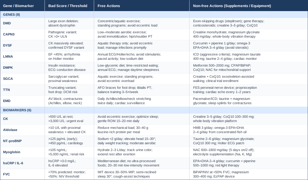
Quick Reference: Genes and Biomarkers at a Glance
What Emerging Muscle Research Gets Right That Most Clinics Don't
One of the most accessible deep dives into the science of muscle physiology comes through the collaboration between Andrew Huberman and Andy Galpin — a muscle physiology researcher — across a series of Huberman Lab episodes covering exercise, muscle fiber types, recovery, and the molecular biology of muscle adaptation. While the series was not designed specifically for muscular dystrophy, several of the core mechanistic insights are directly applicable to how people with MD think about managing residual muscle function.
1. Muscle fiber type ratios matter more than load in damaged muscle
Most MD subtypes preferentially affect type II (fast-twitch) fibers early while sparing type I (slow-twitch, oxidative) fibers longer. Galpin's research into fiber-type-specific training implications suggests that low-intensity, long-duration activities specifically recruit and maintain the type I fiber pool — which is exactly what remains functional longest in many MD subtypes. This means that slow, sustained movement (walking, swimming, gentle cycling) is not just "being careful" — it is strategically exercising the remaining functional pool.
2. Mitochondrial density is trainable even in compromised muscle
One of the most important findings discussed is that mitochondrial biogenesis responds robustly to zone 2 aerobic training (50–65% VO2 max) — even in individuals who cannot perform traditional exercise. This has implications for MD because mitochondrial dysfunction is a secondary consequence of dystrophin and sarcolemmal disruption. Maintaining mitochondrial density in remaining healthy fibers delays the energetic failure that precedes functional decline. The protocol discussed: 3–4 sessions per week, 20–30 minutes at a tolerable moderate pace.
3. Protein synthesis signals degrade without mechanical loading — even partial loading counts
The research reviewed by Galpin and Huberman establishes clearly that even partial weight-bearing and partial-range-of-motion exercises maintain the mTOR signaling that drives muscle protein synthesis. The clinical implication: wheelchair users who can perform seated partial leg extensions or resistance band exercises continue to generate anabolic signals in the muscles being loaded. The minimum effective mechanical signal is lower than most physical therapists historically assumed.
4. Sleep is the primary muscle repair window — and it is frequently compromised in MD
Growth hormone, IGF-1, and anti-inflammatory cytokines peak during slow-wave sleep. The podcast episodes on sleep quality are relevant to MD because sleep-disordered breathing (common in DM1, DMD, and LGMD with respiratory involvement) fragments the sleep architecture precisely when repair processes should be most active. The actionable implication: treating sleep apnea in MD patients is not just a respiratory issue — it is a muscle preservation intervention.
5. Electrolytes govern neuromuscular excitability at the fiber level
Sodium, potassium, and magnesium gradients across the sarcolemma determine action potential generation and calcium release. In MD, where the sarcolemma is already structurally compromised, electrolyte depletion from inadequate intake, heat, or diuretic use worsens weakness disproportionately. The Huberman-Galpin discussion of electrolyte protocols — particularly sodium (1 g/day minimum), potassium (3.5 g/day), and magnesium (400 mg/day) — provides a practical framework for baseline supplementation in MD contexts.
6. Cold water immersion reduces inflammation but impairs muscle adaptation when used immediately after training
This counterintuitive finding is relevant to MD management because many patients use cold baths or ice packs for muscle pain immediately after activity. Galpin's review of the evidence shows that immediate post-exercise cooling reduces the inflammatory signals that drive muscle adaptation — which matters when the goal is preserving remaining muscle mass. The recommendation: if using cold therapy for pain, delay it by 4–6 hours post-exercise rather than applying it immediately.
7. Grip strength correlates with total-body muscle reserve better than any other single test
Regular grip strength measurement (a $15–$30 hand dynamometer) provides a quantitative, trackable proxy for overall neuromuscular status in upper-limb-preserved MD subtypes. More importantly, serial grip strength decline can signal accelerating progression before broader clinical deterioration. The Huberman-Galpin protocols suggest measuring monthly, same time of day, to build a personal baseline — a zero-cost monitoring practice.
8. mTOR and AMPK signaling are inversely regulated — you cannot maximally activate both simultaneously
mTOR drives muscle protein synthesis; AMPK drives mitochondrial biogenesis and fat oxidation. They are partially antagonistic. The practical implication for MD management is that alternating training days — one day targeting low-intensity aerobic exercise (AMPK-dominant) and the next targeting resistance stimulation (mTOR-dominant) — achieves better net outcomes than trying to do both simultaneously. This is a structuring principle, not an all-or-nothing prescription.
9. Breathing mechanics directly affect core muscle activation and spinal loading
The Galpin episodes discuss how diaphragm function is central to trunk stability and spinal mechanics — relevant because many MD patients develop spinal deformity (scoliosis, kyphosis) partly from loss of trunk muscle coordinated activation. Breathing training, specifically diaphragmatic breathing with deliberate core engagement, supports spinal stability without the force demands that overtax weakened muscles.
10. Neurological drive to muscle declines with disuse faster than the muscle itself
One of the most underappreciated findings discussed is that motor neuron firing rate, motor unit recruitment, and neuromuscular junction efficiency all decline rapidly with disuse — and recover with movement, even in the context of underlying muscle disease. This means that some of the functional decline observed in sedentary MD patients may be partly neurological rather than purely mechanical — and therefore partially reversible with reactivation. The prescription is consistent daily movement, at whatever level is possible, rather than extended rest periods between PT sessions.
Complementary Approaches with Evidence in Neuromuscular Disease
The evidence base for complementary therapies in muscular dystrophy is generally modest but not absent. The following approaches were selected based on clinically plausible mechanisms specific to MD, existence of human evidence (trial or high-quality observational data), and practical implementability for people with physical limitations.
Breathing-Based Therapies
Respiratory muscle training is arguably the most evidence-supported non-pharmacological intervention in MD beyond physiotherapy. Because diaphragm and intercostal muscle weakness follows the same trajectory as limb girdle weakness in most subtypes — just delayed — systematic breathing exercise can extend the period before respiratory support is required.
A systematic review published in Cochrane Database of Systematic Reviews examined respiratory muscle training in neuromuscular diseases and found that inspiratory muscle training (IMT) using threshold devices produced measurable improvements in maximal inspiratory pressure (MIP) and, in some subgroups, FVC. The evidence was strongest for conditions with intermediate respiratory muscle weakness — precisely the window where intervention has the greatest potential impact.
Practically: use an IMT device set at 30–40% of measured MIP. Perform 30 breaths per session, twice daily, 5 days per week. Reassess MIP monthly. Complement with pursed-lip breathing exercises and deliberate diaphragmatic breathing during rest periods. Evidence is limited for very advanced respiratory failure, but early and consistent practice during preserved function is well-supported.
Massage Therapy
Manual soft-tissue therapy does not rebuild muscle but addresses several secondary complications that significantly affect quality of life and functional capacity in MD: muscle tightness, trigger points, joint stiffness, postural pain from compensatory loading, and anxiety related to chronic condition management. In the context of MD, massage must be modified — deep tissue techniques that would be appropriate in a healthy adult can cause CK elevation and fiber damage in fragile dystrophic muscle.
A pilot randomized controlled trial in children with DMD (Vignos protocol adaptations) showed that gentle connective tissue massage applied to the lower extremities three times weekly over 8 weeks improved passive joint range of motion and reduced parent-reported pain scores. The mechanism is primarily neuromuscular relaxation and local circulation enhancement, not structural muscle change.
Practically: sessions should use light-to-moderate pressure only. Swedish massage techniques targeting fascial planes rather than deep muscle belly work are most appropriate. Frequency of 1–2 sessions per week is reasonable; self-massage tools (foam rollers, soft therapy balls) allow daily home maintenance. Therapists should be briefed on the specific MD subtype and current CK levels before sessions involving affected muscle groups.
Mindfulness Meditation and MBSR
Living with a progressive condition creates a chronic psychological stress load that is biologically relevant beyond emotional discomfort. Cortisol elevation from unmanaged psychological distress promotes protein catabolism, worsens insulin resistance, suppresses immune regulation, and elevates CRP — all of which directly amplify MD disease burden. Mindfulness-Based Stress Reduction (MBSR) is one of the most rigorously studied behavioral interventions for chronic condition-related distress.
A meta-analysis of MBSR in chronic neurological conditions found significant reductions in anxiety, depression, and pain interference across a range of diagnoses. While MD-specific MBSR trials are lacking, the evidence from related neuromuscular and chronic disability populations is sufficient to support it as a meaningful adjunct.
Practically: the standard MBSR program involves 8 weekly group sessions of approximately 2.5 hours, plus daily 45-minute home practice. For people with fatigue-dominant presentations (common in DM1 and advanced MD), modified shorter protocols — 10–20 minutes of body scan or breath-focused meditation daily — retain most of the stress-reduction benefit with lower fatigue demand. Applications such as Insight Timer or the UCLA Mindful app provide free guided meditations accessible without physical exertion.
Music Therapy
Music therapy in neuromuscular disease operates through multiple mechanisms: rhythmic auditory stimulation supports gait and coordinated movement in people with preserved ambulation, while receptive music therapy reduces anxiety, pain perception, and psychological burden in those with more limited mobility. For pediatric and adolescent MD populations — where disease awareness intersects with developmental identity challenges — music therapy provides a psychosocially meaningful intervention that extends beyond symptom management.
A randomized trial by Wiens et al. in children with chronic medical conditions found that active music therapy (playing instruments adapted for motor limitations) significantly improved quality of life scores and social engagement compared to standard care alone. In DM1 specifically, where cognitive and emotional dysregulation are disease features, music engagement may also support frontal lobe function.
Practically: sessions led by a board-certified music therapist (1–2 per week, 30–45 minutes) are the clinical standard. Home engagement — listening to music with deliberate attention to rhythm and emotional response, or using adapted instruments — provides accessible daily practice. For people with significant upper limb involvement, adapted instruments (foot-pedaled, breath-controlled, or eye-gaze activated) are available through specialized music therapy programs.
Yoga (Adapted)
Standard yoga carries injury risk in MD due to eccentric loading, balance demands, and the instability of many traditional poses. However, adapted yoga — specifically designed for neuromuscular conditions and practiced seated, supine, or with props — provides genuine functional benefit: improved passive joint range of motion, respiratory muscle engagement through ujjayi breathing, parasympathetic nervous system activation, and a structured framework for daily movement that many people with MD report maintaining more consistently than conventional physiotherapy home programs.
A small but well-designed study by Brisebois et al. found that a 12-week adapted yoga program in adults with various LGMD subtypes produced improvements in balance, self-reported fatigue, and upper extremity flexibility without CK elevation or adverse events. The evidence base is small but the safety profile with appropriate adaptation is good.
Practically: classes should be explicitly labeled adaptive, restorative, or chair yoga — not standard flow or power yoga. One session per week with an instructor experienced in neuromuscular conditions, supplemented by 15–20 minutes of home practice 3–4 days per week, is a realistic protocol. Breathing practices (pranayama) can be continued independently on days when physical practice is not tolerable. Yin yoga, with its long-duration passive stretches supported by props, is particularly well-suited to MD because it achieves connective tissue lengthening without demanding muscular effort.
Conclusion
Muscular dystrophy is not one disease and it does not respond to one approach. The genetic subtype shapes everything: which tissues are at risk, which compensatory pathways exist, which biomarkers are most informative, and which interventions are most likely to slow progression. The framework laid out in this article — tracking six key biomarkers, understanding what eight critical genes mean in practice, and layering in evidence-based complementary strategies — does not replace medical care. It makes medical care more specific.
The next smart step is not to change everything at once. It is to identify which of these biomarkers has not been measured recently, which gene variants have been identified (or whether full panel genetic testing has ever been done), and which single intervention has the strongest rationale for your specific situation. A conversation with a neuromuscular specialist who can interpret genetic results in the context of current biomarker findings is the most valuable use of that information. Better data, reviewed by the right clinician, consistently produces better outcomes than generic management alone.
Musculoskeletal: Muscle Conditions
Neurological: Nerve Conditions
Cardiovascular: Heart Conditions Heart Rhythm Conditions
Respiratory: Lung Conditions Sleep & Breathing Disorders