This article contains AI generated content.
· UpdatedMyasthenia Gravis Genes and Biomarkers: 6 Genes and 7 Biomarkers to Track
Introduction
Living with myasthenia gravis means navigating a condition that is unpredictable in ways most diagnoses are not. The same morning can bring near-normal strength and crushing fatigue within hours. Standard follow-up appointments often focus on symptom changes and medication adjustments, but rarely explain the specific immunological events that determine whether your disease is quiet, progressing, or on the edge of a crisis. That gap between what is being managed and what is actually happening is where most people feel lost.
The biology of MG is genuinely varied. Around 85% of patients have antibodies against the acetylcholine receptor, but the remaining 15% have antibodies against MuSK, LRP4, or no detectable antibodies at all. Some people have concurrent thyroid autoimmunity. Some carry genetic variants that amplify immune activation before any treatment has a chance to work. Telling everyone to reduce stress and sleep better is not wrong, but it treats a heterogeneous disease as if it were a uniform one.
This article takes a different approach. Instead of broad recommendations, it focuses on what can be measured, tracked, and acted upon: the specific biomarkers that reveal the most about your disease activity and the genetic variants that shape your immunological baseline. These are not experimental concepts — they are markers already measurable through standard or accessible labs, with a growing body of evidence linking them to MG outcomes.
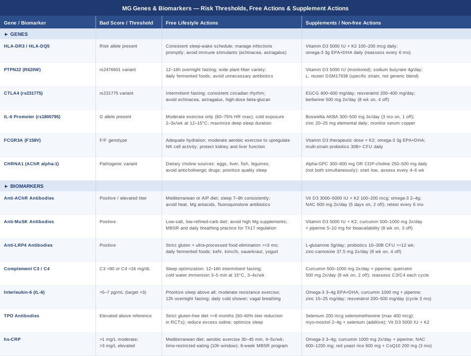
The approach here moves along two parallel tracks. The first — and most actionable — is biomarker monitoring: seven specific blood markers that give a real-time picture of inflammation, antibody load, and immune function. The second is genetic context: six gene variants that help explain why some people have more aggressive disease or respond poorly to standard interventions. Neither track promises a cure. Both offer something more realistic and more useful: better information, and with it, better decisions.
7 Biomarkers That Matter Most in Myasthenia Gravis
Of all the ways to understand MG from the inside, biomarkers are the most actionable. They can be tested, compared over time, and in many cases shifted through specific, targeted interventions. The seven markers below represent the most clinically meaningful data points available — from the core diagnostic antibodies to inflammatory mediators that most MG patients have never been asked about.
1. Anti-AChR Antibody Titers
Why it matters: Antibodies against the acetylcholine receptor (anti-AChR) are the defining marker of the most common form of MG, present in roughly 85% of generalized cases. These antibodies bind to the AChR at the neuromuscular junction and trigger complement-mediated receptor destruction, producing the characteristic fluctuating weakness of MG. Antibody titers do not correlate perfectly with day-to-day symptoms, but they do reflect overall immune activity and can signal disease progression or treatment response when tracked longitudinally.
How to measure it: A standard serum blood test, ordered as an acetylcholine receptor antibody panel. The panel typically includes binding, blocking, and modulating subtypes. Cost ranges from $150 to $400 in the US depending on lab and insurance. Testing should be repeated at baseline, after treatment changes, and at 6–12 month intervals during stable maintenance phases.
If the result is high — without supplements: Reduce systemic inflammatory burden through diet first. A Mediterranean-style pattern or an autoimmune elimination approach (AIP — discussed in the complementary section) has measurable effects on antibody-driven inflammation. Sleep is a direct lever: immune regulation, including B-cell activity and antibody production, is tightly synchronized with sleep. Aim for 7–9 hours in a consistent window. Avoid known MG triggers: extreme heat, magnesium-containing antacids, fluoroquinolone and aminoglycoside antibiotics, and sustained psychological stress. Gentle regular aerobic movement — walking, cycling — reduces systemic inflammation without triggering fatigue-driven exacerbation.
If the result is high — with supplements or equipment: Vitamin D3 (3000–5000 IU daily with vitamin K2 at 100–200 mcg) is the most evidence-supported immune modulator for antibody-mediated autoimmune conditions. Several studies have found inverse relationships between serum vitamin D and autoimmune antibody titers. Test 25-OH vitamin D before supplementing; target 50–70 ng/mL. Omega-3 fatty acids at 2–4g EPA+DHA daily reduce B-cell activation and complement activity — use fish oil or algae-based sources. No cycling needed; reassess every 6 months. N-acetyl cysteine (NAC) at 600 mg twice daily supports glutathione-mediated anti-inflammatory activity; cycle 5 days on, 2 days off to avoid tolerance. Most common side effects: mild GI discomfort, dose-dependent nausea with NAC.
2. Anti-MuSK Antibodies
Why it matters: Muscle-specific kinase (MuSK) antibodies are found in approximately 40% of anti-AChR-negative MG patients, representing a clinically distinct subtype. MuSK-positive MG typically involves more prominent facial, bulbar, and respiratory muscle weakness. Pyridostigmine (the standard acetylcholinesterase inhibitor) is often less effective or even worsening in MuSK+ patients, while rituximab shows unusually strong response rates. Knowing your MuSK status changes the treatment algorithm substantially — and many patients are never tested unless they first come back negative for AChR.
How to measure it: Serum blood test ordered specifically as anti-MuSK antibody. Cost: $200–$500. Not routinely ordered in the initial workup. If you are seronegative for AChR but have clear clinical MG features, explicitly request this test and anti-LRP4 (below) from your neurologist.
If the result is positive — without supplements: MuSK-positive MG is strongly associated with Th17-driven inflammation — a polarization pattern that responds well to low-salt, low-refined-carbohydrate dietary patterns. These reduce the osmotic conditions that promote Th17 differentiation. High magnesium supplementation should be avoided — it can worsen neuromuscular transmission independently. Consistent mind-body practice (MBSR, breathing exercises) has documented effects on T-cell balance in autoimmune conditions.
If the result is positive — with supplements or equipment: Vitamin D3 at therapeutic levels (5000 IU daily under periodic monitoring) suppresses Th17 differentiation across multiple autoimmune models and shifts the immune environment toward Treg-dominant tolerance. Always combine with K2 to reduce hypercalcemia risk. Curcumin at 500–1000 mg twice daily with 5–10 mg piperine for bioavailability suppresses NF-κB signaling upstream of MuSK antibody production. Cycle 8 weeks on, 3 weeks off. Take with food to minimize GI irritation. Main side effects: loose stools at high doses, mild reflux.
3. Anti-LRP4 Antibodies
Why it matters: Low-density lipoprotein receptor-related protein 4 (LRP4) is the third major antibody target in MG, found in a subset of patients who test negative for both AChR and MuSK. LRP4 binds agrin — a molecule essential for clustering acetylcholine receptors at the neuromuscular junction. Anti-LRP4 antibodies disrupt this scaffolding, producing neuromuscular weakness that can look clinically identical to AChR-positive MG but remain undetected on standard panels, delaying diagnosis by months or years.
How to measure it: Serum test with limited availability — typically requires specialized neuromuscular laboratories. Cost: $300–$700. Most useful when clinical presentation strongly suggests MG but both AChR and MuSK are negative.
If the result is positive — without supplements: Anti-LRP4 antibodies appear sensitive to gut-derived antigen leakage in ways that other MG antibodies may not be. A strict elimination trial removing gluten and ultra-processed foods for a minimum of 3 months, combined with fermented food introduction (kefir, kimchi, sauerkraut, live-culture yogurt), is a biologically plausible starting point. The mechanism — reduced mucosal permeability limiting the antigen presentation that drives antibody production — is supported by work on other antibody-mediated autoimmune conditions, though not yet confirmed specifically for LRP4.
If the result is positive — with supplements or equipment: L-glutamine at 5g daily supports gut epithelial tight junction integrity and may reduce antigen-driven antibody stimulation. Multi-strain probiotics (10–30 billion CFU daily) with Lactobacillus rhamnosus and Bifidobacterium longum strains for a minimum 12-week course. No cycling required. Digestive enzymes with each meal if digestive symptoms suggest impaired breakdown. Zinc-carnosine (37.5 mg twice daily, away from meals) has mucosal protective effects specifically studied in gut permeability contexts; cycle 8 weeks on, 4 weeks off.
4. Complement C3 and C4
Why it matters: Complement activation is one of the primary mechanisms by which anti-AChR antibodies destroy neuromuscular junctions. When anti-AChR antibodies bind the receptor, they activate the complement cascade, leading to membrane attack complex formation and progressive receptor loss. C3 and C4 measurements reflect the state of this cascade — consumed (low levels) in active attack, or elevated as general inflammatory reactants. Some patients with high antibody titers but intact complement regulation have a more stable disease course, making this marker important for interpreting antibody levels in context.
How to measure it: Standard chemistry panel add-on. C3 and C4 can be requested alongside any routine blood draw. Optional additions include CH50 (total hemolytic complement) and Bb (alternative pathway activation marker). Cost: $40–$120 combined. C3 normal range: 90–180 mg/dL. C4 normal: 16–47 mg/dL. Low levels in active MG indicate ongoing complement consumption; elevated levels reflect general acute-phase inflammation.
If the result indicates active consumption — without supplements: Sleep optimization is the primary free intervention — complement pathway activity increases measurably with sleep deprivation. Intermittent fasting in the 12–16 hour range reduces complement activation in animal models and has supportive human data in inflammatory conditions. Brief cold water immersion (3–5 minutes at 15°C, 3–4 times weekly) has been shown to transiently reduce inflammatory complement signaling and may support medium-term complement regulation.
If the result is abnormal — with supplements or equipment: Curcumin (500–1000 mg twice daily with piperine) directly inhibits complement activation — this mechanism has been investigated in complement-mediated autoimmune nephritis and may extrapolate to MG. Quercetin at 500 mg twice daily acts synergistically with curcumin through overlapping NF-κB and complement pathway targets. Cycle both for 8 weeks on, 2 weeks off; reassess C3/C4 after each cycle. Take both with food to minimize GI effects. Avoid quercetin with thyroid medications (may reduce absorption).
5. Interleukin-6 (IL-6)
Why it matters: IL-6 sits near the center of MG pathophysiology. It promotes B-cell differentiation into the antibody-secreting plasma cells that produce anti-AChR antibodies, drives Th17 polarization, and amplifies the systemic inflammatory state that worsens disease activity. Elevated serum IL-6 is associated with more severe MG presentations, greater crisis risk, and poorer response to first-line immunosuppression. Crucially, IL-6 responds to lifestyle faster than almost any other immune marker — making it one of the highest-leverage targets for behavioral and nutritional intervention.
How to measure it: Serum IL-6 (high-sensitivity). Not a standard order — specifically request it. Cost: $50–$150. Measure fasting, in the morning, and at least 48 hours away from infections, vaccinations, or high-intensity exercise (all acutely elevate IL-6). Target: below 3 pg/mL. In an MG monitoring context, values above 5–7 pg/mL are clinically significant.
If the result is high — without supplements: Sleep deprivation is the single most reliable IL-6 elevator — even one poor night raises IL-6 measurably. Correcting sleep comes first. Resistance exercise at moderate intensity (not high-intensity) chronically suppresses resting IL-6 while only transiently elevating it acutely. Consistent 12-hour overnight fasting reduces IL-6 within 2–4 weeks in multiple clinical studies. Cold exposure — a 2–5 minute cold shower at 15°C or less, daily — blunts the IL-6 stress response over time. Diaphragmatic breathing practice activates the vagal anti-inflammatory reflex, which directly suppresses IL-6 production in macrophages.
If the result is high — with supplements or equipment: Omega-3 fatty acids at 3–4g EPA+DHA daily are the most studied IL-6 suppressors, with measurable effects in 6–8 weeks in randomized trials. Curcumin (1000 mg with piperine, twice daily) directly inhibits IL-6 transcription through NF-κB suppression. Zinc at 15–25 mg daily with food is a reliable and underused IL-6 regulator — do not exceed 40 mg/day without monitoring serum copper. Resveratrol at 200–500 mg daily (with a small meal for absorption) has shown IL-6 reduction in clinical trials in inflammatory conditions; cycle 3 months on, 1 month off. Avoid resveratrol in pregnancy or combined with anticoagulants (mild blood-thinning effect).
6. Thyroid Peroxidase (TPO) Antibodies
Why it matters: Autoimmune thyroid disease — primarily Hashimoto's thyroiditis — co-occurs with MG at rates well above chance. Approximately 15–20% of MG patients have concurrent thyroid autoimmunity, and the relationship is bidirectional: shared HLA haplotypes and regulatory T-cell dysfunction underlie both conditions simultaneously. High TPO antibodies signal ongoing thyroid inflammation that amplifies total autoimmune burden, complicates the fatigue picture (thyroid-related fatigue and MG fatigue are clinically nearly identical), and can worsen overall immune dysregulation. TPO testing is almost never included in standard MG monitoring panels — it should be.
How to measure it: Thyroid antibody panel: TPO antibodies and anti-thyroglobulin (anti-TG) antibodies. TSH and free T3/T4 should accompany them. Cost: $30–$80 as an add-on. Annual testing is appropriate for most MG patients. If thyroid-related symptoms develop (cold intolerance, unexplained weight change, cognitive fog, hair thinning), test more frequently.
If the result is high — without supplements: A strict gluten-free diet has the most consistent published evidence for reducing TPO antibody titers over time — multiple randomized controlled trials in Hashimoto's patients demonstrate 50–60% reductions in TPO titers after 6–12 months of adherence. Excess iodine intake (high-dose iodine supplements, excessive seaweed) can worsen TPO antibody production — reduce if relevant. Sleep optimization reduces thyroid autoantibody production through its effects on T-cell regulation.
If the result is high — with supplements or equipment: Selenium at 200 mcg daily as selenomethionine is the most studied single intervention for TPO antibody reduction. A meta-analysis in European Journal of Endocrinology found significant and consistent reductions with 6 months of supplementation across multiple trials. Do not exceed 400 mcg/day — selenosis risk (hair and nail brittleness, GI symptoms, neurological effects at very high doses). Myo-inositol at 2–4g daily combined with selenium shows additive TPO-reducing effects in published Italian studies. Vitamin D3 (5000 IU with K2) reduces thyroid-directed autoimmunity by supporting Treg activity across multiple autoimmune thyroid studies.
7. High-Sensitivity C-Reactive Protein (hs-CRP)
Why it matters: hs-CRP is a general marker of systemic inflammation, produced by the liver in response to IL-6 and other upstream cytokines. In MG, elevated hs-CRP reflects the total inflammatory load driving antibody production, complement activation, and T-cell dysregulation. It is not MG-specific, but its value lies in sensitivity and responsiveness — hs-CRP changes within days to weeks of lifestyle or treatment changes, making it the fastest feedback loop available without specialized testing. Peter Attia has consistently positioned hs-CRP as one of the most important routine monitoring markers in any inflammatory chronic condition, and that principle holds directly in MG.
How to measure it: Standard lab add-on, available at virtually every clinical laboratory. Cost: $15–$50. Target: below 1 mg/L. Between 1–3 mg/L is moderate; above 3 mg/L is elevated and warrants action. Measure fasting, without recent infection, injury, or high-intensity exercise within 48 hours — all cause transient CRP elevation unrelated to chronic inflammation.
If the result is high — without supplements: A Mediterranean dietary pattern consistently reduces CRP by 30–40% in clinical trials — the effect is dose-responsive and appears within 4–8 weeks. Regular moderate aerobic exercise (30–45 minutes, 4–5 times per week at 60–70% max heart rate) reduces CRP independently of body weight changes. Time-restricted eating within a 10-hour window reduces CRP through multiple metabolic pathways. An 8-week MBSR program produces measurable CRP reductions in randomized trials — the mechanism involves cortisol regulation and reduced sympathetic-driven inflammatory signaling.
If the result is high — with supplements or equipment: Omega-3s at 3–4g EPA+DHA daily are the most evidence-backed CRP reducers available without prescription. Curcumin (1000 mg twice daily with piperine) reduces CRP across multiple randomized controlled trials in inflammatory conditions — the effect is consistent and appears within 8 weeks. NAC at 600–1200 mg daily provides supplementary antioxidant and anti-inflammatory action through glutathione-dependent pathways; cycle 5 days on, 2 days off. Red yeast rice at 600 mg twice daily can reduce CRP via mild statin-like mechanisms — only if not already on statins, and always pair with CoQ10 at 200 mg daily to protect against muscle-related side effects. Cycle 3 months on, 1 month off with liver function monitoring.
Tracking these seven biomarkers systematically — rather than relying on symptoms alone — gives a far more precise and actionable picture of MG activity over time. Each one is a lever. Understanding what it measures makes the lever meaningful.
The Genetic Architecture Behind Myasthenia Gravis
Genetics do not determine your MG outcome, but they do define the terrain. Six gene variants stand out in current MG research for their roles in immune regulation, autoantibody production, and neuromuscular junction maintenance. Testing these variants (through direct-to-consumer panels like those from major clinical genetics labs, or through whole-exome sequencing) provides context that can sharpen the interpretation of your biomarker results and guide more targeted interventions.
HLA-DR3 and HLA-DQ5
Human leukocyte antigen (HLA) genes on chromosome 6p21 represent the most strongly established genetic risk factors in MG. The specific associations depend on subtype: HLA-DR3 and HLA-B8 are linked to early-onset generalized MG (more common in women under 40), while HLA-DR7 and HLA-DQ5 appear in late-onset disease. These genes determine how immune cells present peptide fragments to T cells — if your HLA configuration happens to present acetylcholine receptor peptides in a way that activates autoreactive helper T cells, the structural risk for breaking tolerance is elevated from the outset.
If the variant is present — without supplements: The gene cannot be changed, but its expression of risk is modifiable. Avoid cumulative immune over-activation: manage infections early and aggressively, avoid unnecessary immune stimulants (high-dose echinacea, astragalus), maintain regular sleep-exercise patterns that support immunological tolerance rather than chronic immune priming. Consistent circadian rhythm is particularly important — HLA-linked T-cell responses vary by time of day and are disrupted by irregular sleep schedules.
If the variant is present — with supplements: Vitamin D3 at therapeutic levels (5000 IU daily, monitored via 25-OH vitamin D testing every 3–6 months) directly modulates HLA-mediated T-cell activation and is the most documented natural lever for this. Target 50–70 ng/mL. Combine with K2 (100–200 mcg daily) to prevent soft tissue calcification at higher D3 doses. Omega-3s at 3g EPA+DHA daily reduce HLA-linked inflammatory cytokine amplification through membrane phospholipid modulation. Neither requires cycling; reassess blood levels every 6 months.
PTPN22 (R620W Variant)
PTPN22 encodes a tyrosine phosphatase that regulates T-cell receptor signaling strength. The R620W variant (rs2476601) reduces the activity of regulatory T cells (Tregs) — the immune cells responsible for suppressing self-reactive responses. Carriers have an effectively weakened immune brake and are at elevated risk for multiple autoimmune diseases including MG, rheumatoid arthritis, systemic lupus, and type 1 diabetes. The same basic mechanism underlies all of these: Treg insufficiency allows autoreactive T and B cells to persist and act.
If the variant is present — without supplements: Fasting and caloric restriction have documented Treg-expanding effects in multiple published studies, partially compensating for PTPN22-related Treg dysfunction. Even consistent 12–16 hour overnight fasting supports Treg expansion through butyrate production and metabolic shifts in the gut microbiome. Dietary diversity — wide variety of plant fibers, fermented foods — is the strongest modifiable environmental input for Treg function outside of medication. Avoid unnecessary antibiotic courses, which acutely reduce butyrate-producing bacteria and temporarily collapse Treg support.
If the variant is present — with supplements: Vitamin D3 (5000 IU daily under monitoring) is the single most potent natural Treg inducer available. Sodium butyrate at 4g daily (or equivalent via high-fiber intake and resistant starch) promotes Treg differentiation in the colon — start at 1–2g and increase over 4 weeks to minimize bloating. Lactobacillus reuteri DSM17938 specifically has demonstrated Treg-promoting effects in human clinical studies — this strain is distinct from generic probiotic blends and should be selected specifically.
CTLA4 (rs231775 Variant)
CTLA4 (cytotoxic T-lymphocyte antigen 4) is an immune checkpoint protein that normally limits T-cell activation after an initial immune response — it is the natural brake on T-cell persistence. Variants in CTLA4, particularly rs231775, reduce expression or functional efficiency of this checkpoint, allowing T cells to remain active longer. The therapeutic relevance is direct: abatacept (a CTLA4-Ig fusion protein) is used in autoimmune conditions and is under early-stage investigation in MG specifically. Carriers of loss-of-function CTLA4 variants may need more aggressive checkpoint support.
If the variant is present — without supplements: Intermittent fasting shows early evidence for upregulating CTLA4 expression on T cells in animal models. Consistent circadian alignment — regular sleep-wake timing — appears to support immune checkpoint gene expression cycles. Deliberately avoid immune-stimulating supplements that further activate T cells (echinacea, astragalus, beta-glucan in high doses) — these strategies are counterproductive if CTLA4 function is already reduced.
If the variant is present — with supplements: EGCG (standardized green tea extract at 400–600 mg daily) has shown early evidence for immune checkpoint modulation through pathway interactions with CTLA4 signaling. Resveratrol at 200–400 mg daily (with a small amount of dietary fat for absorption) influences checkpoint-adjacent pathways in early human studies; cycle 3 months on, 1 month off, GI effects mild — take with meals. Berberine at 500 mg twice daily acts via AMPK activation with downstream immune checkpoint modulation; cycle 8 weeks on, 4 weeks off. Avoid combining resveratrol with anticoagulants at therapeutic doses.
IL-6 Gene Promoter Variants (rs1800795)
The -174G>C polymorphism in the IL-6 gene promoter region influences the baseline output of IL-6 in response to inflammatory stimuli. Carriers of the G allele tend to produce more IL-6 in response to the same level of immune activation. In MG, this translates to greater B-cell activation, more aggressive antibody production, and a more inflammatory disease course — the same mechanism described in the biomarker section, but baked in genetically rather than acquired through lifestyle alone.
If the variant is present — without supplements: High-intensity exercise transiently spikes IL-6 — G-allele carriers may have greater and more prolonged post-exercise elevations. Shift training toward moderate intensity: 60–75% max heart rate, 30–45 minutes, 3–5 times per week. Cold exposure at 2–3 sessions per week (3–5 minute cold shower or immersion at 12–15°C) blunts the IL-6 response to subsequent stressors. Sleep is the most impactful free lever for this variant specifically — each additional hour of deep sleep has measurable effects on resting IL-6 in carriers.
If the variant is present — with supplements: All IL-6-targeting strategies from the biomarker section apply here with higher priority. Add boswellic acids (Boswellia serrata extract standardized to AKBA, 300–500 mg three times daily) — boswellia inhibits leukotriene synthesis and reduces IL-6 as a secondary effect; cycle 3 months on, 1 month off. Zinc at 20–25 mg elemental zinc daily directly suppresses IL-6 transcription — monitor serum copper with long-term use and add copper at 1–2 mg if taking zinc beyond 3 months.
FCGR3A (F158V Variant, rs396991)
Fc gamma receptor IIIa (FCGR3A) mediates the cellular immune response to IgG antibodies by binding their constant (Fc) region. The F158V variant reduces receptor affinity for IgG, affecting how efficiently the immune system clears immune complexes and how effectively NK cells respond to IgG-coated targets. In MG, this influences clearance of circulating anti-AChR immune complexes and — importantly — affects treatment response to IgG-based therapies like IVIG and rituximab. The F/F genotype may correlate with reduced rituximab response, which has practical implications for treatment planning.
If the variant is present — without supplements: Support immune complex clearance indirectly through maintaining healthy kidney function (adequate hydration, moderate dietary protein), liver health (limited alcohol, adequate micronutrient status), and overall complement function. Regular moderate aerobic exercise upregulates NK cell activity and partial functional compensation for reduced FCGR3A affinity is plausible through this mechanism.
If the variant is present — with supplements: Vitamin D3 (therapeutic dosing) upregulates Fc receptor expression on macrophages — this is documented specifically for FcγR expression. Omega-3s at 3g daily enhance NK cell cytotoxicity through cell membrane modulation, providing some compensation for reduced FCGR3A function. Probiotics (multi-strain, minimum 30 billion CFU) support the gut-associated immune complex clearance that complements systemic Fc receptor function.
CHRNA1 (Acetylcholine Receptor Alpha-1 Subunit)
CHRNA1 encodes the alpha-1 subunit of the nicotinic acetylcholine receptor — the very molecule that anti-AChR antibodies target. While most MG is acquired through antibody production against normal CHRNA1-encoded protein, rare variants in CHRNA1 alter receptor structure in ways that may affect immunogenicity (how readily the immune system forms antibodies against the receptor) and acetylcholine binding efficiency even before antibody interference. More commonly relevant in familial and some early-onset presentations.
If the variant is present — without supplements: Optimize endogenous acetylcholine production through dietary choline: eggs (particularly yolks), liver, fish, and legumes provide the precursors for synthesis. Avoid anticholinergic medications when alternatives exist — many commonly used drugs have significant anticholinergic burden (certain antihistamines, bladder medications, tricyclic antidepressants). Quality sleep supports AChR recycling and neuromuscular junction maintenance — this is one of the clearest free interventions for receptor-level function.
If the variant is present — with supplements: Alpha-GPC at 300–600 mg daily or CDP-choline at 250–500 mg daily are the most bioavailable choline precursors for supporting acetylcholine synthesis. Do not combine both simultaneously. Start at the lower dose and assess tolerance — dose-dependent GI symptoms, vivid dreams, and occasional headache are the most common effects. Reassess every 4–6 weeks; take breaks if cholinergic side effects (nausea, bradycardia, excessive sweating) develop.
What Neuroscience and Acetylcholine Research Reveal About Managing MG
Andrew Huberman, professor of neurobiology and ophthalmology at Stanford School of Medicine, has built a significant body of publicly accessible work on acetylcholine — its role in neuromuscular function, learning, focus, and inflammation. While his work addresses neuroscience broadly rather than MG specifically, the mechanisms he describes are directly relevant to anyone managing a disease that disrupts cholinergic signaling at the neuromuscular junction. Here are ten of the most impactful insights applicable to MG management.
1. Acetylcholine Is the Molecular Currency of Muscle Control and Focus
Huberman describes acetylcholine as the modulator that "gates" both muscular contraction and focused attention. In MG, the postsynaptic receptor is the primary target of attack — but the presynaptic synthesis of acetylcholine and the broader health of the junction remain meaningful targets. Maximizing choline intake and protecting cholinergic signaling everywhere it is accessible is worthwhile even when receptor numbers are reduced.
2. Deep Sleep Is When Neuromuscular Junction Repair Occurs
Slow-wave sleep is when synaptic homeostasis takes place — the structural maintenance and pruning of synaptic connections. Huberman cites work showing that sleep deprivation rapidly degrades cholinergic signaling and disrupts junction maintenance. For MG, sleep is not a lifestyle recommendation — it is a direct biological intervention for neuromuscular junction function.
3. Moderate Exercise Upregulates Acetylcholine Receptor Sensitivity
Short bouts of deliberate physical effort followed by recovery temporarily increase acetylcholine receptor sensitivity in muscle tissue. For MG patients not in active exacerbation, calibrated gentle exercise — never to the point that triggers ptosis or diplopia — appears to support receptor responsiveness over time. The key is ending exercise well before fatigue, not pushing through it.
4. Most People Are Chronically Choline-Deficient
Daily adequate intake for adults is 425–550 mg, but average dietary intake is closer to 250–350 mg. Eggs remain the most efficient single food source. For MG patients, adequate choline does not restore lost receptors — but it maximizes the acetylcholine available to compete for the receptors that remain functional.
5. Chronic Stress Depletes Cholinergic Tone Directly
The stress response reduces vagal (parasympathetic/cholinergic) tone. Huberman describes the well-documented inverse relationship between sustained sympathetic activation and cholinergic function. In MG, chronic psychological stress does not just worsen immune dysregulation — it actively reduces the cholinergic environment that neuromuscular junction function depends on, compounding the receptor-level deficit.
6. The Vagal Cholinergic Pathway Suppresses IL-6 and TNF-Alpha
Huberman has covered the vagal anti-inflammatory reflex extensively: acetylcholine released via the vagus nerve signals macrophages to suppress production of TNF-alpha, IL-6, and IL-1 — the key inflammatory cytokines driving MG disease activity. Stimulating the vagus (through slow breathing, cold water face immersion, humming, gargling) activates this suppressive pathway through a cholinergic mechanism that is directly relevant to every elevated biomarker discussed in this article.
7. Morning Light Regulates the Immune Circadian Clock
Outdoor light exposure within 30 minutes of waking sets the circadian rhythm that governs immune cell timing, cytokine release cycles, and the daily maintenance windows of synaptic structures. Huberman has discussed how circadian disruption alters B-cell and T-cell function in ways that promote autoimmunity. Ten to twenty minutes of outdoor morning light is the most accessible free circadian anchor available.
8. Non-Sleep Deep Rest Restores Cholinergic Reserves
Huberman popularized NSDR — non-sleep deep rest based on yoga nidra protocols — as a tool for neurotransmitter recovery after cognitively demanding periods. The principle applies to acetylcholine as well as dopamine: a 10–20 minute NSDR session after physically or cognitively demanding activity accelerates acetylcholine circuit recovery. For MG patients managing limited daily energy, building short rest windows into the day may reduce the accumulation of cholinergic fatigue.
9. Deliberate Motor Practice Maintains Receptor Density
Focused, precise motor practice — as opposed to exhausted or passive movement — upregulates nicotinic acetylcholine receptor density at neuromuscular junctions over time. This is the mechanistic basis of motor learning. For MG patients, this suggests that gentle physical therapy exercises performed during the best-function window of the day (typically mornings for many MG patients) may maintain neuromuscular junction architecture more effectively than unfocused activity at any time.
10. Omega-3s Support Membrane Fluidity at the Neuromuscular Junction
Huberman has referenced omega-3 fatty acids not only for their anti-inflammatory effects but for their role in maintaining the physical integrity of neuronal and neuromuscular membranes. Phosphatidylcholine — found in egg yolks and sunflower lecithin — is a structural component of these membranes and a direct precursor to acetylcholine itself. Membrane fluidity and receptor packing density are directly relevant to how the junction performs under reduced receptor numbers.
Complementary Approaches With Meaningful Evidence in MG
Standard medical management of MG remains the non-negotiable foundation of treatment. However, several complementary approaches have published human evidence for reducing the immune dysregulation, systemic inflammation, and stress burden that drive MG activity. The four below are selected specifically for their biological relevance to MG mechanisms and for the quality of supporting evidence.
The Autoimmune Protocol — Sarah Ballantyne
The Autoimmune Protocol (AIP), developed by Dr. Sarah Ballantyne (PhD), is a structured elimination and reintroduction dietary framework designed to reduce gut permeability, calm immune activation, and lower autoimmune antibody production. The elimination phase removes grains, legumes, dairy, eggs, nightshades, nuts, seeds, alcohol, and processed foods, then systematically reintroduces foods to identify individual triggers. In MG — an antibody-mediated autoimmune disease — the AIP directly targets several upstream drivers discussed throughout this article: gut-derived antigen leakage, B-cell activation, and systemic inflammatory cytokine production.
A pilot study by Konijeti et al. published in Inflammatory Bowel Diseases demonstrated significant reductions in inflammatory markers and clinical improvement in an autoimmune gut condition after 6 weeks of AIP. The immunological mechanisms studied — reduced intestinal permeability, reduced antigen-driven B-cell activation, and lower inflammatory cytokine output — are directly applicable to MG pathophysiology even though the condition studied was different. Dr. Ballantyne's full protocol is detailed in The Paleo Approach and accessible through her research platform.
For MG: begin with a minimum 30-day strict elimination phase. Many patients report clearest early benefit from removing gluten and dairy first if full AIP feels overwhelming. Work with a registered dietitian to prevent nutritional gaps during the elimination phase — it is nutrient-dense but requires planning. Do not reduce or discontinue MG medications during this period. The AIP is a complement to standard immunotherapy, not a replacement.
Mindfulness-Based Stress Reduction (MBSR)
MBSR is a structured 8-week program developed by Jon Kabat-Zinn combining body scanning, sitting meditation, and gentle mindful movement. Its relevance to MG is mechanistic and specific: the program has documented effects on cortisol regulation, IL-6 suppression, and vagal tone — each of which corresponds to a named biomarker or pathway discussed in this article. Psychological stress is one of the most reliable and consistent MG exacerbation triggers, and reducing chronic stress signaling changes the immunological environment in measurable ways.
A randomized controlled trial published in Brain, Behavior, and Immunity (Witek-Janusek et al.) demonstrated that MBSR reduced pro-inflammatory cytokines and improved NK cell function in a clinical population. In autoimmune conditions broadly, 8-week MBSR programs have been shown to reduce relapse rates and improve quality of life measures in multiple studies. For MG specifically, the target is stress-triggered exacerbation prevention and chronic IL-6 reduction.
Practice: 45–60 minutes daily during the 8-week structured program, then 20–30 minutes for ongoing maintenance. The free Palouse Mindfulness program online delivers the full MBSR curriculum. Body scan practice is particularly well-suited to MG because it builds proprioceptive awareness of early fatigue onset — allowing patients to rest before weakness escalates into exacerbation. Neurological effects require consistent daily practice, not occasional sessions.
Biofeedback
Biofeedback involves learning to consciously influence physiological variables — heart rate variability (HRV), muscle tension, respiratory rate — through real-time physiological feedback. For MG, HRV biofeedback is the most biologically relevant application. HRV directly reflects vagal tone, and as detailed in the Huberman section, the vagal cholinergic anti-inflammatory pathway is a measurable lever for reducing IL-6, TNF-alpha, and complement activity — the same biomarkers tracked throughout this article.
A meta-analysis published in Applied Psychophysiology and Biofeedback (Gevirtz) demonstrated that HRV biofeedback reliably improves autonomic balance and reduces inflammatory markers across multiple clinical populations. Hardware options range from free-tier phone apps with compatible HR chest straps (Elite HRV) to dedicated clinical-grade devices (HeartMath Inner Balance, Lief) at $100–$400. Clinical biofeedback with a certified therapist runs $100–$250 per session.
A realistic protocol for MG: 10–15 minutes daily of slow-paced HRV biofeedback breathing (typically 4–6 breath cycles per minute). Begin with a certified biofeedback therapist for 4–6 sessions to establish correct technique, then continue independently. Benefits are measurable within 4–6 weeks as resting HRV improves. The main risk is incorrect technique producing hyperventilation — professional guidance for initial sessions is warranted.
Breathing-Based Therapies
Breathing exercises are directly relevant to MG on two overlapping levels: respiratory muscle weakness is a serious MG complication where targeted training has demonstrated benefit, and slow breathing patterns are one of the most accessible tools for vagal activation, IL-6 reduction, and stress-triggered exacerbation prevention. These two applications are distinct and complement each other.
A randomized controlled trial by Fregonezi et al. published in Chest studied inspiratory muscle training (IMT) specifically in MG patients and found measurable improvements in maximal inspiratory pressure after 3 months of resistance-based training. This is condition-specific evidence that targeted respiratory muscle training — not generic breathing relaxation — may help preserve respiratory reserve in MG.
Practical protocol: IMT using a threshold resistance device (Threshold IMT by Philips Respironics, approximately $35) at 30% of measured maximal inspiratory pressure, 30 breaths per session, once daily for 8 weeks. Reassess respiratory function with your neurologist before and after. Never train breathing during active exacerbation or acute weakness. Separately — and entirely without equipment — practice slow breathing at 4–6 cycles per minute for 10 minutes daily as a daily vagal activation and anti-inflammatory practice. These two protocols address different mechanisms and can be done on different schedules.
Conclusion
Myasthenia gravis is a condition where greater precision consistently yields better outcomes. The disease varies too much — in antibody type, genetic background, inflammatory load, and treatment response — for generic management to be sufficient. The framework laid out in this article gives you concrete tools: seven biomarkers to track, six genetic variants to contextualize, and complementary approaches backed by actual human evidence.
The next smart step is not to attempt everything at once. Choose one or two biomarkers you have not recently tested, bring them to your next appointment, and begin tracking changes as you implement even one or two targeted interventions. Discuss genetic testing with your neurologist if it is not yet part of your evaluation. Ask questions about your antibody profile that your standard visits may not have addressed. Better information, acted on consistently, is the most reliable path toward more stable disease management.
Musculoskeletal: Muscle Conditions
Neurological: Nerve Conditions
Endocrine & Metabolic: Thyroid Conditions
Autoimmune: Inflammatory Conditions