This article contains AI generated content.
· UpdatedMyositis Ossificans — 5 Genes And 6 Biomarkers To Track
Introduction
If you are managing myositis ossificans — or watching a post-injury site that will not fully recover — you already know the frustration. The standard advice focuses on rest, anti-inflammatories, and imaging follow-ups. What it rarely addresses is the biological process unfolding beneath the surface: an inflammatory and osteogenic cascade that can be active for weeks before a scan confirms anything. By the time a mass is visible, the window for early intervention has often narrowed considerably.
The challenge is that myositis ossificans is not a single, uniform process. It ranges from a contained traumatic response in an athlete's thigh muscle to a relentlessly progressive condition driven by rare genetic mutations. The difference between those two scenarios is not always clear from symptoms alone. That gap is where objective biology becomes useful. Biomarkers and genetic data do not replace clinical judgment, but they can reveal things that imaging, pain scores, and physical examination miss entirely.
Generic recovery protocols are often too broad because they treat heterotopic ossification as a purely mechanical problem. They do not account for individual differences in inflammatory signaling, bone remodeling capacity, vitamin D metabolism, or cytokine gene variants that amplify or dampen the osteogenic response. Two people with the same injury site and the same MRI report may have completely different underlying biologies — and therefore respond very differently to the same treatment.
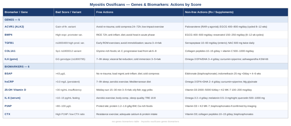
This article takes a more targeted approach. The first section covers the six most informative biomarkers for tracking the activity and trajectory of myositis ossificans, including how to measure each one, what a bad score means practically, and which interventions — with and without supplements — have reasonable evidence behind them. The second section covers five genes whose variants are increasingly linked to heterotopic ossification risk and severity. Understanding even one or two of these can change the conversation with your clinician and open up more personalized strategies. Neither section promises a cure; both offer better information — which, in a condition this poorly understood at the individual level, can itself be a meaningful advantage.
6 Biomarkers That Can Track What Imaging Often Misses
Biomarkers matter in myositis ossificans for a specific reason: the condition moves through phases — an early inflammatory window, then an active osteogenic phase, then a maturation and calcification phase — and different interventions are appropriate at different points. Blood and urine markers can give you a real-time signal about where in that cycle you are, something a single X-ray or MRI snapshot cannot.
The six markers below were selected for clinical relevance, accessibility, and practical actionability. They are not all routinely ordered together, but any of them can be requested and interpreted with the help of a knowledgeable physician or sports medicine specialist.
1. Bone-Specific Alkaline Phosphatase (BSAP)
Why it matters: Alkaline phosphatase is one of the oldest and most reliable markers of osteoblast activity. The bone-specific isoform (BSAP) is more precise than total ALP because it filters out liver and intestinal contributions. In heterotopic ossification, BSAP rises significantly during active bone formation and peaks before the lesion is fully mineralized. In traumatic myositis ossificans, elevated BSAP in the weeks following injury correlates with the pace and extent of ectopic bone formation. In progressive forms, persistently elevated BSAP signals ongoing osteogenic activity that has not resolved.
How to measure it: A serum blood draw ordered as "bone-specific alkaline phosphatase" or "BSAP." Standard labs perform this routinely. Cost ranges from approximately $30 to $90 depending on location and whether it is bundled with a bone turnover panel. Total ALP is a cheaper proxy ($10–$30) but is less specific. Reference ranges typically place normal adult BSAP between 14 and 43 µg/L, though labs vary. Tracking trends over 4–8 weeks is more informative than a single reading.
If the score is elevated — without supplements: Prioritize eliminating repeated mechanical trauma to the site. Each re-injury reactivates inflammatory and osteogenic signaling. Strict load management, splinting or bracing if appropriate, and avoiding aggressive massage in the active phase are the most evidence-aligned non-pharmacological steps. Anti-inflammatory dietary approaches (reduced refined carbohydrates, increased omega-3 rich foods, elimination of processed seed oils) address the upstream inflammatory environment that drives osteoblast recruitment. Cold application in the acute phase (10–20 minutes, 2–3 times per day) has practical rationale though direct evidence specific to MO-related BSAP reduction is limited.
If the score is elevated — with supplements or medical intervention: Bisphosphonates, particularly etidronate, are the most studied pharmacological agents for reducing heterotopic bone formation and have been used post-surgery and post-trauma. These require a prescription and carry side effects including GI distress and, with long-term use, atypical femur fractures and osteonecrosis of the jaw. Short-term, low-dose NSAIDs (indomethacin is the most studied) have moderate evidence for inhibiting osteoblast differentiation and can reduce MO progression when started early. Indomethacin 25 mg three times daily for 4–6 weeks is the most commonly referenced regimen, with GI protection recommended alongside it. See relevant research on PubMed.
2. High-Sensitivity C-Reactive Protein (hsCRP)
Why it matters: hsCRP is the most accessible systemic inflammation marker and is particularly relevant in the first six to eight weeks following the triggering injury. Myositis ossificans initiates with a florid inflammatory response; macrophages, mast cells, and inflammatory cytokines flood the damaged tissue. That inflammatory environment is not merely a symptom — it is the biological permissive state that allows osteoprogenitor cells to differentiate into bone-forming osteoblasts. Monitoring hsCRP gives you a rough read on the intensity of that permissive window and how quickly it is resolving.
How to measure it: A routine blood test, widely available and inexpensive ($15–$40). Many annual physicals include it. The relevant threshold for metabolic and inflammatory risk is generally below 1.0 mg/L (low risk), 1–3 mg/L (moderate), and above 3.0 mg/L (high). In the acute post-injury phase, values can temporarily spike to 10–40 mg/L, which is expected. The concern is persistent elevation at weeks four to eight when it should be resolving.
If the score remains elevated — without supplements: Sleep is among the most potent modifiable factors for CRP. Seven to nine hours per night has dose-response evidence for reducing systemic inflammation. Sustained aerobic exercise (when medically appropriate and not aggravating the injury site) reduces CRP over weeks to months. An anti-inflammatory diet pattern — Mediterranean-style with ample fatty fish, olive oil, vegetables, and limited refined sugar — consistently shows CRP-lowering effects in controlled trials. Smoking cessation, if relevant, produces measurable CRP reductions within weeks.
If the score remains elevated — with supplements: Omega-3 fatty acids (EPA and DHA combined, 2–4 g/day) have consistent human trial evidence for reducing CRP in inflammatory states. Standard dosing is 2–4 weeks to see effect; long-term use at higher doses carries minor bleeding risk and should be discussed with a physician if on anticoagulants. Curcumin (with piperine for absorption, 500–1000 mg twice daily) has moderate evidence for CRP reduction; it is generally well-tolerated but can interfere with certain drug metabolisms. Magnesium glycinate (300–400 mg/day) is associated with lower CRP in observational data; it is inexpensive and has a favorable safety profile.
3. 25-Hydroxyvitamin D (25-OH Vitamin D)
Why it matters: Vitamin D is not just a bone-building nutrient — it is a powerful immunomodulatory hormone that directly influences macrophage polarization and the balance between pro-inflammatory and anti-inflammatory signaling. In the context of myositis ossificans, low vitamin D status is associated with exaggerated inflammatory responses, impaired muscle healing, and dysregulation of calcium-phosphate homeostasis that can facilitate ectopic mineralization. Vitamin D receptor expression is present on osteoblasts, osteoclasts, and immune cells — all central players in the MO process. Multiple studies on heterotopic ossification after arthroplasty and spinal cord injury document higher HO rates in vitamin D-deficient patients. See relevant research.
How to measure it: A serum 25-OH vitamin D test, available at any routine lab ($30–$80, sometimes covered by insurance). The clinically meaningful threshold for bone and immune function is generally considered to be 40–60 ng/mL (100–150 nmol/L). Values below 30 ng/mL represent insufficiency; below 20 ng/mL, deficiency. The upper tolerable limit before toxicity risk begins is approximately 100 ng/mL — a wide therapeutic window.
If the score is low — without supplements: Consistent midday sun exposure (face, arms, and legs without sunscreen for 15–30 minutes, 3–5 times per week depending on skin type and latitude) can raise serum vitamin D meaningfully. Foods alone cannot realistically correct significant deficiency, but oily fish (salmon, mackerel, sardines), egg yolks, and fortified dairy contribute modestly.
If the score is low — with supplements: Vitamin D3 supplementation at 2000–5000 IU daily is widely used and well-tolerated in adults. For significant deficiency (below 20 ng/mL), short-term higher doses (5000–10,000 IU/day for 8–12 weeks) under medical supervision can restore levels faster, after which a maintenance dose of 2000–4000 IU is typical. Always pair with vitamin K2 (100–200 mcg/day, MK-7 form) to direct calcium toward bones rather than soft tissue — this combination is particularly relevant in a condition involving ectopic calcification. Re-test at 8–12 weeks to calibrate. Side effects of excess vitamin D are rare at these doses but include hypercalcemia, particularly in those with granulomatous diseases or primary hyperparathyroidism.
4. Interleukin-6 (IL-6)
Why it matters: IL-6 is among the most important cytokines in the myositis ossificans process. It bridges the inflammatory phase and the osteogenic response by activating STAT3 signaling, which promotes the differentiation of mesenchymal progenitor cells into osteoblasts. In patients with spinal cord injury and severe burns — two populations with very high heterotopic ossification rates — IL-6 is consistently elevated and tracks with HO severity. Measuring IL-6 in traumatic MO is less routinely done but provides a more direct read on the biological driver than CRP, which is a downstream acute-phase reactant. See relevant research.
How to measure it: Serum IL-6 can be measured via ELISA-based panels, available through functional medicine labs and some academic medical centers. Cost: $60–$150, less commonly covered by insurance. It is often included in cytokine panels (TNF-alpha, IL-1β, IL-6, IL-10). Normal fasting serum IL-6 is typically below 7 pg/mL; values above 10–15 pg/mL in a non-acute context suggest ongoing low-grade inflammatory activity.
If elevated — without supplements: Vigorous aerobic exercise is one of the most reliably documented IL-6 reducers over time, paradoxically — muscle-derived IL-6 during exercise is transient and beneficial, while chronic elevated baseline IL-6 is pathological. Prioritize body composition (excess adipose tissue, especially visceral fat, is a major source of chronic IL-6). Sleep quality directly affects cytokine regulation; fragmented sleep increases IL-6 the following day. Time-restricted eating (16:8 pattern) has emerging evidence for cytokine modulation.
If elevated — with supplements: Omega-3 fatty acids at 2–4 g/day reduce IL-6 in clinical trials. Melatonin (0.5–3 mg taken 30–60 minutes before sleep) has anti-inflammatory effects including IL-6 modulation and is well-tolerated; it is best cycled (5 days on, 2 off) to preserve receptor sensitivity. Quercetin (500–1000 mg/day with food) inhibits STAT3 signaling downstream of IL-6 and has preliminary human evidence; GI side effects are possible at higher doses. Boswellia serrata (400–500 mg twice daily, standardized to 65% boswellic acids) is a well-tolerated anti-inflammatory with some cytokine evidence and can be cycled at 8–12 week intervals.
5. P1NP (Procollagen Type 1 N-Terminal Propeptide)
Why it matters: P1NP is a direct byproduct of type 1 collagen synthesis — which means it rises specifically when new bone matrix is being laid down. It is considered one of the most sensitive bone formation markers available, recommended by major endocrinology societies for monitoring bone turnover. In the context of myositis ossificans, elevated P1NP indicates active new bone formation within the ectopic site. It is particularly useful for tracking whether the ossification process is still progressing after an initial injury, and whether interventions are having any measurable effect on the pace of bone formation. Peter Attia emphasizes P1NP alongside CTX as the two most practical bone turnover markers for routine monitoring in his clinical framework.
How to measure it: A serum test ordered as "P1NP" or "procollagen type 1 N-terminal propeptide." Blood should ideally be drawn fasting in the morning, as it shows diurnal variation. Cost: $60–$120. Reference ranges vary by lab and age; in adults, values above 80–100 µg/L suggest elevated bone formation activity beyond normal remodeling. Trends over 4–8 weeks are more meaningful than a single point in time.
If elevated — without supplements: Weight-bearing physical activity (when safe for the injury site) normalizes bone turnover rather than suppressing it. The priority here is protecting the site from further reinjury, which perpetually re-elevates formation markers by triggering new osteogenic signaling. An anti-inflammatory dietary pattern, adequate protein intake (1.2–1.6 g/kg body weight), and sufficient calcium from food sources (dairy, leafy greens, sardines) help regulate the bone formation environment without excessive pharmaceutical intervention.
If elevated — with supplements or intervention: The same vitamin D3/K2 stack mentioned above is directly relevant here — vitamin K2 as MK-7 activates osteocalcin, which helps direct calcium into appropriate bone sites rather than ectopic ones. If bisphosphonate therapy is being considered medically, P1NP is the ideal marker to confirm it is actually reducing bone formation activity. Re-testing at 8–12 weeks after any intervention gives a clean read on effect.
6. CTX (C-Terminal Telopeptide of Type 1 Collagen)
Why it matters: While P1NP tracks bone formation, CTX tracks bone resorption — the breakdown of existing bone matrix by osteoclasts. In healthy bone remodeling, formation and resorption stay in balance. In myositis ossificans, the osteogenic phase involves excessive formation without a corresponding resorption signal at the ectopic site. Tracking CTX alongside P1NP gives you a formation-to-resorption ratio — an imbalance tilted heavily toward formation (high P1NP, low or normal CTX) confirms an active ossification process. Over time, as the MO lesion matures and the body begins to partially resorb it, CTX may rise modestly. This paired approach is the most clinically informative use of bone turnover markers.
How to measure it: A fasting morning serum test (CTX is highly sensitive to food intake — it drops after meals, so morning fasting is essential for accurate results). Cost: $50–$100. Normal fasting CTX in adults is approximately 0.1–0.5 ng/mL; values vary by age and sex. The ratio of P1NP to CTX, or simply tracking both over time, is more meaningful than either alone. Thomas Dayspring and Peter Attia both recommend paired P1NP/CTX monitoring in protocols for tracking bone health comprehensively.
If ratio is unfavorable (high formation, low resorption) — without supplements: Resistance exercise promotes healthy remodeling balance over time — it stimulates both osteoblasts and osteoclasts in proportion, unlike the uncoupled formation seen in ectopic ossification. Adequate dietary calcium and protein support both sides of bone remodeling. Estrogen status matters significantly for CTX regulation in women; perimenopausal transitions can dysregulate this balance and should be evaluated if relevant.
If ratio is unfavorable — with supplements or intervention: Vitamin D3 (as above) improves both formation and resorption coupling. Strontium ranelate (available in some countries as a prescription) has evidence for simultaneously stimulating formation and reducing resorption, though it is not approved in all markets and has cardiovascular contraindications. Collagen peptides (10–15 g/day) support the matrix environment for healthy remodeling and have a favorable safety profile. Bisphosphonates, if prescribed, primarily suppress CTX (resorption) and are most appropriate when imaging confirms mature, progressive heterotopic ossification rather than early reactive formation.
The Genetic Factors That May Shape Your Risk and Recovery
Genetics does not determine your outcome in myositis ossificans — but it helps explain why some people develop it after relatively minor trauma while others escape after major injury, why certain individuals progress from traumatic MO to a more recurrent or severe phenotype, and why some respond poorly to standard anti-inflammatory approaches. The five genes below represent the most evidence-supported contributors to heterotopic ossification biology, ranging from highly penetrant rare variants to common single-nucleotide polymorphisms (SNPs) that modulate baseline risk.
Gene 1: ACVR1 (ALK2) — The BMP Receptor Gate
What it does: ACVR1 encodes the activin receptor type 1 (ALK2), a type I bone morphogenetic protein (BMP) receptor. In its normal function, this receptor mediates BMP signaling that controls bone and cartilage development during embryogenesis and remains involved in tissue repair throughout life. Gain-of-function mutations in ACVR1 are the defining genetic cause of fibrodysplasia ossificans progressiva (FOP), the most severe form of progressive heterotopic ossification. But beyond FOP, research has identified ACVR1 variants that lower the threshold for osteogenic BMP signaling in non-FOP individuals, potentially explaining heightened risk of traumatic MO in certain populations. See relevant research.
If the gene is problematic — plan without supplements: Any sport or occupation involving repeated muscle trauma deserves extra protective precaution: appropriate warm-up and cool-down, protective padding over high-risk muscle groups, and strict avoidance of aggressive manual manipulation of post-injury sites. Early and consistent cold compression in the first 24–72 hours of any muscle injury limits the inflammatory window that activates BMP signaling. A low-impact exercise emphasis reduces cumulative microtrauma.
If the gene is problematic — plan with supplements or targeted approaches: ACVR1 overactivation responds to BMP pathway inhibition. Dorsomorphin analogs (research-stage compounds) and the investigational drug palovarotene (a retinoic acid receptor gamma agonist) are currently in trials for FOP and show promise for dampening aberrant BMP signaling. For non-FOP individuals with ACVR1 variants, practical approaches include vitamin A management (maintain adequate but not excessive retinol levels, since retinoid signaling interacts with BMP pathways) and anti-BMP dietary strategies: green tea extract (EGCG, 400–800 mg/day) has shown BMP pathway modulation in preclinical studies; human evidence is preliminary. Cycling recommended at 8–12 weeks on, 4 weeks off. Avoid high-dose vitamin A supplementation (above 10,000 IU/day) which can paradoxically activate osteogenic pathways.
Gene 2: BMP4 — The Osteogenic Signal Amplifier
What it does: Bone morphogenetic protein 4 is a secreted growth factor that triggers the differentiation of mesenchymal progenitor cells into osteoblasts and chondrocytes. BMP4 overexpression has been documented in the muscle tissue surrounding heterotopic ossification lesions in both traumatic MO and FOP. Common promoter variants that increase BMP4 expression are associated with higher ectopic bone formation responses to injury. BMP4 also interacts with the inflammatory microenvironment — TNF-alpha and IL-1β can upregulate BMP4 locally, creating a feed-forward loop that links the inflammatory cascade directly to osteogenic signaling. See relevant research.
If the gene variant suggests high expression — plan without supplements: Aggressive management of the inflammatory window after injury is the highest-leverage intervention. This means consistent, protocol-driven acute care: RICE (rest, ice, compression, elevation) strictly followed for the first 72 hours after any significant muscle injury; avoiding heat in early phase (heat accelerates BMP4-mediated differentiation); and a diet pattern that minimizes chronic low-grade inflammation (thereby reducing TNF-alpha and IL-1β that would otherwise amplify BMP4). Regular aerobic exercise maintains appropriate BMP4 regulation in non-injury contexts.
If the gene variant suggests high expression — plan with supplements: Noggin (a natural BMP inhibitor) cannot be supplemented directly, but its downstream activity is supported by adequate folate and B12 status — relevant because these affect methylation and gene regulation broadly. EGCG (green tea extract, 400–600 mg/day) has experimental evidence for modulating BMP4-driven osteoblast differentiation. Resveratrol (150–250 mg/day with food) shows anti-BMP4 signaling activity in cell studies; human bone evidence is limited but safety profile is favorable for 8–12 week cycles. Standard side effect profile: mild GI symptoms at high doses; resveratrol can interact with anticoagulants.
Gene 3: TGFB1 — The Fibrosis and Ossification Driver
What it does: Transforming growth factor beta 1 regulates both fibrosis and bone formation. In the context of muscle injury, TGF-β1 is released from platelets and macrophages immediately following trauma. It promotes fibroblast activation, collagen deposition, and over time, the myofibroblast phenotype that characterizes fibrotic scar tissue — tissue that can serve as a template for ectopic mineralization. Common TGFB1 promoter polymorphisms (including the rs1800469 C/T variant) are associated with higher TGF-β1 production and a tendency toward exaggerated fibrotic healing responses. Individuals with these variants may show more extensive and faster-progressing myositis ossificans after trauma.
If the variant is unfavorable — plan without supplements: Physical therapy focused on mobility and tissue extensibility (when the inflammatory phase has subsided, typically after 4–6 weeks post-injury) is the most direct non-pharmaceutical strategy. Gentle range-of-motion exercises prevent fibrous adhesion and reduce the fibrotic scaffold available for mineralization. Avoiding prolonged immobilization is important for the same reason. Sauna use (far-infrared or traditional, 3–4 sessions per week, 20–25 minutes at 80–100°C or 55–60°C for infrared) has emerging evidence for reducing TGF-β1 levels systemically and improving tissue remodeling.
If the variant is unfavorable — plan with supplements: Pirfenidone and nintedanib are prescription anti-fibrotic agents used in pulmonary fibrosis and have shown TGF-β1 pathway inhibition; these are not routinely used for MO but represent a pharmacological option in severe or recurrent cases. Supplement-level options include: Serrapeptase (10–60 mg/day, enteric coated) — a proteolytic enzyme with clinical evidence for reducing tissue fibrosis and inflammation; cycle 4 weeks on, 1 week off; avoid with anticoagulants. NAC (N-acetylcysteine, 600 mg twice daily) reduces TGF-β1 signaling through antioxidant and glutathione pathways; well-tolerated and inexpensive with a favorable long-term safety record.
Gene 4: COL1A1 — The Collagen Scaffold Variable
What it does: COL1A1 encodes the alpha-1 chain of type 1 collagen, the predominant structural protein of bone, tendon, and ligament. The Sp1 binding site polymorphism in COL1A1 (rs1800012) alters the ratio of alpha-1 to alpha-2 collagen chains, affecting both bone mineral density and the mechanical properties of soft tissue scaffolds. In the context of myositis ossificans, COL1A1 variants influence the quality of the collagen matrix laid down during healing — a poor-quality scaffold accelerates disorganized ossification, while a well-regulated matrix allows for more orderly resolution. These variants are also associated with altered response to mechanical load and injury frequency, which directly affects cumulative MO risk.
If the variant is unfavorable — plan without supplements: Collagen synthesis is supported by adequate protein intake (especially glycine-rich foods: bone broth, gelatin, poultry skin) and consistent vitamin C status. Progressive loading — carefully dosed resistance training that does not aggravate the injury site — stimulates collagen remodeling and improves the structural quality of healed tissue. High-impact loading in the acute phase is contraindicated; low-impact progressive loading from week 6–8 onward is supported by evidence.
If the variant is unfavorable — plan with supplements: Hydrolyzed collagen peptides (10–15 g/day, taken 30–60 minutes before exercise with vitamin C) have level 1 evidence for stimulating collagen synthesis in connective tissue. Vitamin C (500–1000 mg/day) is required for hydroxylation of proline and lysine in collagen assembly — deficiency directly compromises collagen quality. Copper (2–4 mg/day from food or low-dose supplement) is a cofactor for lysyl oxidase, the enzyme that cross-links collagen. Silicon in food form (oats, barley, green beans) supports collagen matrix organization. These are all low-risk interventions appropriate for long-term daily use.
Gene 5: IL6 — The Inflammatory Thermostat
What it does: The IL6 gene encodes interleukin-6, which as discussed in the biomarkers section is a central mediator of the osteogenic inflammatory cascade. But the gene itself matters independently: the rs1800795 promoter polymorphism (G/C variant) is among the most studied common variants in inflammatory biology. The G allele is associated with higher IL-6 transcription and consistently elevated baseline IL-6 levels in multiple population studies. Individuals homozygous for the G allele (GG genotype) have a significantly higher inflammatory set point and, in the context of muscle injury, may mount a more intense and prolonged IL-6 response — expanding the osteogenic permissive window that allows ectopic bone formation to initiate and progress. See relevant research.
If the variant suggests high IL-6 expression — plan without supplements: All the lifestyle factors that reduce systemic IL-6 apply with even more weight here: sleep optimization (consistent 7–9 hour sleep window, dark environment, stable sleep/wake timing), visceral fat reduction through dietary and exercise strategies, and stress management (chronic psychological stress independently elevates IL-6 via HPA axis activation). Cold water immersion (10–15 minutes at 10–15°C, 3–5 times weekly) has emerging evidence for reducing baseline inflammatory cytokines including IL-6 over a training period.
If the variant suggests high IL-6 expression — plan with supplements: The IL-6/STAT3 signaling axis is a legitimate pharmaceutical target: tocilizumab (a biologic IL-6 receptor blocker) is used for severe inflammatory conditions and has been studied in HO contexts, though not as standard MO treatment. At the supplement level: Omega-3 EPA/DHA (3–4 g/day), curcumin with piperine, and melatonin (as above under the IL-6 biomarker section) represent the most evidence-supported options. Ashwagandha (KSM-66 extract, 300–600 mg/day) has human trial evidence for reducing inflammatory markers including IL-6 in stressed individuals; cycle 8 weeks on, 4 weeks off; mild sedative effect possible at higher doses.
The biomarker and genetic data above translate directly to each other — the IL6 gene variant predicts your IL-6 biomarker trajectory, and the ACVR1 or BMP4 variant helps explain why some patients show rapid BSAP or P1NP elevation after injury while others do not. Using both lenses together is where real personalization becomes possible.
What Peter Attia's Outlive Reveals About Bone Metabolism, Inflammation, and Long-Term Recovery
Outlive: The Science and Art of Longevity by Peter Attia, MD (2023) does not address myositis ossificans directly — but it contains one of the most rigorous, clinically-grounded frameworks for understanding bone turnover, inflammation biology, and biomarker-based health management available in a mainstream book. For anyone navigating a condition that sits at the intersection of all three, several of Attia's frameworks are directly applicable and challenge assumptions commonly held in standard rehabilitation settings.
1. The Bone Turnover Marker Framework Changes the Conversation
Attia argues that bone health is not captured by DEXA scans alone — bone density tells you about the current structure, not the dynamic process of formation and resorption that determines where you are heading. He recommends tracking P1NP and CTX as paired turnover markers precisely because they reveal the biological momentum, not just the static state. In myositis ossificans, where the concern is aberrant bone formation momentum, this reframes monitoring from "wait and scan" to "measure, intervene, re-test."
2. Inflammatory Biomarkers Require Trend Analysis, Not Single Points
One of Attia's core clinical methodologies is serial biomarker measurement — no single lab value is diagnostic, but the direction of change over 8–12 weeks under a specific intervention tells you whether you are moving the biology or not. This is particularly important for CRP and IL-6 in MO, where normal post-injury elevation can mask the persistence of a pro-osteogenic inflammatory state. He emphasizes measuring CRP quarterly at minimum, and treating anything above 1.0 mg/L as a signal worth investigating rather than dismissing.
3. Visceral Fat Is an Underestimated Driver of Cytokine Dysregulation
Attia dedicates significant attention to visceral adiposity as a source of chronic low-grade inflammation — not through BMI, but through direct fat depot measurement (DEXA or MRI-based visceral fat assessment). Visceral fat is metabolically active, secreting IL-6, TNF-alpha, and resistin continuously. For someone with an IL6 GG genotype who has accumulated visceral fat, this means their inflammatory baseline is doubly amplified before any injury even occurs.
4. Protein Intake Is Systematically Underestimated for Tissue Repair
Attia challenges the conventional wisdom on protein requirements, citing evidence that 1.6–2.2 g/kg lean body mass per day — substantially above standard dietary guidelines — is optimal for maintaining muscle mass, supporting collagen synthesis, and enabling healing from musculoskeletal injuries. In myositis ossificans recovery, adequate protein supports the collagen matrix remodeling that allows orderly healing, reduces fibrotic scaffolding, and preserves muscle mass around the injury site.
5. Vitamin D Is Among the Most Actionable Supplementation Decisions
Attia is direct: most adults in northern latitudes are insufficient in vitamin D, and the consequences extend well beyond bone density. He targets 40–60 ng/mL for his patients, always paired with K2, and checks levels twice yearly to adjust dose. This precise, target-driven approach contrasts with the "just take 1000 IU" recommendation that leaves many patients in the 20–30 ng/mL range — insufficient for optimal immune and inflammatory regulation.
6. Zone 2 Cardio Reduces Systemic Inflammatory Markers Over Time
Attia's training framework places heavy emphasis on Zone 2 aerobic exercise (conversational pace, approximately 60–70% max heart rate, 3–4 sessions per week, 45–60 minutes each) as the most evidence-supported intervention for reducing systemic inflammation, improving mitochondrial density, and enhancing metabolic flexibility. For MO patients in whom the inflammatory phase has resolved, systematic Zone 2 training is one of the most powerful long-term strategies for normalizing CRP, IL-6, and overall inflammatory burden.
7. Sleep Quality Is a Non-Negotiable Inflammatory Lever
Attia draws from Matthew Walker's research and his own clinical experience to establish sleep as the single most impactful daily intervention for inflammatory regulation. Fragmented or shortened sleep (below 7 hours) elevates CRP and IL-6 acutely; chronic sleep restriction compounds this. In a condition where minimizing the osteogenic permissive inflammatory environment is the goal, treating sleep architecture as a medical intervention — not a lifestyle preference — changes how patients prioritize their recovery.
8. Fasting Insulin and Metabolic Status Affect Inflammatory Gene Expression
Metabolic dysfunction (insulin resistance, even without frank diabetes) amplifies inflammatory gene expression including IL-6, TNF-alpha, and CRP. Attia uses fasting insulin (target below 6 µIU/mL) as a key metabolic health marker, arguing it is more sensitive than fasting glucose for detecting early insulin resistance. Elevated fasting insulin correlates with increased inflammatory cytokine output — relevant for anyone with IL6 gene variants that already predispose to high baseline IL-6.
9. The Omega-6 to Omega-3 Ratio Matters More Than Total Fat Intake
Attia emphasizes the importance of EPA and DHA specifically — not just "healthy fats" — for resolving inflammation rather than merely reducing it. Resolvins and protectins, derived from EPA and DHA, are the biological molecules that actively terminate the inflammatory cascade. In a condition where incomplete inflammatory resolution drives persistent osteogenic signaling, this distinction — inflammation resolution versus inflammation suppression — is clinically meaningful.
10. The Muscle Preservation Mandate — Especially Around Injured Sites
Attia argues that the primary aging- and injury-related functional deficit is muscle loss, and that preserving and rebuilding skeletal muscle is one of the most protective investments in long-term health outcomes. In myositis ossificans, the muscle tissue surrounding the ectopic bone site is at risk of atrophy, fibrosis, and functional loss. A systematic approach to muscle preservation — resistance training when safe, adequate protein, and early carefully-dosed mobilization — is not cosmetic but structural, determining the long-term function of the limb.
Complementary Approaches Worth Exploring
Standard care for myositis ossificans relies primarily on watchful waiting, NSAIDs, and sometimes surgery or bisphosphonates for severe or progressive cases. The modalities below do not replace that framework — but for pain management, inflammation regulation, and quality of life, several have meaningful clinical evidence and fit sensibly alongside conventional treatment.
Low-Level Laser Therapy / Photobiomodulation
Low-level laser therapy (LLLT), also called photobiomodulation (PBM), uses near-infrared and red light to stimulate mitochondrial function in tissue, reduce pro-inflammatory cytokine production, and promote tissue repair. It is non-invasive and can be applied directly to injury sites. In the context of myositis ossificans, its most relevant mechanism is the reduction of IL-1β and TNF-alpha in inflamed muscle tissue — the same inflammatory environment that fuels osteogenic BMP signaling. Several controlled trials in soft tissue injuries, post-surgical recovery, and musculoskeletal inflammation support its analgesic and anti-inflammatory effects.
A 2017 meta-analysis published in the BMJ Open found that photobiomodulation reduced pain and improved functional recovery in musculoskeletal conditions, with effects maintained at follow-up. For heterotopic ossification specifically, human trial data is limited but mechanistically coherent; animal models show reduced HO with PBM applied in the early post-injury window. See relevant research.
In practice: PBM devices suitable for home or clinic use (850 nm or 810 nm near-infrared, 30–100 mW/cm²) applied to the injury site for 10–20 minutes per session, 3–5 times per week for 4–8 weeks, represent a realistic protocol. Importantly, timing matters: PBM applied during the active inflammatory phase (first 1–3 weeks post-injury) has the strongest mechanistic rationale. It should not be applied directly over growing or actively mineralizing lesions without clinician guidance, as the evidence in established ectopic bone is less clear. Side effects are minimal at standard doses — mild transient warmth is the most reported.
Mindfulness Meditation and MBSR
Mindfulness-based stress reduction (MBSR) is a structured 8-week program developed by Jon Kabat-Zinn that combines body scan practices, seated meditation, and mindful movement. Its relevance to myositis ossificans is primarily through pain modulation and neuroimmune pathways. Chronic pain associated with MO lesions, particularly near joints, creates a central sensitization pattern that amplifies pain perception beyond the structural problem. MBSR has level 1 evidence for reducing chronic pain intensity and pain catastrophizing, and has documented effects on reducing IL-6 and CRP in controlled studies of chronic pain populations.
A randomized controlled trial (Creswell et al., 2016, PMID 26479070) found that MBSR training reduced loneliness-related gene expression of inflammatory markers including NF-κB-regulated cytokines in older adults. For conditions with inflammatory gene expression components — including those driven by IL6 variants — this represents a meaningful non-pharmacological lever.
Practically: attending an in-person or online MBSR program (8 weeks, 2.5 hours per week plus daily 45-minute home practice) is the evidence-based format. For those who cannot commit to the full program, consistent daily meditation of 20–30 minutes using a structured app (Waking Up, Ten Percent Happier) provides real but more modest effects. The evidence specifically for MO is indirect — but the pain management and inflammatory regulation benefits are well-documented and appropriate to apply here.
Breathing-Based Therapies
Controlled breathing interventions — including slow diaphragmatic breathing (4–7–8 pattern or 5.5-second inhale/5.5-second exhale as described in James Nestor's Breath) and the Wim Hof method — activate the parasympathetic nervous system, reduce cortisol, and have documented effects on reducing inflammatory cytokines. Wim Hof breathing specifically (30 deep breaths followed by breath retention, 3–4 rounds) produced measurable suppression of TNF-alpha and IL-6 in response to endotoxin challenge in a controlled trial at Radboud University Medical Center.
That study (PMID 24799686) demonstrated that trained Wim Hof practitioners could voluntarily influence their innate immune response — an unprecedented finding. While this does not directly translate to MO outcomes, the cytokine pathways involved (TNF-alpha, IL-6) are identical to those driving ectopic ossification. For individuals with IL6 gene variants or persistently elevated IL-6 biomarker values, systematic breathing practice represents an accessible, low-cost adjunct.
In practice: 10–15 minutes of controlled breathing practice daily is achievable. Start with simple box breathing (4 seconds in, 4 hold, 4 out, 4 hold) to build parasympathetic tone, then explore the Wim Hof method through guided instruction. Contraindications include cardiovascular conditions and pregnancy. The Wim Hof hyperventilation protocol should not be performed in water or while operating machinery due to hypocapnia and potential syncope risk. Standard slow-breathing protocols carry no meaningful safety concerns.
Conclusion
Myositis ossificans is a condition where the underlying biology — inflammatory signaling, osteogenic cascades, genetic predisposition — often moves faster than the clinical response. Waiting for imaging to confirm what blood markers and genetic data could have revealed weeks earlier is a gap worth closing.
The six biomarkers covered here — BSAP, hsCRP, 25-OH vitamin D, IL-6, P1NP, and CTX — give you a measurable, trackable picture of what is happening at the tissue level. The five genes — ACVR1, BMP4, TGFB1, COL1A1, and IL6 — help explain why your biology responds the way it does and point toward targeted interventions that generic protocols miss.
The most useful next step is a straightforward one: bring a targeted lab panel request to your physician, share the clinical rationale from the biomarker section, and establish a baseline before committing to any specific intervention. If genetic data is accessible through a service like 23andMe or a clinical genetics panel, reviewing the relevant variants with a knowledgeable clinician adds another layer of personalization. Better information does not guarantee a better outcome — but in a condition as individually variable as myositis ossificans, it consistently improves the quality of the decisions that shape one.
Musculoskeletal: Bone Conditions Muscle Conditions Sports Injuries
Autoimmune: Inflammatory Conditions Connective Tissue Conditions