This article contains AI generated content.
· UpdatedMyotonic Dystrophy Genes and Biomarkers – 4 Genes and 6 Biomarkers to Track
When the Diagnosis Raises More Questions Than Answers
Myotonic dystrophy is not one thing. It is a cascade — genetic, metabolic, cardiac, hormonal, and neurological — that unfolds differently in each person. Some patients experience primarily muscle stiffness and weakness; others are hit harder by fatigue, cognitive changes, or arrhythmias. If you have tried to fit your experience into a single clinical category and found that it does not quite map, that frustration has a physiological basis.
Standard care often focuses on the most visible symptoms: managing myotonia with medication, monitoring the heart, referring to physical therapy. These are not wrong approaches, but they tend to leave the underlying biology unaddressed. Without tracking the specific biomarkers and understanding the genes driving the condition, it is difficult to make decisions that go beyond symptom management.
This article takes two complementary approaches. The first looks at six key biomarkers that are consistently disrupted in myotonic dystrophy — measurable, affordable to test, and responsive to targeted actions. The second examines the four genes at the core of DM biology: understanding them will not change your genetic sequence, but it clarifies which downstream pathways are most worth supporting. A third layer looks at ten research insights that challenge conventional assumptions, and a final section covers the complementary therapies with the most meaningful human evidence for this condition.
No single intervention reverses myotonic dystrophy. But the gap between passive management and tracking the right numbers and acting on them is real and meaningful. Better information consistently leads to better decisions, and that is what this article aims to provide.
6 Biomarkers Worth Monitoring in Myotonic Dystrophy
The value of biomarkers in a condition like myotonic dystrophy is not just diagnostic — it is longitudinal. A single measurement tells you where you are; repeated measurements tell you whether things are improving, stable, or drifting. The six biomarkers below are not the only ones worth watching, but they are among the most informative for understanding the multi-system involvement that makes DM so complex to manage.
Biomarker 1: Creatine Kinase (CK)
Why it matters
Creatine kinase is an enzyme found primarily in muscle tissue, and elevated blood levels indicate active muscle damage. In myotonic dystrophy, CK is typically elevated above normal but usually less dramatically than in Duchenne muscular dystrophy. It is a useful baseline marker and a rough proxy for how much muscle turnover is occurring. Tracking CK over time — rather than treating a single elevated result as alarming — gives a cleaner picture of whether disease activity is stable or changing.
How to measure it
CK is part of a standard metabolic panel or can be ordered separately. Most labs charge $15–50 for a standalone CK test. Serum CK values below 200 U/L are typically within normal range; in myotonic dystrophy, values of 200–600 U/L are common and do not necessarily indicate an acute problem. Values significantly above 1,000 U/L warrant closer attention and discussion with a neurologist.
If the score is bad, the plan without supplements
The first lever is exercise modulation. Eccentric loading — the lengthening phase of muscle contraction — causes the most muscle fiber microtrauma. For someone with elevated CK, reducing high-intensity eccentric work (heavy downhill walking, plyometrics, heavy negative-phase lifting) and replacing it with concentric or isometric forms of movement can reduce ongoing muscle damage. Heat exposure should also be moderated, as myotonia often worsens with heat and post-exercise temperature rises. Sleep is another free intervention: growth hormone is released primarily during deep sleep, and consistent 7–9 hours of sleep supports muscle repair at no cost.
If the score is bad, the plan with supplements or equipment
CoQ10 has modest evidence for reducing exercise-induced oxidative stress in muscle tissue; doses of 200–300 mg/day with food are typical, with no established cycling protocol for this population. Magnesium glycinate (300–400 mg/day) supports muscle membrane stability and is broadly safe. Vitamin E at 400 IU/day has been used in some muscular dystrophy research, though evidence is limited. Working with a physical therapist trained in neuromuscular conditions is the most evidence-based non-supplement investment for managing CK through appropriate exercise prescription.
Biomarker 2: Fasting Insulin and HOMA-IR
Why it matters
Insulin resistance is one of the most underappreciated complications of myotonic dystrophy type 1. The mechanism is unusually direct: the DMPK repeat expansion produces toxic CUG RNA that sequesters MBNL proteins, which in turn disrupts the splicing of the insulin receptor (INSR) gene. This produces an embryonic (IR-A) isoform of the insulin receptor instead of the adult (IR-B) form, reducing metabolic signaling efficiency in muscle and liver. Savkur et al. (2001, Nature Genetics) first described this splicing defect and directly linked it to insulin resistance in DM1. This means insulin resistance in DM1 is not simply lifestyle-driven — it is genetically programmed at the splicing level. Nonetheless, it is not fixed, and lifestyle interventions can meaningfully improve insulin sensitivity.
How to measure it
You need two numbers: fasting glucose (standard in most metabolic panels) and fasting insulin (often not included automatically, so request it explicitly). Cost: $30–80 combined. HOMA-IR is calculated as (fasting insulin × fasting glucose) / 405 in US units. A score below 1.0 is optimal; above 1.5 is a sign of emerging resistance; above 2.0 indicates established insulin resistance. Peter Attia has consistently flagged HOMA-IR above 1.5 as a meaningful action threshold — this applies at least as strongly in DM1.
If the score is bad, the plan without supplements
Time-restricted eating (TRE) — compressing meals into an 8–10 hour window — reduces fasting insulin in most people who try it, including those with metabolic conditions. A 10-hour eating window (e.g., 8am–6pm) is achievable and avoids the energy deficit risks of more aggressive fasting protocols, which are poorly suited to individuals with muscle weakness. Walking after meals — even 10 minutes — activates a non-insulin-dependent glucose uptake mechanism through GLUT4 translocation in muscle, which is partially preserved in DM. A low-glycemic, carbohydrate-quality approach (replacing refined grains and sugars with legumes, vegetables, and whole fruit) is more sustainable than strict carbohydrate restriction for most DM patients.
If the score is bad, the plan with supplements or equipment
Berberine (500 mg, 2–3x/day with meals) activates AMPK and has shown insulin-sensitizing effects comparable to metformin in multiple trials. Cycling is advisable: 8 weeks on, 2–4 weeks off. Side effects include GI discomfort at higher doses. Myo-inositol (2–4 g/day) improves insulin signaling and is well tolerated. A continuous glucose monitor (CGM) such as Dexcom or Libre provides real-time feedback on how specific meals affect glucose — one of the most instructive and actionable tools available. A two-week CGM trial is affordable ($35–60 for a sensor) and quickly identifies personal glycemic triggers.
Biomarker 3: IGF-1 (Insulin-Like Growth Factor 1)
Why it matters
IGF-1 is the primary mediator of growth hormone's anabolic effects on muscle. In myotonic dystrophy, both the DMPK gene abnormality and secondary metabolic changes can depress IGF-1 levels, reducing the signal that drives muscle protein synthesis and repair. Low IGF-1 in DM does not produce the same presentation as classical growth hormone deficiency, but it contributes to the muscle atrophy and recovery impairment that many patients experience. Tracking IGF-1 is particularly useful as a downstream indicator of sleep quality, protein intake adequacy, and overall anabolic status.
How to measure it
Serum IGF-1 is a single blood test, typically $60–120. Reference ranges are age-adjusted. For adults aged 20–40, values below 100 ng/mL are generally considered low; for adults 40–60, the lower range shifts downward. Peter Attia has consistently flagged IGF-1 below the 25th percentile for age as a meaningful risk marker for muscle and metabolic health.
If the score is bad, the plan without supplements
Sleep architecture is the most powerful free lever. Growth hormone — and consequently IGF-1 — is secreted predominantly during slow-wave sleep in the first half of the night. Consistent bedtime before midnight, room temperature below 68°F, complete darkness, and avoidance of alcohol all measurably improve slow-wave sleep and downstream GH secretion. Protein intake is the second lever: 1.6–2.0 g/kg/day of complete protein, distributed across meals rather than concentrated in one sitting, is the evidence-based target for supporting muscle anabolism. Adapted resistance training — even moderate load, 2–3x/week — stimulates GH/IGF-1 release directly.
If the score is bad, the plan with supplements or equipment
Zinc (25 mg elemental/day with food) is required for GH receptor signaling and is frequently depleted in DM1 patients. Vitamin D3 (2,000–4,000 IU/day depending on baseline) improves androgen and growth factor signaling. Magnesium glycinate (300–400 mg before bed) improves sleep quality and has a modest effect on GH pulse amplitude. These three can be combined safely. Sleep trackers (Oura Ring, Garmin, Whoop) provide objective slow-wave sleep data, which is more actionable than subjective sleep ratings.
Biomarker 4: Full Thyroid Panel (TSH, Free T3, Free T4)
Why it matters
Thyroid dysfunction is significantly more common in myotonic dystrophy than in the general population. Both hypothyroidism and subclinical thyroid disruption affect energy metabolism, cardiac rate, muscle function, and cognitive clarity — all domains already under pressure in DM. The mechanism is not fully clarified but likely involves the same RNA splicing dysregulation affecting multiple organ systems. A TSH alone is insufficient to capture the full picture; free T3 and free T4 together reveal whether the conversion and delivery of thyroid hormones to tissues is functioning properly.
How to measure it
A full thyroid panel (TSH + free T3 + free T4) costs $50–150 depending on the lab. Optimal TSH is generally considered 1.0–2.5 mU/L by integrative practitioners (versus the standard 0.4–4.0 clinical range), with free T3 ideally in the upper half of the reference range. Low-normal free T3 with a high-normal TSH is a pattern worth discussing with an endocrinologist, particularly in someone with significant fatigue.
If the score is bad, the plan without supplements
Selenium-rich foods support the enzyme that converts T4 to the active T3 form. Brazil nuts (1–2 per day), sardines, and tuna are practical sources. Avoiding goitrogenic foods in excess (raw cruciferous vegetables) matters primarily when iodine intake is already marginal. Stress management is underrated: chronically elevated cortisol suppresses TSH and reduces T4-to-T3 conversion. Structured relaxation — even 10 minutes of slow breathing daily — measurably reduces cortisol over time.
If the score is bad, the plan with supplements or equipment
Selenium supplementation at 100–200 mcg/day (as selenomethionine) is the most evidence-backed thyroid-adjacent intervention and carries low risk at this dose. Excess selenium above 400 mcg/day becomes toxic — do not exceed this threshold. Iodine supplementation is only warranted when deficiency is confirmed; supplementing without deficiency can paradoxically worsen thyroid function. If free T3 is consistently low despite normal T4, discuss T3 or combination therapy with an endocrinologist — this is beyond lifestyle optimization and requires medical oversight.
Biomarker 5: NT-proBNP (Cardiac Strain Marker)
Why it matters
Cardiac involvement in myotonic dystrophy is not a rare complication — it is a central feature. Conduction defects, arrhythmias, and cardiomyopathy affect the majority of DM1 patients over time and account for a significant proportion of DM-related mortality. NT-proBNP (N-terminal pro-B-type natriuretic peptide) is a sensitive marker of cardiac wall stress and early cardiomyopathy. Tracking it longitudinally allows early detection of cardiac deterioration before symptoms become obvious — which matters because the arrhythmias of DM can be sudden and serious.
How to measure it
NT-proBNP is a single blood test, typically $80–200. Values below 125 pg/mL are generally normal across ages; values above 300 pg/mL in a younger adult with DM warrant cardiac evaluation. Cardiologists familiar with DM will also order a 12-lead ECG, Holter monitor, and echocardiogram — NT-proBNP is most useful as a screening and monitoring tool, not a standalone diagnostic.
If the score is bad, the plan without supplements
Sodium restriction (below 2,000 mg/day) reduces cardiac preload and is one of the most directly effective dietary interventions for elevated BNP. Low-to-moderate intensity aerobic exercise — walking, swimming, cycling — improves cardiac function without the arrhythmia risk of high-intensity effort. Alcohol should be minimized or eliminated, as it is directly cardiotoxic and worsens conduction problems. Wearable ECG monitors (Apple Watch Series 4+, KardiaMobile) are genuinely useful for detecting symptomatic arrhythmias in real time.
If the score is bad, the plan with supplements or equipment
Omega-3 fatty acids (2–4 g/day of combined EPA+DHA from fish oil) have the best evidence base for reducing cardiac inflammation and modestly lowering NT-proBNP in heart failure populations. CoQ10 (200–400 mg/day with a fat-containing meal) has shown benefit for cardiac function in multiple trials on non-ischemic cardiomyopathy. Magnesium glycinate (300–400 mg/day) reduces arrhythmia risk — it is one of the most relevant supplements for cardiac stability in DM. These are adjuncts, not replacements for cardiology monitoring, which is non-negotiable in DM with elevated NT-proBNP.
Biomarker 6: Total and Free Testosterone
Why it matters
Testicular atrophy and hypogonadism are well-documented features of DM1 in males, affecting a substantial minority of men with the condition. Testosterone decline in DM is not simply age-related — it can occur in men in their 30s and 40s. Low testosterone in this context affects muscle mass preservation, mood regulation, cognitive function, and energy levels — all of which are already under pressure in DM. In women with DM1, polycystic ovarian-like features have been reported, and sex hormone evaluation is worthwhile if menstrual irregularity or other symptoms are present.
How to measure it
Total testosterone plus free testosterone (or SHBG to calculate free T) is the standard panel, $40–120. In males, total testosterone below 300 ng/dL is clinically low; values in the 300–500 range with symptoms suggest functional hypogonadism worth investigating. Free testosterone below 50 pg/mL in men under 50 is worth attention even if total T appears normal. Test in the morning (8–10 am) when testosterone peaks.
If the score is bad, the plan without supplements
Sleep optimization has the largest free effect on testosterone. A single night of 5 hours of sleep reduces testosterone by 10–15% in clinical studies. Consistent 7–9 hours of sleep is the highest-leverage free action. Resistance training — even adapted resistance training 2–3x/week — stimulates LH and FSH production which drives testicular function. Reducing alcohol and processed foods, managing visceral fat, and minimizing endocrine disruptor exposure (BPA in plastics, phthalates in personal care products) are lower-impact but cumulative contributors.
If the score is bad, the plan with supplements or equipment
Zinc (25 mg/day with food) is directly required for testosterone synthesis and is commonly depleted. Vitamin D3 at 2,000–5,000 IU/day is significantly correlated with testosterone in deficient populations. Ashwagandha (KSM-66 extract, 300–600 mg/day) has multiple clinical trials showing modest testosterone increases and cortisol reduction — cycling 8 weeks on, 4 weeks off is reasonable. DHEA (25–50 mg/day) is a precursor to both testosterone and estrogen; it works in some men, less reliably in others, and should be used with monitoring. For confirmed hypogonadism in DM1, testosterone replacement therapy (TRT) is a legitimate medical option — the decision involves weighing benefits against infertility implications and requires an endocrinologist with neuromuscular disease experience.
Building on the biomarker picture, understanding the four genes driving this cascade makes the interventions above more logical — and reveals why some symptoms are mechanistically inevitable while others remain modifiable.
The Genetic Architecture: 4 Key Genes in Myotonic Dystrophy
Understanding the genetic basis of myotonic dystrophy does not change the sequence of your DNA. What it does is clarify which biological pathways are most disrupted and therefore which downstream interventions are most worth targeting. The four genes below capture the core molecular cascade of both DM1 and DM2.
DMPK — The Root Cause of DM1
What it does and what goes wrong
DMPK (Dystrophia Myotonica Protein Kinase) encodes a serine-threonine kinase expressed in muscle, heart, and brain. The gene sits on chromosome 19q13.3. In healthy individuals, the CTG repeat in the 3' untranslated region of DMPK has 5–37 copies. In DM1 patients, this repeat expands — sometimes to hundreds or thousands of copies. The length of the expansion broadly correlates with disease severity and age of onset. Importantly, the DMPK protein itself may not be the primary problem: the expanded CUG RNA transcripts accumulate in the nucleus as toxic foci that sequester RNA-binding proteins. GeneReviews provides the authoritative overview of DMPK and DM1 genetics.
If the gene is expanded, the plan without supplements
No lifestyle intervention changes CTG repeat length. The strategy is to manage downstream consequences as directly as possible. Heat worsens myotonia — this is well established — so avoiding thermal stress before physical activity is practical. Cognitive stimulation — reading, learning new skills, musical practice — supports brain resilience in the context of the neurocognitive involvement of DM1. Structuring activity around the warm-up phenomenon (muscle stiffness reduces after initial use) means scheduling physically demanding tasks after 20–30 minutes of gentle movement, not immediately upon waking.
If the gene is expanded, the plan with supplements or equipment
No currently available supplement directly inhibits toxic CUG RNA foci — that remains the target of antisense oligonucleotide (ASO) trials currently in progress. However, targeting the downstream consequences is meaningful. The DMPK expansion drives insulin resistance (via INSR splicing), muscle fiber splicing defects, and calcium dysregulation. Supporting insulin sensitivity (berberine, myo-inositol), calcium homeostasis (magnesium, vitamin D), and oxidative stress reduction (NAC 600 mg twice daily) addresses the pathways downstream of DMPK dysfunction. None of these change the mutation — but each can reduce the physiological burden it creates.
CNBP — The Root Cause of DM2
What it does and what goes wrong
CNBP (CCHC-type zinc finger nucleic acid binding protein, previously called ZNF9) is located on chromosome 3q21. In DM2, a CCTG tetranucleotide repeat in intron 1 of CNBP expands — sometimes to 11,000 repeats. As with DMPK in DM1, the toxic mechanism is RNA-mediated: the expanded CCUG RNA foci sequester MBNL proteins and disrupt downstream splicing. DM2 generally produces a milder phenotype than DM1 — congenital or infantile onset does not occur, and anticipation across generations is less predictable. But cardiac involvement, insulin resistance, and cataracts remain relevant in DM2.
If the gene is expanded, the plan without supplements
The proximal muscle weakness pattern of DM2 — predominantly affecting hips, thighs, and elbow flexors — makes adapted aquatic exercise particularly practical. Water reduces impact and thermal load while allowing resistance training at a safe intensity. One practically important point: avoiding statin medications when possible is worth discussing with a physician. Statins can worsen myopathy in myotonic dystrophy, and the need to prescribe them should be weighed carefully in DM2 patients with only modest cardiovascular risk.
If the gene is expanded, the plan with supplements or equipment
The same downstream support strategy as DM1 applies: insulin sensitivity support, magnesium for muscle and cardiac stability, CoQ10 for mitochondrial function. Because DM2 patients are typically older at onset, the age-related decline in endogenous CoQ10 production is more relevant — 200–400 mg/day is reasonable and has a good safety profile. Pain management is often a greater concern in DM2 than in DM1; omega-3 fatty acids (2–4 g/day EPA+DHA) have an anti-inflammatory effect that may reduce myalgia, and this is among the more evidence-supported supplements for musculoskeletal pain.
MBNL1 — The Silenced Regulator
What it does and what goes wrong
MBNL1 (Muscleblind-like splicing regulator 1) is not mutated in myotonic dystrophy — it is captured. MBNL1 protein binds to the expanded CUG/CCUG repeat RNA in nuclear foci, rendering it functionally absent even though the gene sequence is normal. MBNL1's normal job is to regulate the alternative splicing of dozens of pre-mRNAs during development, ensuring adult splice patterns are maintained. When it is sequestered, many genes revert to embryonic splice patterns — including the chloride channel CLCN1 (causing myotonia), the insulin receptor INSR (causing insulin resistance), cardiac troponin T, and BIN1 (affecting muscle fiber organization). MBNL1 depletion is arguably the central molecular event connecting the genetic defect to the clinical phenotype.
If the gene's function is depleted, the plan without supplements
Because MBNL1 loss affects chloride channel splicing — which is directly responsible for myotonia — temperature management and gradual warm-up before activity are the most immediately practical free strategies. For hands specifically: soaking in warm water 3–5 minutes, then performing light open-and-close exercises before any fine motor demand, reduces grip myotonia significantly for many patients. Using muscles slowly and deliberately before demanding tasks allows the warm-up phenomenon to work in your favor. Sustained low-level activity throughout the day, rather than burst patterns followed by long rest, is more metabolically compatible with MBNL1-driven myotonia.
If the gene's function is depleted, the plan with supplements or equipment
Mexiletine (a sodium channel blocker used as a prescription anti-myotonic) is the most evidence-based pharmaceutical intervention for MBNL1-driven chloride channel splicing defects — it treats the symptom (myotonia) rather than the mechanism and is worth discussing with a neurologist. For research context: MBNL1 overexpression in mouse models of DM1 rescues myotonia and many other features — this is the rationale behind ASO and gene therapy approaches in clinical trials. NAC (N-acetyl cysteine, 600 mg twice daily) reduces oxidative stress that exacerbates RNA foci formation in some models; its human evidence in DM is limited but its safety profile is excellent.
CELF1 — The Overactivated Counterpart
What it does and what goes wrong
CELF1 (CUG-BP Elav-like family member 1, also called CUG-BP1) is the counterpart to MBNL1 in the splicing regulatory system. Under normal circumstances, MBNL1 and CELF1 compete to regulate alternative splicing, and their balance shifts across development. In myotonic dystrophy, CELF1 is hyperactivated through protein kinase C (PKC)-mediated phosphorylation triggered by the CUG repeat RNA. The result is a double disruption: MBNL1 is depleted and CELF1 is excessive. Many of the embryonic splice patterns in adult DM tissue — including aberrant cardiac troponin T splicing — reflect CELF1 overactivity as much as MBNL1 loss.
If the gene's activity is elevated, the plan without supplements
CELF1 hyperactivity is driven partly by PKC activation, which is downstream of inflammatory and metabolic stress signals. Anti-inflammatory dietary patterns — Mediterranean diet, reducing linoleic acid from seed oils, increasing omega-3 sources — reduce chronic PKC activation. Managing postprandial glucose spikes through meal composition and timing reduces the metabolic stress that drives inflammatory signaling and, downstream, CELF1 overactivation. These are indirect effects, but they converge with all the other metabolic strategies described for HOMA-IR and CK.
If the gene's activity is elevated, the plan with supplements or equipment
Omega-3 fatty acids (EPA+DHA, 2–4 g/day) are the most evidence-supported anti-inflammatory supplement and directly relevant to reducing PKC-driven CELF1 hyperactivation. Curcumin with piperine (500–1,000 mg/day of curcumin with 20 mg piperine for absorption) is a PKC and NF-κB inhibitor with a reasonable body of human anti-inflammatory data. Cycling 8 weeks on, 2 weeks off is reasonable. Quercetin (500–1,000 mg/day) has similar mechanistic logic. These three form a coherent anti-inflammatory supplement stack for DM that targets CELF1 overactivation indirectly and is accessible without a prescription.
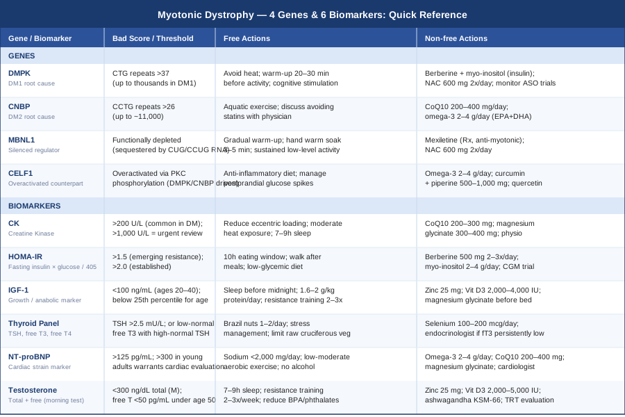
The table below consolidates all four genes and six biomarkers into a practical reference with action options at a glance.
10 Research Insights That Could Change How You Approach Myotonic Dystrophy
Most of what follows reflects the growing body of DM research and goes beyond what patients typically hear in routine neurology consultations. The perspective here is grounded in evidence — but challenges several assumptions that still shape standard care.
1. RNA toxicity, not just protein loss, is the real driver
For years, myotonic dystrophy was described primarily as a problem of DMPK protein deficiency. Current evidence — consolidated across dozens of studies since 2000 — makes clear that the expanded repeat RNA itself is the toxic agent. The sequestration of MBNL proteins by nuclear RNA foci better explains the multi-system clinical picture than protein loss alone. This distinction matters because it redirects therapeutic focus: the target is not restoring DMPK kinase activity but dismantling or blocking the toxic RNA foci.
2. Insulin resistance in DM1 is mechanistically unique
The insulin resistance of DM1 is not the same as metabolic syndrome. It arises from a specific splice form switch in the insulin receptor gene — one of the best-characterized examples of how splicing defects create real clinical disease. This means standard dietary interventions for insulin resistance work partly but do not address the receptor-level problem. It also means that INSR isoform testing — not yet widely available clinically — could eventually become a useful molecular severity marker for DM1.
3. Repeat length does not perfectly predict severity
The number of CTG repeats in DMPK broadly correlates with disease severity, but the relationship is imprecise. Somatic mosaicism — different cells carrying different repeat lengths within the same individual — creates significant complexity. Two people with similar repeat lengths can have very different phenotypes. Epigenetic factors, repeat configuration, and modifier genes all contribute. Genetic testing provides important but incomplete prognostic information.
4. The brain is more affected than most patients are told
Cognitive and personality changes in DM1 — executive dysfunction, increased apathy, hypersomnia — are direct neurological features of the disease, not secondary reactions to disability or fatigue. White matter changes on MRI are common and well-documented. The same splicing dysregulation that affects muscle affects neuronal function. This matters because it shapes how fatigue should be interpreted and managed: central fatigue in DM has a biological substrate distinct from peripheral muscle fatigue.
5. Mexiletine is underused for myotonia
Mexiletine is a sodium channel blocker with two randomized controlled trials demonstrating significant reduction of myotonia in DM1. It is not widely prescribed, partly due to cardiologist caution about sodium channel blockade in a population with cardiac involvement. The evidence supports its use at low doses (150 mg three times daily) with cardiac monitoring — and it can meaningfully improve daily function. Many patients who would benefit from it have never been offered it.
6. Sleep disorders are a biological feature, not a lifestyle problem
Excessive daytime sleepiness and abnormal sleep architecture in DM1 reflect central nervous system involvement — specifically in the brain regions that regulate sleep-wake cycles. Modafinil and methylphenidate have evidence for improving wakefulness in DM1 specifically. Sleep studies are appropriate for patients with significant hypersomnia. Treating this as a behavioral problem (better sleep hygiene alone) without considering the neurological substrate is a common clinical oversight.
7. Cardiac risk is present before symptoms develop
In DM1, subclinical conduction abnormalities on ECG are common before any cardiac symptoms appear. Annual 12-lead ECG and consideration of Holter monitoring is standard in guidelines, but not universally applied in practice. Pacemaker implantation for high-degree AV block can be lifesaving. The threshold for aggressive cardiac monitoring in asymptomatic DM1 patients should be lower than in the general population, full stop.
8. Antisense oligonucleotides are moving through clinical trials
Multiple companies are conducting Phase 1/2 trials of ASO and antibody-oligonucleotide conjugate (AOC) therapies targeting the expanded CUG RNA in DM1 muscle. Early results have shown CUG foci reduction and MBNL protein release in muscle biopsies. These are not approved treatments yet, but the pipeline is more active than in most rare diseases. Monitoring clinicaltrials.gov for open trials is a meaningful step for motivated patients.
9. Physical activity must be adapted, not avoided
The old advice to rest and avoid strenuous exercise in muscular dystrophy has been substantially revised. Moderate-intensity exercise — particularly aerobic and low-resistance activities — is safe and beneficial in DM. The evidence supports maintaining regular movement, with the caveat that high-intensity eccentric loading and exercise to failure should be avoided. Supervised physical therapy 1–2x/week is among the most evidence-supported ongoing interventions currently available.
10. Multi-disciplinary care gaps are the rule, not the exception
DM1 requires co-management by neurology, cardiology, pulmonology, endocrinology, ophthalmology, and physiotherapy. Most patients do not receive all of these routinely. Annual respiratory function testing (FVC, MIP, MEP) catches respiratory decline before crisis. Ophthalmology for cataracts is often overlooked. Endocrinology for testosterone, thyroid, and insulin resistance is rarely initiated from neurology. Being a proactive advocate for multi-disciplinary review — using the biomarker framework in this article as a starting point — closes this gap more effectively than waiting for any single specialist to see the whole picture.
Complementary Approaches With Meaningful Evidence
The following three modalities have relevant human clinical evidence for myotonic dystrophy or closely related neuromuscular and fatigue-dominant conditions. None replace medical management, and evidence quality varies — but each addresses a dimension of DM that standard care often underserves.
Breathing-Based Therapies
Respiratory muscle weakness is a defining feature of advanced DM1 and one of its most serious complications — respiratory failure is among the leading causes of DM-related mortality. Breathing-based therapies in this context are not relaxation techniques; they are targeted respiratory muscle training (RMT) — a structured approach to strengthening the diaphragm and accessory breathing muscles using threshold resistive devices. The physiological rationale is direct: DM1 causes progressive respiratory muscle weakness, and like other skeletal muscles, respiratory muscles respond to resistance training within their available capacity.
A randomized controlled trial by Rassler et al. (2011) demonstrated that inspiratory muscle training in patients with muscular dystrophy improved respiratory endurance and reduced perceived exertion without adverse events. Threshold devices (such as Threshold IMT from Respironics) provide adjustable resistance — 30% of maximal inspiratory pressure is a standard starting point, 5 sets of 5 breaths twice daily, progressing over weeks. Spirometry monitoring (FVC, PImax) is used to track progress and calibrate intensity.
For practical application: begin with formal spirometry to establish a baseline FVC and maximal inspiratory pressure. Start at the lowest device resistance setting, progressing only after two weeks at each level. Avoid training on days of respiratory illness. Annual pulmonary function tests are standard in DM1 care and provide the longitudinal data needed to calibrate training intensity over time. RMT does not reverse the underlying disease but can delay the point at which assisted ventilation becomes necessary — a meaningful clinical outcome.
Mindfulness Meditation and MBSR
Fatigue in myotonic dystrophy is multidimensional. Peripheral muscle fatigue, central nervous system hypersomnia, and the psychological weight of a progressive genetic condition all contribute. Mindfulness-Based Stress Reduction (MBSR) — an 8-week structured program of meditation, body scan, and mindful movement — addresses the central and psychological dimensions of fatigue without the physical demands that can exacerbate muscular symptoms. Its evidence base in chronic neurological conditions and fatigue syndromes is well established.
A systematic review by Grossman et al. (2004) in the Journal of Psychosomatic Research documented significant improvements in pain, fatigue, and quality of life across chronic illness populations using MBSR. For neuromuscular conditions specifically, a pilot study in multiple sclerosis — a condition with overlapping central fatigue mechanisms — showed significant improvements in fatigue and mental health. The 8-week standard protocol involves one 2.5-hour session per week plus daily home practice of 30–45 minutes; a reduced-intensity version (20 minutes daily) has shown similar effects in some populations and is more manageable for DM patients.
For DM patients, MBSR is particularly accessible because it requires no physical exertion, can be practiced lying down during high-fatigue periods, and digital programs (MBSR on apps such as Waking Up, Insight Timer, or the original Jon Kabat-Zinn recordings) require no out-of-pocket cost. The realistic target outcomes in DM are reduced fatigue catastrophizing, improved sleep quality, lower perceived pain, and better emotional regulation — none of which are exceptional claims, but all of which meaningfully contribute to daily function over time.
Biofeedback
Biofeedback uses real-time physiological monitoring — most commonly heart rate variability (HRV), surface electromyography (sEMG), or respiratory rate — to help individuals consciously influence states they would otherwise not have direct access to. In myotonic dystrophy, two applications are particularly relevant: HRV biofeedback for autonomic dysregulation and cardiac monitoring, and sEMG biofeedback for improving neuromuscular control in targeted movement rehabilitation.
HRV is well established as a marker of cardiac autonomic function, and autonomic neuropathy is documented in DM1. Daily HRV monitoring using a chest strap (Polar H10) or a validated wearable (Oura Ring, Garmin) provides actionable data on recovery status and cardiac load. A randomized controlled trial by Lehrer et al. (2010, Applied Psychophysiology and Biofeedback) demonstrated that HRV biofeedback training improved autonomic balance and reduced cardiopulmonary symptoms in patients with cardiac and pulmonary conditions. The protocol involves resonance-frequency breathing at approximately 5–6 breaths per minute for 20 minutes daily, guided by HRV feedback.
For DM patients, starting with a low-cost HRV app (Elite HRV, HRV4Training) and a compatible chest strap provides immediate, practical biofeedback data at under $100. More advanced sEMG biofeedback for hand and forearm muscles — useful in grip myotonia rehabilitation — is available through specialized physical therapists. Evidence is limited in DM specifically; the recommendation here is based on mechanism and adjacent condition evidence, and formal DM-specific trials are still needed. For any biofeedback protocol in a patient with known cardiac involvement, cardiologist input before starting is sensible.
Moving Forward
Myotonic dystrophy is a condition where the biology is genuinely complex and the clinical picture varies enormously between individuals. The six biomarkers covered here — CK, HOMA-IR, IGF-1, thyroid panel, NT-proBNP, and testosterone — give you a concrete, measurable framework that is accessible, affordable, and directly actionable. The genetic layer — DMPK, CNBP, MBNL1, and CELF1 — provides the mechanistic context that explains why these biomarkers move in the wrong direction and which downstream pathways are worth supporting.
The most useful next step is rarely the most complex one. Starting with a comprehensive blood panel that includes the biomarkers above, reviewing results with a physician who understands DM, and identifying the two or three numbers most out of range — then acting on those specifically — is more valuable than trying to implement every intervention simultaneously. Small, targeted adjustments to diet, sleep, and movement, supported where appropriate by well-chosen supplements, do not cure myotonic dystrophy. But they are how you take genuine control of the factors that remain within reach, and they are how better information becomes better outcomes.
Men's Health Neurological Endocrine & Metabolic
Musculoskeletal: Muscle Conditions
Neurological: Memory & Cognitive Conditions
Cardiovascular: Heart Conditions Heart Rhythm Conditions
Respiratory: Sleep & Breathing Disorders
Endocrine & Metabolic: Diabetes & Blood Sugar Thyroid Conditions