This article contains AI generated content.
· UpdatedNeonatal Lupus Genes and Biomarkers — 5 Genes And 6 Biomarkers To Track
Introduction
Being told that your newborn has neonatal lupus — or discovering during pregnancy that you carry anti-Ro antibodies — tends to produce two simultaneous experiences: a clinical explanation of what is happening, and a frustrating sense that you still don't know what to do about it. Clinicians describe the mechanism, outline a monitoring schedule, and reassure you that most cases resolve on their own. That is true, and it matters. But it leaves a significant gap between understanding and action.
Neonatal lupus is not a hereditary disease in the conventional sense. Infants don't develop it independently — they carry it temporarily through antibodies borrowed from their mothers. Those antibodies, primarily anti-Ro/SSA and sometimes anti-La/SSB, are IgG immunoglobulins that cross the placenta during the second and third trimesters and temporarily disrupt the infant's immune balance. For most affected newborns, the consequences — skin rashes, mild platelet drops, transient liver enzyme elevation — resolve completely within six months as maternal antibodies clear. But for a small subset, those same antibodies interfere with the electrical conduction of the fetal heart during a narrow developmental window, and that interference can be permanent.
The risk is not evenly distributed, and that's what generic information tends to miss. Specific antibody subtypes, specific titers, specific maternal gene variants — these factors meaningfully influence who is at higher risk, who needs more intensive monitoring, and where targeted intervention is justified. A woman with high anti-Ro52 titers who carries HLA-DR3 and has a prior affected pregnancy is not in the same risk category as someone with an incidentally detected weak positive anti-Ro result. Treating them the same way is imprecise at best.
Two complementary strategies run in parallel here. The first — the more clinically immediate — maps out six biomarkers that matter in neonatal lupus, covering what each reveals, what it costs to measure, and what can realistically be done when results are unfavorable. The second examines five maternal genetic variants that shape anti-Ro antibody production and immune reactivity, with specific protocols for each. Beyond those, a section on Ben Lynch's framework for actionable genetics and a complementary approaches section complete a picture that standard clinical guidance rarely offers in one place.
Summary
This article walks through the six most clinically important biomarkers in neonatal lupus — anti-Ro/SSA subtypes (Ro52 and Ro60), anti-La/SSB, complement C3 and C4, complete blood count, liver function tests, and cardiac assessment. For each, you'll find what it reveals, how to measure it with cost ranges, and what to do when results are unfavorable — with and without supplements or equipment. The genetics section then explains five maternal gene variants (HLA-DR3, IRF5, STAT4, the C4A null allele, and FCGR2A) that shape antibody production and immune reactivity, each with targeted lifestyle and supplement protocols. A summary of Ben Lynch's Dirty Genes translates epigenetics research into ten practical takeaways. The complementary section covers Sarah Ballantyne's Autoimmune Protocol, mindfulness-based stress reduction, and microbiome-directed strategies — all grounded in human clinical evidence. If "monitor and wait" has been your only plan, this article offers a more detailed map of what is happening and where it can be influenced.
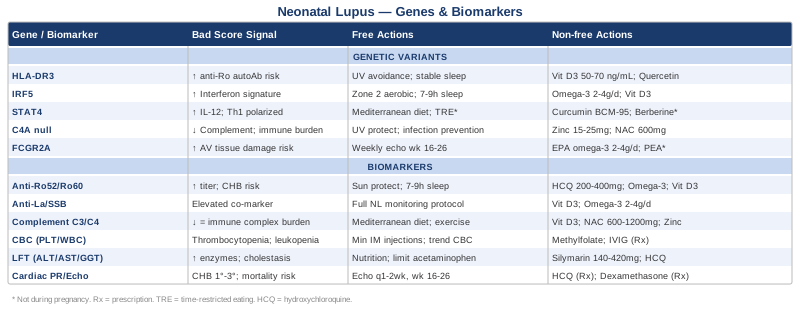
6 Biomarkers Worth Tracking Closely
Tracking the right biomarkers is the most immediately actionable step available to pregnant women with known autoantibodies and to parents of infants with neonatal lupus. These six markers collectively cover the cardiovascular, hematologic, hepatic, and immunological dimensions of the condition. Not all are relevant at every stage — timing and clinical context shape which to prioritize — but understanding each one enables better conversations with your care team and more informed decisions throughout.
1. Anti-Ro/SSA Antibodies — Ro52 and Ro60 Subtypes
Why It Matters
Anti-Ro/SSA antibodies are the central driver of neonatal lupus. They are present in approximately 95 percent of confirmed cases and are found in the maternal serum of virtually every woman whose infant develops congenital heart block (CHB) — the most serious and potentially permanent manifestation of the condition. What makes this biomarker particularly important is that the Ro52 and Ro60 subtypes do not behave the same way. Ro52, encoded by the TRIM21 gene, is more strongly associated with cardiac injury. High titers of Ro52-specific antibodies represent a meaningful independent risk signal for CHB, separate from overall anti-Ro levels. Most clinical laboratories still report a combined anti-Ro/SSA result, but requesting subtype differentiation provides substantially more information.
Maternal antibody titer matters as well. Higher titers correlate with higher probability of neonatal manifestations, and the recurrence risk in subsequent pregnancies — approximately 15 to 18 percent after one affected child — is highest in women with persistently elevated Ro52 titers. Tracking changes in titer across a pregnancy adds a time dimension to risk stratification.
How to Measure It
Anti-Ro/SSA antibodies are measured via ELISA or multiplex immunoassay from a standard blood draw. Available through all major commercial laboratories and most hospital systems. Cost range: $100–$300 as part of an autoantibody panel that typically includes anti-La, ANA, and sometimes anti-dsDNA. Subtype-specific testing (Ro52 vs. Ro60 separately) may require a specialized rheumatology or immunology reference lab and can add $50–$150 to the panel. In a known anti-Ro positive woman, retesting each trimester is reasonable to track trajectory.
If the Score Is Bad: The Plan Without Supplements
A high anti-Ro52 titer in a pregnant woman warrants structured fetal cardiac surveillance. The evidence-based protocol is fetal echocardiography every one to two weeks from gestational weeks 16 through 26 — the critical window during which CHB almost exclusively develops. PR interval prolongation (first-degree block) is detectable before it progresses to complete block, and early detection allows for closer monitoring and faster clinical response.
Beyond surveillance, UV light is a reproducible trigger for anti-Ro antibody flares. Consistent sun protection — high-SPF mineral sunscreen, protective clothing, minimizing midday sun exposure — reduces one of the most modifiable environmental drivers of antibody production. Sleep quality is a less discussed but equally meaningful lever. Chronic sleep disruption increases systemic interferon signaling, which amplifies antibody production pathways. Protecting 7–9 hours of quality sleep per night is not a soft lifestyle recommendation — it operates through documented neuroimmune pathways.
If the Score Is Bad: The Plan With Supplements or Equipment
Hydroxychloroquine (HCQ) is the intervention with the strongest evidence base for reducing CHB risk in anti-Ro positive pregnancies. The PATCH study (Izmirly PM et al., 2020, Journal of the American College of Cardiology) demonstrated a significant reduction in CHB recurrence in anti-Ro positive women treated with HCQ compared to historical controls. Standard dosing is 200–400 mg/day under rheumatologic supervision. This is a prescription medication, not a supplement, but knowing about it means you can initiate an informed conversation with your specialist.
For adjunctive support: vitamin D3 at 2,000–4,000 IU/day (guided by serum 25-OH vitamin D levels, targeting 50–70 ng/mL) has documented immunomodulatory effects including downregulation of autoreactive B cell populations. Omega-3 fatty acids at 2–4 g EPA+DHA per day reduce pro-inflammatory eicosanoids and have shown modest anti-Ro titer reduction in small observational studies. Both are considered safe during pregnancy at these doses. Take omega-3s with food to minimize GI discomfort; no cycling required. Allow 8–12 weeks to see measurable anti-inflammatory effect.
2. Anti-La/SSB Antibodies
Why It Matters
Anti-La/SSB antibodies are found in 50 to 75 percent of neonatal lupus cases, almost always alongside anti-Ro. They are rarely the sole antibody present. Their clinical interpretation has evolved: historically considered a straightforward marker of disease severity, more nuanced analyses suggest that the presence of anti-La may in some cases be associated with a different risk profile than anti-Ro52 alone — with some data suggesting a possible moderating effect when both are co-present at high titers. The picture is not fully settled, but knowing your anti-La status completes the antibody profile in ways that can influence clinical decision-making.
How to Measure It
Anti-La/SSB is measured by the same ELISA or multiplex panel as anti-Ro, typically reported together. No additional cost when bundled in the standard autoantibody panel ($100–$300). Results are expressed as a titer or as positive/negative depending on the laboratory. Semi-quantitative values (expressed in U/mL or IU/mL) are more useful than binary positive/negative for tracking over time.
If the Score Is Bad: The Plan Without Supplements
Elevated anti-La alongside elevated anti-Ro52 reinforces the rationale for the full neonatal lupus monitoring protocol. In the newborn, this includes: complete skin examination at birth, CBC and differential in the first 24–48 hours, liver function panel, and ECG. In the mother during pregnancy, it supports the case for the fetal echocardiography surveillance schedule described above.
No intervention is specifically and exclusively targeted at anti-La alone — management is driven by the overall antibody profile and clinical risk assessment.
If the Score Is Bad: The Plan With Supplements or Equipment
The same supplementation framework as for anti-Ro applies: vitamin D3 optimization, omega-3 at 2–4 g/day, and HCQ under medical supervision where appropriate. No supplement has specific evidence for selectively reducing anti-La titers. Prioritize the interventions with the broadest autoimmune modulating evidence, starting with vitamin D and omega-3 as the safest and most supported combination. Begin preconception if possible, continue through the third trimester.
3. Complement C3 and C4
Why It Matters
The complement system is a network of immune proteins responsible for clearing immune complexes — clusters of antibodies bound to their targets. In autoantibody-driven conditions like neonatal lupus, complement proteins are consumed at elevated rates, causing serum levels of C3 and C4 to fall. Low complement levels reflect ongoing immune activation and immune complex burden.
C4 carries a dual significance here. It can be low due to active disease (consumption exceeding production), but it can also be genetically low — the C4A null allele, discussed in the genetics section, produces a structural deletion that results in a reduced baseline C4 level even in the absence of active disease. Knowing whether your C4 is low at baseline before pregnancy or dropping during pregnancy helps distinguish these two entirely different situations. In the infant, low complement supports the clinical diagnosis and helps quantify disease activity during the first weeks of life.
How to Measure It
C3 and C4 are measured from a serum blood draw, available at all commercial and hospital laboratories. Cost range: $40–$100 per marker; ordered together as a complement panel. Normal ranges: C3 typically 90–180 mg/dL; C4 typically 16–47 mg/dL. For women with known anti-Ro antibodies, establishing a preconception baseline is particularly useful so that pregnancy-related changes can be interpreted accurately.
If the Score Is Bad: The Plan Without Supplements
Consistently low complement in the mother during pregnancy is a signal to intensify monitoring rather than to treat directly. Factored alongside antibody titers, it strengthens the clinical picture of active immune complex formation. From a lifestyle standpoint, the factors that most directly reduce complement consumption are those that reduce systemic inflammation: Mediterranean-pattern eating (abundant vegetables, olive oil, fatty fish, limited ultra-processed foods), adequate sleep, and moderate regular exercise. These are not peripheral suggestions — they reduce the immunological demand on the complement system by lowering the antigen-antibody complex burden.
If the Score Is Bad: The Plan With Supplements or Equipment
Vitamin D3 (at levels sufficient to reach 50–70 ng/mL serum concentration) supports complement regulatory pathways and reduces antigen-driven immune activation more broadly. N-acetyl cysteine (NAC) at 600–1,200 mg/day is an antioxidant precursor to glutathione with immunomodulatory properties that may help reduce complement consumption in autoimmune settings; a reasonable cycling protocol is 8 weeks on followed by a 2-week break, with GI discomfort as the main side effect. Zinc glycinate or zinc picolinate (15–25 mg/day with food) supports complement protein synthesis; cycle 5 days on, 2 days off to prevent copper displacement, or co-supplement with 1–2 mg copper if sustained use is needed. Evidence for these specific supplement effects on complement in neonatal lupus is preliminary — they reflect mechanistic plausibility supported by autoimmune and immune biology research rather than neonatal lupus-specific trials.
4. Complete Blood Count With Differential
Why It Matters
Hematologic manifestations are among the most common clinical findings in neonatal lupus, estimated to affect 10 to 15 percent of confirmed cases. The key abnormalities are thrombocytopenia (low platelet count), leukopenia (low white blood cell count), and hemolytic anemia. These occur because maternal anti-Ro antibodies bind to surface antigens on blood cells and trigger their destruction through immune mechanisms.
The reassuring part is that these abnormalities are almost universally transient — they resolve within 4 to 6 months as maternal antibodies clear the infant's circulation. The concerning part is that severe thrombocytopenia (platelet count below 50,000/µL) carries a bleeding risk that warrants prompt clinical attention. Catching the trend early through regular CBC monitoring is what prevents a manageable finding from becoming an emergency.
How to Measure It
Standard CBC with differential from a routine blood draw — the most widely available and lowest-cost lab test in clinical medicine. Cost range: $25–$80. In infants born to anti-Ro positive mothers, best practice is a CBC at birth, repeated weekly or biweekly until counts normalize or a clear trend toward normal is established. Platelet count and the white blood cell differential (particularly neutrophil and lymphocyte counts) are the primary targets of interest in this context.
If the Score Is Bad: The Plan Without Supplements
For infants with thrombocytopenia: minimize unnecessary intramuscular injections, avoid any medications that further impair platelet function (including standard NSAIDs and aspirin unless otherwise clinically indicated), and maintain close monitoring. For platelet counts above 50,000/µL without active bleeding, watchful monitoring is the standard approach. Below that threshold, or with active bleeding symptoms, escalate to pediatric hematology. IVIG or short-course corticosteroids may be initiated at this threshold — these are medical decisions made in a clinical setting, not home strategies, but knowing they exist helps you advocate for timely evaluation.
For the mother before delivery: confirm all medications with your obstetric and rheumatology team, particularly anything with anticoagulant or antiplatelet properties.
If the Score Is Bad: The Plan With Supplements or Equipment
For the mother preconception: ensuring sufficiency in iron (ferritin), B12 (methylcobalamin preferred over cyanocobalamin for absorption efficiency), and folate (RBC folate, not just serum) supports healthy hematopoiesis. If MTHFR variants are present (see genetics section), switching from folic acid to methylfolate (400–800 mcg/day) is a straightforward adjustment that may meaningfully improve folate utilization. Standard prenatal vitamins cover these bases for most people, but pre-testing levels before relying on them is worth the marginal cost.
Maternal IVIG during late pregnancy has been studied as a strategy to reduce transplacental antibody transmission to the infant. Evidence is mixed — some studies showed modest antibody level reduction, others showed no significant benefit. Discuss this option with a maternal-fetal medicine specialist in documented high-risk cases, specifically those with prior hematologic involvement in a previous affected infant.
5. Liver Function Tests (ALT, AST, GGT, Bilirubin)
Why It Matters
Hepatic involvement is a less-publicized but clinically significant dimension of neonatal lupus, estimated to affect 10 to 25 percent of confirmed cases depending on the diagnostic criteria used. Presentations range from mild, asymptomatic enzyme elevation to cholestatic hepatitis, where bile flow from the liver is impaired. Like the hematologic findings, hepatic involvement is almost always transient — it resolves completely by 6 to 12 months of age in the vast majority of cases.
The markers to track in the infant are ALT and AST for hepatocellular injury, GGT as a sensitive indicator of bile duct involvement, and total and direct bilirubin to characterize the pattern of jaundice when present. Together, these form a sufficient hepatic profile for tracking neonatal lupus-related liver involvement.
How to Measure It
Comprehensive metabolic panel (CMP) or a dedicated liver function panel from a standard blood draw. Cost range: $40–$100. In infants with confirmed neonatal lupus or high-titer maternal antibodies, liver function is checked at birth and repeated at 4–6 weeks if initial values are elevated, then monthly until normalization. Abdominal ultrasound ($200–$500) may be added to assess liver size, echogenicity, and biliary anatomy if enzyme elevation is significant or prolonged.
If the Score Is Bad: The Plan Without Supplements
For the affected infant: ensure adequate nutrition and hydration to support hepatic recovery. Breastfeeding is generally appropriate and not contraindicated by neonatal lupus liver involvement. Avoid unnecessary hepatic stressors — acetaminophen should be used at the minimum necessary dose and duration; no herbal supplements without explicit pediatrician approval. Recheck liver enzymes every 4 to 6 weeks until normalization is confirmed.
For cholestatic presentations (elevated GGT and direct bilirubin), a pediatric gastroenterologist or hepatologist should be consulted. Ursodeoxycholic acid (UDCA) is sometimes prescribed to facilitate bile flow in symptomatic cholestasis — a clinical decision that belongs in a pediatric hepatology context.
If the Score Is Bad: The Plan With Supplements or Equipment
For the mother during pregnancy: silymarin (milk thistle extract, standardized to 70–80% silymarin, 140–420 mg/day) has a documented hepatoprotective profile in human studies and is generally considered low-risk in pregnancy, though obstetric confirmation is appropriate before starting. The mechanism — antioxidant and anti-inflammatory effects on hepatocytes — is relevant to the maternal liver stress that can accompany autoimmune conditions during pregnancy.
More directly, maternal HCQ use during pregnancy appears to reduce the overall severity of neonatal lupus manifestations including hepatic involvement. The research on HCQ in this context suggests a broader protective effect beyond cardiac outcomes, which is one more reason to discuss it with your rheumatologist if you are anti-Ro positive. For the infant, there is no over-the-counter supplement protocol appropriate to add to the clinical monitoring plan — watchful support and close follow-up are the cornerstones.
6. Cardiac Assessment: PR Interval and Echocardiogram
Why It Matters
This is the most consequential biomarker area in neonatal lupus monitoring, and the one where timing matters most acutely. Congenital heart block (CHB) occurs when anti-Ro antibodies — particularly anti-Ro52 — bind to L-type calcium channels and cause inflammation in the atrioventricular (AV) node of the fetal heart, disrupting electrical conduction between the atria and ventricles. First-degree heart block (mildly prolonged PR interval) can be transient or reversible. Third-degree (complete) heart block is irreversible in nearly all cases and may require a permanent pacemaker. Mortality in complete CHB, without pacing, is approximately 20 percent.
The critical window is narrow and well-defined: CHB almost exclusively develops between 16 and 26 weeks of gestation. Surveillance concentrated in this window is what makes a real clinical difference.
How to Measure It
Two complementary modalities:
Fetal echocardiogram: performed by a maternal-fetal medicine specialist or pediatric cardiologist with obstetric echocardiography expertise. Evaluates the mechanical PR interval (time from atrial contraction to ventricular contraction), fetal heart rate, and structural cardiac anatomy. Cost range: $800–$3,000 per exam, varying significantly by facility and geography. Most insurance plans cover this with appropriate documentation of anti-Ro positivity. Recommended frequency in high-risk pregnancies: every 1–2 weeks between weeks 16 and 26, then monthly if results are normal throughout that window.
Neonatal ECG: a 12-lead ECG or rhythm strip in the newborn to assess postnatal PR interval and AV conduction. Cost range: $50–$200. All infants born to anti-Ro positive mothers should receive an ECG within the first 24–48 hours of life, regardless of how the fetal echos looked.
If the Score Is Bad: The Plan Without Supplements
Detection of first-degree heart block on fetal echo (mechanical PR interval above the 98th percentile for gestational age) warrants immediate escalation to weekly surveillance and pediatric cardiology consultation. Any medications that slow AV conduction should be reviewed and ideally discontinued if clinically safe to do so.
Complete heart block detected in utero or postnatally should prompt urgent pediatric electrophysiology referral. The decision about timing of pacemaker implantation is complex and depends on ventricular rate, hemodynamic stability, and gestational age — this is a decision made by specialists, but knowing to advocate for that referral quickly matters. Pacing in infants with CHB has dramatically improved long-term outcomes.
If the Score Is Bad: The Plan With Supplements or Equipment
Hydroxychloroquine again anchors the pharmacological approach. The cumulative evidence — from multiple observational cohorts and the PATCH study — is strong enough that HCQ in anti-Ro positive pregnancy is becoming the standard of care at major academic rheumatology centers, particularly in women with prior CHB history. The recurrence risk reduction with HCQ is estimated at approximately 50 percent in documented high-risk cases.
Dexamethasone (a fluorinated corticosteroid that crosses the placenta) has been used in cases of second-degree block detected on fetal echo. The PRIDE observational study found limited overall benefit from dexamethasone in reversing established second-degree block, and it carries significant fetal and maternal side effects with prolonged use. Most current expert consensus reserves it for carefully selected cases — another reason this conversation must happen with a maternal-fetal medicine specialist, not as a self-directed decision.
Omega-3 fatty acids at 2–4 g EPA+DHA/day are a reasonable, risk-free adjunct throughout pregnancy in anti-Ro positive women. The mechanism — reduction of AV node inflammatory signaling — is biologically plausible and carries no fetal risk at these doses.
---
The six biomarkers above map the current state: what antibodies are circulating, how the immune system is responding, and what tissues are affected. What they don't explain is why some women produce these antibodies in the first place — and why the risk varies so substantially between individuals with similar antibody profiles. That question is where genetics enters the picture.
5 Genetic Variants That Shape the Risk Landscape
Genetics in neonatal lupus is primarily a story about the mother. The variants that matter most are those that predispose a woman to producing anti-Ro antibodies, to maintaining a more reactive immune baseline, and to generating a stronger interferon response when triggered. Understanding these variants doesn't change the clinical picture that biomarkers reveal, but it explains the underlying terrain — and opens targeted lifestyle and nutritional levers that are different from generic immune support.
Gene 1: HLA-DRB1*03:01 (HLA-DR3)
What It Affects
The human leukocyte antigen system controls how the immune system distinguishes self from non-self. HLA-DRB1*03:01 — commonly called HLA-DR3 — is the single strongest genetic predictor of anti-Ro antibody production. It is the same allele that drives primary Sjögren's syndrome susceptibility, and it functions by shaping how T cells present self-antigens, including Ro52 and Ro60, to B cells. Women carrying HLA-DR3 have substantially elevated lifetime probability of developing anti-Ro autoantibodies, particularly when combined with environmental or hormonal triggers. Roughly 90 percent of women with primary Sjögren's and anti-Ro antibodies carry this allele.
HLA type is available through clinical HLA typing (often ordered in transplant or rheumatology contexts) or through consumer-facing genomic testing with clinical interpretation. HLA-DR3 is typically reported in the DRB1 gene results.
If the Gene Is Bad: The Plan Without Supplements
HLA type cannot be modified, but its downstream effects can be meaningfully managed. UV avoidance is the highest-impact non-pharmacological strategy for HLA-DR3 carriers: UV light drives Ro antigen upregulation in keratinocytes, making them available targets for autoantibody binding. Year-round mineral sunscreen (SPF 50+), UV-protective clothing, and minimizing midday sun exposure reduce one of the most reproducible triggers of anti-Ro flares.
Estrogen amplifies the autoantibody-producing pathways that HLA-DR3 sensitizes. The transition off hormonal contraception before attempting conception may be worth timing carefully with your rheumatologist, particularly if antibody titers have risen during hormonal use. Circadian rhythm stability — consistent sleep-wake timing, morning bright light exposure, minimal blue light after 9 PM — is an underappreciated regulator of dendritic cell activity, the cell type where HLA-DR3's effects are most pronounced.
If the Gene Is Bad: The Plan With Supplements or Equipment
Vitamin D deficiency is particularly consequential in HLA-DR3 carriers. This allele is associated with impaired 1,25-dihydroxyvitamin D-mediated immune tolerance. Rather than targeting the conventional "normal" cutoff of 30 ng/mL, target 25-OH vitamin D at 50–70 ng/mL, which typically requires 4,000–6,000 IU/day in deficient individuals. Test every 3 months during dose adjustment; toxicity is uncommon below 10,000 IU/day with monitoring. No specific cycling needed.
Quercetin (500–1,000 mg/day, cycling 8 weeks on, 2–4 weeks off) has documented effects on antigen presentation pathways and dendritic cell activation that are mechanistically relevant to HLA-DR3-driven autoantibody production. Evidence is limited to cell culture and early human studies — it is a biologically plausible adjunct, not an established treatment.
Gene 2: IRF5 (Interferon Regulatory Factor 5)
What It Affects
IRF5 encodes a transcription factor that drives type I interferon (IFN-α, IFN-β) production — the immune cytokines that are chronically elevated in lupus, Sjögren's, and related autoimmune conditions. The risk variants rs2004640 and rs10954213 are among the most replicated genetic associations with systemic lupus erythematosus and Sjögren's syndrome in genome-wide association studies. When IRF5 is constitutively overactive — which these variants promote — the interferon signature (a measurable pattern of gene expression across hundreds of interferon-stimulated genes) is amplified, creating an immune environment where autoreactive B cells are more easily generated, expanded, and maintained.
Importantly, IRF5 activity is not genetically fixed. It responds to environmental inputs including omega-3 fatty acid levels, vitamin D, gut microbiome signals, sleep, and exercise — making it one of the more actionable genetic risk factors in this condition.
If the Gene Is Bad: The Plan Without Supplements
Zone 2 aerobic exercise (30–45 minutes, 4–5 days per week) consistently downregulates IRF5 expression in peripheral blood mononuclear cells and reduces circulating type I interferon levels in autoimmune populations. Excessive intensity (ultra-endurance training, very high-volume training) can paradoxically spike interferon signaling — moderate, consistent aerobic work is the target. Strength training 2–3 days per week adds complementary anti-inflammatory benefit through myokine release (particularly IL-6 in the post-exercise, anti-inflammatory context).
Sleep is the most reliable lever on IRF5 activity. IRF5 and its downstream interferon production peak acutely with sleep disruption. Chronic short sleep (below 6 hours) is associated with measurably elevated interferon signatures in autoimmune populations. Prioritizing sleep above other lifestyle interventions is not an overcorrection — it is mechanistically justified.
If the Gene Is Bad: The Plan With Supplements or Equipment
Omega-3 fatty acids (EPA+DHA, 2–4 g/day) are the most evidence-backed nutritional modifier of IRF5-driven interferon production. EPA and DHA compete with arachidonic acid for metabolism and generate resolvins and protectins that directly inhibit IRF5 transcriptional activity in macrophages and plasmacytoid dendritic cells. Take with food to reduce GI effects; no cycling required; allow 8–12 weeks for measurable effect.
Vitamin D3 (targeting 50–70 ng/mL as above) directly inhibits IRF5 transcription through the vitamin D receptor — this is one of the more mechanistically specific vitamin D effects in autoimmunity, supported by both in vitro and human observational data. Low-dose naltrexone (LDN, 1.5–4.5 mg at bedtime) is an off-label intervention with growing autoimmune use that modulates endorphin receptor signaling to reduce interferon-related immune activation. It requires a prescription from a physician experienced in autoimmune management and is not appropriate during pregnancy.
Gene 3: STAT4 (Signal Transducer and Activator of Transcription 4)
What It Affects
STAT4 mediates signaling downstream of IL-12 and type I interferons — the same pathway nodes that IRF5 variants dysregulate upstream. The risk SNP rs7574865 (the T allele) is strongly associated with systemic lupus, Sjögren's, rheumatoid arthritis, and anti-Ro antibody production. In women who carry risk variants in both IRF5 and STAT4, the combined effect on interferon pathway hyperactivity is additive. Knowing STAT4 status alongside IRF5 completes a more precise genetic risk profile for anti-Ro antibody production.
STAT4 also mediates Th1 immune polarization — biasing the adaptive immune response toward the cell-mediated arm that, when dysregulated, contributes to autoantibody persistence.
If the Gene Is Bad: The Plan Without Supplements
Dietary quality is the most accessible non-supplement lever on STAT4-driven IL-12 and interferon signaling. Mediterranean-pattern eating — high in olive oil, fatty fish, diverse vegetables, legumes, and low in ultra-processed foods — consistently reduces IL-12 production and Th1 polarization in multiple human studies. This is not a generic dietary recommendation; the mechanism is specific to the STAT4 pathway.
Time-restricted eating (16:8 pattern — eating within an 8-hour window) has documented effects on STAT4-related immune signaling through AMPK activation and mTOR inhibition. This is not appropriate during pregnancy and warrants caution while breastfeeding but may be useful preconception or postpartum as a metabolic immune-regulating strategy.
If the Gene Is Bad: The Plan With Supplements or Equipment
Curcumin (as phospholipid-complexed BCM-95 or nanoparticle theracurmin, 500–1,000 mg/day) has reasonably documented inhibitory effects on STAT4 signaling through NF-κB suppression. Bioavailability is the critical variable — standard curcumin powder has very poor absorption; choose a bioavailable formulation. Cycle 8–12 weeks on, 2–4 weeks off. GI sensitivity is the main side effect; take with food.
Resveratrol (250–500 mg/day) shows STAT4 pathway modulation through similar NF-κB mechanisms in autoimmune models. Evidence is predominantly preclinical. Cycle 8 weeks on, 4 weeks off; avoid in pregnancy due to limited safety data. Berberine (500 mg, 2–3 times daily with meals) shows meaningful immunomodulatory effects on STAT4 and related inflammatory transcription factors and additionally benefits gut barrier integrity — a relevant combination given the gut-immune connection in autoimmunity. Not appropriate in pregnancy; cycle 8 weeks on, 2–4 weeks off.
Gene 4: C4A Null Allele
What It Affects
The C4A null allele is a structural gene variant — a copy number deletion in the C4A locus — that results in reduced baseline serum levels of complement component C4. Normally, C4 plays a critical role in the classical complement pathway for clearing immune complexes. When C4 is genetically low, immune complexes accumulate more readily, promote antibody-driven inflammation, and impair the tolerogenic signals that normally suppress autoreactive B cells. C4A null alleles are significantly enriched in lupus and Sjögren's populations compared to the general population.
This variant directly links two layers of this article: it explains why some women have persistently low C4 as a baseline trait (not a disease marker), and it predicts a more permissive environment for the anti-Ro antibody production that causes neonatal lupus.
If the Gene Is Bad: The Plan Without Supplements
Establishing a pre-pregnancy C4 baseline is the most practically important step for a known C4A null carrier. A genetically low C4 (stable low baseline) is clinically different from a pathologically falling C4 (reflecting active immune complex consumption). That distinction changes how physicians interpret the biomarker during pregnancy.
Reducing the immune complex burden that the compromised C4 system must manage is the lifestyle focus: consistent UV protection (UV drives immune complex deposition in skin), infection prevention (infections are major drivers of complement-consuming antibody flares), and stress management (chronic low-grade cortisol dysregulates complement regulatory protein expression). These are all meaningful levers operating through documented complement biology.
If the Gene Is Bad: The Plan With Supplements or Equipment
Supporting the remaining complement pathway function is the supplemental focus. Vitamin D3 at sufficient serum levels supports complement regulatory expression broadly. Zinc glycinate or zinc picolinate (15–25 mg/day with food) is required for complement protein synthesis — zinc deficiency is associated with reduced complement components across the board. Cycle 5 days on, 2 days off, or supplement with 1–2 mg copper if sustained daily zinc use exceeds 4–6 weeks.
NAC (600–900 mg/day, cycling 8 weeks on, 2 weeks off) supports glutathione availability, which protects complement proteins from oxidative inactivation — a less-cited but mechanistically valid connection.
Gene 5: FCGR2A (Fc Gamma Receptor IIA)
What It Affects
FCGR2A encodes an Fc gamma receptor expressed on monocytes, neutrophils, and platelets that binds to the Fc region of IgG antibodies — including the anti-Ro IgG that drives neonatal lupus. The rs1801274 polymorphism (H131R) affects the binding efficiency of this receptor to IgG subclasses. The R131 variant (arginine) binds IgG2 less efficiently and alters the effector function of cells that encounter anti-Ro-coated targets. At the fetal tissue level, this may influence how severely maternal anti-Ro antibodies damage AV node cardiomyocytes — affecting not whether CHB occurs but potentially how severe the resulting damage is.
FCGR2A genotype is available through clinical exome sequencing panels and some autoimmune genomic testing services.
If the Gene Is Bad: The Plan Without Supplements
The primary implication of FCGR2A risk variants in this context is that they support the rationale for more intensive fetal cardiac surveillance — knowing this variant is present adds one more reason to stay consistent with the weekly echocardiography protocol during weeks 16 to 26. Chronic inflammation increases FCGR2A expression on immune effector cells, amplifying its effects. All inflammation-reducing lifestyle strategies — dietary quality, sleep, moderate exercise — remain relevant here, not as FCGR2A-specific interventions but as modulators of the inflammatory context in which FcγR-mediated effects play out.
If the Gene Is Bad: The Plan With Supplements or Equipment
EPA-rich omega-3 supplementation (2–4 g/day) has in vitro evidence of modulating Fc gamma receptor expression and activity on immune effector cells. At these doses with food, this is a safe and well-supported adjunct for any anti-Ro positive pregnancy.
Palmitoylethanolamide (PEA, 600–1,200 mg/day) is an endogenous fatty acid amide with anti-inflammatory properties that operates partly through PPAR-α activation, which dampens FcγR-mediated inflammatory cascades in mast cells and macrophages. Evidence comes primarily from pain and neuroinflammation studies; direct FCGR2A-specific data are limited. No cycling required; well tolerated. This is a reasonable adjunct for women managing complex autoimmune inflammation who are not pregnant — consult a physician for pregnancy safety assessment.
---
Knowing your genes and your biomarkers provides a detailed biological map. What Ben Lynch's work on genetic variants adds is a framework for what to do with that map in practical terms — organized around the principle that genetic risk is modifiable, not fixed.
What Dirty Genes by Ben Lynch Gets Right About Immune Variants
Ben Lynch's 2018 book Dirty Genes argues that genetic variants are not destiny — they are defaults that respond to environmental inputs including nutrition, sleep, stress, and toxin load. Lynch calls variants with clear functional consequences "dirty genes" and proposes lifestyle and nutritional protocols to "clean" them. While the book focuses primarily on genes like MTHFR, COMT, and MAOA, the framework extends naturally to immune-relevant variants like IRF5 and STAT4 that shape neonatal lupus risk. The book synthesizes a large amount of biochemistry and genomic research into actionable, practical protocols — which is what makes it genuinely useful in a clinical context rather than purely academic.
Below are the ten most impactful insights from Lynch's work as applied to the immune gene variants most relevant in neonatal lupus.
1. Gene Variants Shift the Odds, They Don't Lock the Outcome
Lynch's core argument is that variants like HLA-DR3, IRF5, and STAT4 represent a higher-probability pathway, not a guaranteed destination. Every one of these variants has a large population of carriers who never develop significant autoimmune disease — because their environment did not sufficiently trigger the pathway. Understanding this reframes the entire conversation from "I have a bad gene" to "I have a gene that requires me to pay attention to specific inputs."
2. MTHFR Affects Far More Than Folate
The MTHFR gene — which affects conversion of folic acid to the active methylfolate — is relevant to neonatal lupus risk through its role in methylation, the process by which gene expression is regulated. Impaired methylation (from MTHFR variants C677T or A1298C) can reduce the body's ability to silence inflammatory genes, including immune regulatory sequences affected by the IRF5 and STAT4 variants discussed above. Lynch's recommendation: switch from folic acid to methylfolate (400–800 mcg/day) and from standard B12 to methylcobalamin or adenosylcobalamin. This is particularly important during pregnancy, where folic acid supplementation is standard but MTHFR carriers may not be converting it adequately.
3. Glutathione Is the Master Antioxidant for Dirty Immune Genes
Lynch emphasizes that oxidative stress "dirties" multiple genes simultaneously — including immune-regulatory variants that perform differently under oxidative conditions than under reduced conditions. Glutathione (or its precursor NAC at 600–1,200 mg/day) is his central tool for multiple dirty genes. In the neonatal lupus context, this is relevant because oxidative stress amplifies AV node inflammation triggered by anti-Ro antibodies. Liposomal glutathione (250–500 mg/day) is a more bioavailable alternative to oral glutathione. Cycle 8 weeks on, 2–4 weeks off.
4. The Gut Microbiome Modifies Gene Expression Through Short-Chain Fatty Acids
Lynch devotes significant attention to how butyrate — produced by gut bacteria fermenting dietary fiber — turns off inflammatory gene expression through histone deacetylase (HDAC) inhibition. This is not abstract: specific bacterial species that produce butyrate (Faecalibacterium prausnitzii, Roseburia spp.) are consistently reduced in lupus and Sjögren's patients. Increasing dietary fiber (30–40 g/day from diverse plant sources) and considering targeted butyrate supplementation (sodium butyrate or tributyrin, 300–600 mg/day with meals) is a meaningful epigenetic lever on multiple inflammatory gene variants.
5. Sleep Is the Most Powerful Cleaner for Dirty Immune Genes
Lynch is emphatic that no supplement protocol fixes a dirty gene if sleep is inadequate. The immune regulatory functions of deep sleep include DNA repair, cytokine calibration, and IRF5/STAT4 expression reset. He references research showing that chronic short sleep measurably worsens multiple immune gene variants' functional expression. This is not a throwaway lifestyle recommendation — it sits at the top of his intervention hierarchy because it is the most powerful systemic modifier.
6. Omega-3s Clean Multiple Dirty Immune Genes Simultaneously
Lynch identifies omega-3 fatty acids as the single supplement that modifies the most relevant gene pathways simultaneously — including the IRF5, STAT4, and NF-κB networks that drive anti-Ro antibody production. His protocol: 2–4 g EPA+DHA/day from triglyceride-form fish oil, taken with food. The triglyceride form has meaningfully better bioavailability than ethyl ester forms. He recommends testing omega-3 index (target above 8%) to confirm adequate tissue incorporation — a biomarker worth knowing about if you're uncertain whether supplementation is achieving its intended effect.
7. Environmental Toxins Are a Major Driver of Dirty Gene Expression
Heavy metals (mercury, lead, cadmium) and persistent organic pollutants activate inflammatory gene expression in immune cells — including the IRF5 and NF-κB pathways. Lynch recommends periodic toxin assessment (urinary heavy metals panel, $100–$250) and attention to primary sources: large predatory fish (limit tuna, swordfish), non-stick cookware, unfiltered tap water. Water filtration (activated carbon + reverse osmosis) and choosing lower-mercury fish (sardines, anchovies, salmon) are the accessible modifications. For significant metal burden, a physician-supervised chelation protocol may be warranted.
8. Stress Methylates Inflammatory Genes ON — Permanently If Chronic
Lynch draws on epigenetics research showing that chronic psychological stress causes lasting methylation changes that leave inflammatory gene pathways more accessible — essentially turning dirty genes dirtier through a mechanism that outlasts the stressor. His position: stress management is not optional for anyone managing immune gene variants. Heart rate variability (HRV) biofeedback, breathwork (specifically slow-paced breathing at 5–6 breaths per minute), and MBSR are the interventions with documented epigenetic effects. Twenty minutes per day of structured relaxation practice is the minimum he considers meaningful.
9. Vitamin D Is the Most Impactful Single Nutrient for Multiple Dirty Immune Genes
Lynch places vitamin D3 above all other nutrients for its breadth of effect on immune gene variants — it inhibits IRF5 transcription, suppresses STAT4 pathway signaling, modulates HLA-mediated antigen presentation, and reduces NF-κB activation. His target: 50–70 ng/mL serum 25-OH vitamin D, achieved through individualized supplementation (not a single universal dose). Test first, supplement to target, retest at 3 months.
10. Genetic Testing Should Inform, Not Overwhelm
Lynch is careful to note that broad genetic panels generate anxiety more than insight if not interpreted through a clear framework. His advice: focus on two to three variants most relevant to your condition, understand their mechanisms, and apply targeted interventions for those — rather than trying to "fix" a list of fifty variants simultaneously. For neonatal lupus, that focused set is IRF5, STAT4, HLA-DR3, C4A, and FCGR2A — the five discussed in this article. Work with those specifically before expanding scope.
---
With biomarkers, genetics, and nutritional epigenetics covered, the following section adds the complementary modalities with meaningful human clinical evidence in autoimmune and maternal-fetal contexts — approached with the same specificity rather than as generic wellness additions.
Complementary Approaches With Relevant Clinical Evidence
The Autoimmune Protocol (Sarah Ballantyne)
Sarah Ballantyne's Autoimmune Protocol (AIP) — detailed in her book The Paleo Approach and the subsequent The Autoimmune Protocol Elimination and Reintroduction Guide — is directly relevant here: neonatal lupus is driven by maternal autoimmune antibody production, and reducing the inflammatory and gut-barrier drivers of that production is a legitimate upstream strategy.
The AIP is a structured elimination and reintroduction dietary approach that removes grains, legumes, dairy, eggs, nuts, seeds, nightshades, coffee, alcohol, and all food additives during an initial 30–90 day elimination phase. The reintroduction phase systematically reintroduces foods to identify individual triggers. Simultaneously, the protocol emphasizes nutrient density — organ meats, bone broth, fermented vegetables, and a wide variety of non-nightshade vegetables — to address the micronutrient deficiencies common in autoimmune conditions. A 2017 pilot study by Konijeti et al. in Inflammatory Bowel Diseases showed significant clinical and endoscopic remission in 11 patients with active IBD following the AIP, providing early human evidence for the protocol's anti-inflammatory effects. Subsequent case series and observational studies in Hashimoto's thyroiditis have shown measurable thyroid antibody reductions, providing additional autoimmune-specific evidence (PubMed search: AIP and autoimmune antibodies).
For practical application in neonatal lupus: the AIP is best begun preconception rather than during pregnancy, as dietary restriction during pregnancy requires careful nutritional monitoring. A modified version that eliminates the most inflammatory food categories (gluten, industrial seed oils, processed foods, nightshades) while maintaining caloric and micronutrient adequacy is more appropriate for pregnant women. Work with a dietitian familiar with autoimmune nutrition to adapt the protocol safely. The goal is to reduce the gut-driven inflammatory signals that amplify anti-Ro antibody production — not to achieve perfection, but to meaningfully reduce load.
Mindfulness-Based Stress Reduction (MBSR)
Psychological stress is a documented driver of autoimmune flares, including increases in anti-Ro antibody titers and complement consumption. The mechanism involves cortisol dysregulation, elevated sympathetic tone, and direct effects on immune cell trafficking and cytokine production. MBSR — the structured 8-week program developed by Jon Kabat-Zinn involving body scan, sitting meditation, and mindful movement — has been tested directly in autoimmune populations.
A randomized controlled trial by Greco et al. (2004, Arthritis & Rheumatism) examined MBSR in a cohort of patients with systemic lupus erythematosus and found significant improvements in perceived health quality and psychological distress markers. More broadly, systematic reviews of mind-body interventions in autoimmune conditions (Bower et al., 2019) have documented reductions in pro-inflammatory cytokines including TNF-α and IL-6 with regular mindfulness practice — cytokines that are upstream of the interferon and STAT4 signaling pathways most relevant in neonatal lupus (PubMed search: MBSR and lupus inflammation).
For practical application: the formal 8-week MBSR program is available through hospital integrative medicine programs, community mindfulness centers, and online platforms. An accessible starting point is 15–20 minutes of daily slow-paced breathing (4 seconds in, 4 seconds hold, 6 seconds out) combined with a 10-minute body scan in the evening. During pregnancy, avoid prolonged lying flat on the back — supported reclined or seated postures work well. The goal is consistent daily practice rather than technique perfection.
Microbiome-Directed Therapies
The gut microbiome is increasingly recognized as a significant regulator of systemic immune activity — including the B cell pathways that produce anti-Ro antibodies. Studies in lupus and Sjögren's syndrome populations have consistently identified gut dysbiosis: reduced diversity, lower abundance of butyrate-producing bacteria (Faecalibacterium prausnitzii, Roseburia intestinalis), and increased abundance of pro-inflammatory species. This dysbiosis is not merely associated with the conditions — experimental transfer of lupus-patient microbiota to germ-free mice reproduces aspects of the autoimmune phenotype, establishing a functional role.
A 2020 study by Hevia et al. in mBio characterized significant differences in microbiome composition between primary Sjögren's patients (the population most closely linked to maternal anti-Ro antibody production) and healthy controls, with reduced Lactobacillus and Bifidobacterium and increased pathobiont species. Targeted probiotic supplementation with Lactobacillus rhamnosus GG and Bifidobacterium longum has demonstrated modest reductions in pro-inflammatory cytokines in autoimmune populations in small clinical trials (PubMed search: probiotics and lupus microbiome).
For practical application: high-dose, multi-strain probiotic supplementation (10–100 billion CFU/day, strains including Lactobacillus and Bifidobacterium species) combined with a high-fiber, diverse plant-based diet is the most accessible microbiome intervention with a plausible benefit in anti-Ro antibody reduction. Both are safe during pregnancy when using well-characterized probiotic strains (check with your obstetrician). Fermented foods — kefir, yogurt, kimchi, sauerkraut, tempeh — provide additional live bacteria and fermentation byproducts with complementary immunomodulatory effects. Prebiotic fiber (specifically inulin, FOS, and resistant starch at 10–20 g/day from food or supplement sources) feeds the butyrate-producing bacteria whose output directly modulates inflammatory gene expression.
Conclusion
Neonatal lupus is a condition where the gap between clinical diagnosis and useful, actionable information tends to be wide. The six biomarkers covered here — anti-Ro/SSA subtypes, anti-La/SSB, complement C3 and C4, CBC, liver function tests, and cardiac assessment — are the most clinically relevant data points available for monitoring risk, tracking progression, and guiding intervention in both mother and infant. The five genetic variants — HLA-DR3, IRF5, STAT4, C4A null allele, and FCGR2A — explain why risk varies between individuals with similar antibody profiles and open targeted lifestyle and nutritional levers. The epigenetics framework from Lynch's work reinforces that none of these genes operate in a fixed, predetermined way — they respond to what you eat, how you sleep, how you manage stress, and what you expose yourself to over time.
Better information does not guarantee better outcomes, but it enables better decisions. The next smart step is not to implement everything here at once — it is to pick the two or three most relevant biomarkers or gene variants to your specific situation, discuss them with your rheumatologist or maternal-fetal medicine specialist, and build an individualized monitoring plan around them. Track, adjust, and repeat. That is how meaningful improvement happens in any complex condition.
Cardiovascular: Heart Conditions
Digestive: Liver & Gallbladder Conditions
Autoimmune: Inflammatory Conditions
Women's Health: Pregnancy Conditions