This article contains AI generated content.
· UpdatedNeuromyelitis Optica Genes Biomarkers - 5 Genes And 7 Biomarkers To Track
Introduction
Living with neuromyelitis optica spectrum disorder (NMOSD) means navigating a condition that most clinicians rarely see and most people have never heard of. The attacks arrive with brutal speed — sudden vision loss, ascending paralysis, intractable nausea — and they leave damage behind even when treated aggressively. What often goes unaddressed is the period between attacks: the uncertainty, the incomplete recovery, the unanswered question of what is happening inside the nervous system right now.
Most of the information available focuses on diagnosing the condition and managing acute relapses, which is genuinely essential. But it stops short of something increasingly within reach: understanding the molecular signals that reflect your disease activity on a day-to-day basis, and the genetic context that shapes how your immune system is likely to behave. Generic autoimmune advice tends to miss this specificity. Not all inflammation is the same, and NMOSD has mechanisms — the aquaporin-4 antibody, complement activation, IL-6-driven plasmablast survival — that call for a more targeted lens.
This article takes a sharper approach. It examines the 7 biomarkers most worth tracking in NMOSD — what they measure, how to get tested, and what to do when the results are unfavorable — and then explores the 5 genetic factors most strongly linked to disease susceptibility and behavior, with practical implications for each. Neither layer is a substitute for disease-modifying therapy, but together they offer a more precise way to monitor, respond, and make better-informed decisions.
Better information changes outcomes. Understanding what your immune system is doing, and why, is not a passive act. It is one of the few areas where an engaged patient can meaningfully influence the trajectory of their care.
Summary
This article covers 7 actionable biomarkers for NMOSD — starting with AQP4-IgG and MOG-IgG (the antibodies that define disease subtype), then moving into damage markers like serum GFAP and NfL, the central inflammatory driver IL-6, complement proteins that are directly targeted by the newest approved therapies, and vitamin D (cheap, overlooked, and consistently relevant). For each biomarker, you will find how it is measured, what a bad result signals, and what to do about it — both with and without supplements. The genetics section maps 5 key genes — HLA-DRB1, AQP4, IL6R, FCGR3A, and BAFF — with practical action plans for each variant. Beyond that: a deep look at a protocol that reversed a researcher's own progressive neurological autoimmune disease, plus five evidence-supported complementary approaches, including the Autoimmune Protocol (AIP) from Dr. Sarah Ballantyne, mindfulness-based interventions, and microbiome-directed strategies with specific NMO research behind them.
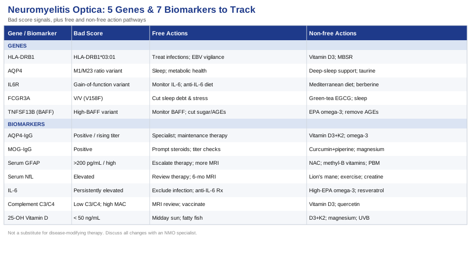
7 Biomarkers Worth Tracking in Neuromyelitis Optica
Biomarkers in NMOSD serve two distinct purposes: establishing and refining the diagnosis, and monitoring disease activity between attacks. The ones listed here were selected because they have genuine clinical relevance, are measurable in practice, and produce actionable information. A few are standard of care in specialist settings. Others are emerging tools that the most proactive neurologists are beginning to incorporate. All of them are worth understanding in depth.
Biomarker 1 — AQP4-IgG: The Defining Antibody
Why it matters
Anti-aquaporin-4 IgG is the immunological hallmark of the most common form of NMOSD. Aquaporin-4 is a water channel protein expressed at high density on astrocytes in the central nervous system — particularly around the blood-brain barrier, the optic nerves, and the spinal cord. When the immune system generates IgG antibodies against AQP4, those antibodies trigger complement activation and astrocyte destruction, producing the selective CNS lesions characteristic of NMO attacks. AQP4-IgG is detectable in approximately 70–80% of people who meet clinical criteria for NMOSD, and its presence in the right clinical context defines the diagnosis and predicts high relapse risk if maintenance therapy is not initiated.
Beyond diagnosis, AQP4-IgG titer changes carry prognostic meaning. Rising titers during apparent clinical stability have been associated with upcoming relapse in some patients, while titer reductions following therapy correlate with treatment response. Serial monitoring adds value over single time-point testing.
How to measure it
The gold-standard test is the cell-based assay (CBA), which has largely replaced older ELISA methods due to its markedly superior sensitivity and specificity. Testing is performed on serum. Reference laboratories including Mayo Clinic Laboratories and Quest Diagnostics in the US offer AQP4-IgG CBA testing; European equivalents exist in most academic neurology centers.
Cost range: $150–$400 depending on the laboratory and insurance status. In most national health systems, this test is covered when ordered in an appropriate clinical context.
If the score is positive or rising: the plan without supplements
A confirmed positive AQP4-IgG in a compatible clinical picture should prompt: - Immediate or re-assessed specialist referral (an NMO neurologist, not a general neurologist, is strongly preferred) - Initiating or optimizing one of the four approved maintenance therapies: eculizumab, inebilizumab, satralizumab, or rituximab - Establishing a clear acute relapse action plan with on-call access to high-dose IV methylprednisolone and a low threshold for plasma exchange - Identifying and minimizing relapse triggers: intercurrent infection (the most consistent trigger), significant psychological stress, and extreme heat - Serial titer monitoring every 6–12 months or after any clinical change
If the score is positive or rising: the plan with supplements or equipment
No supplement directly reduces AQP4-IgG titers. However, adjunct interventions can reduce the inflammatory amplification that converts antibody presence into tissue damage: - Vitamin D3 (5,000–10,000 IU/day) paired with K2 MK-7 (100–200 mcg/day): A potent immune modulator that supports T-regulatory cell function and reduces Th17 inflammatory pathways. Low vitamin D is associated with higher relapse rates across autoimmune neurological diseases. Target serum 25-OH vitamin D between 50–80 ng/mL. No cycling required. Retest every 6 months. Long-term safety is well-established at these doses with K2 co-administration. - Omega-3 fatty acids (EPA + DHA, 2–4 g/day in triglyceride form): Downregulate pro-inflammatory cytokines including IL-6 and TNF-alpha. Choose enteric-coated or triglyceride-form products for better absorption. No cycling needed; long-term daily use is safe and broadly supported. - Sleep optimization (7–9 hours, consistent schedule, dark and cool room): Sleep deprivation activates IL-6, TNF-alpha, and NF-kB pathways that amplify autoimmune activity. This is among the lowest-risk, highest-impact lifestyle adjustments for any autoimmune disease.
Biomarker 2 — MOG-IgG: The Other Side of the Diagnosis
Why it matters
MOG-associated disease (MOGAD) is now recognized as a clinically distinct entity from AQP4-positive NMOSD, despite presenting with strikingly similar attacks: optic neuritis, transverse myelitis, and area postrema episodes. Anti-myelin oligodendrocyte glycoprotein antibodies are found in approximately 20–40% of patients who test negative for AQP4-IgG but have an NMO-like clinical presentation. The distinction is clinically critical: MOGAD often carries a better prognosis, produces different MRI lesion patterns, and does not respond identically to all of the same treatments.
Equally important, MOG-IgG titer is prognostically meaningful. Persistently high titers after an attack predict higher relapse risk, while titers that decline to seronegative over months may indicate a monophasic course — potentially allowing gradual reduction of maintenance therapy in discussion with a specialist.
How to measure it
The preferred method is the live cell-based assay (CBA). Older ELISA-based MOG tests had poor specificity and generated numerous false positives; only CBA should be used. Both MOG-IgG and AQP4-IgG CBA testing are typically ordered simultaneously when evaluating suspected inflammatory CNS disease.
Cost range: $150–$400. Often bundled in a single inflammatory CNS antibody panel.
If the score is positive: the plan without supplements
- MOGAD does not uniformly require the same aggressive chronic immunosuppression as AQP4-positive NMOSD — the decision depends on titer trend, relapse frequency, and severity - Rituximab, mycophenolate mofetil, and azathioprine are used but with different evidence profiles than in AQP4-positive disease - Steroid responsiveness is typically good; IV methylprednisolone should be given promptly for any acute attack - Serial titer monitoring every 3–6 months is important — declining to seronegative may justify a supervised taper discussion
If the score is positive: the plan with supplements or equipment
- Curcumin with piperine (1,000–2,000 mg curcumin/day, with 10–20 mg piperine for bioavailability): Inhibits NF-kB and reduces multiple inflammatory cytokines relevant to MOGAD. Evidence is mechanistic and from other autoimmune conditions, not MOGAD-specific. Cycling: 8 weeks on, 2 weeks off is reasonable. Avoid at high doses during pregnancy. Monitor for gastrointestinal tolerance. - Magnesium glycinate (300–400 mg nightly): Supports nerve conduction, reduces neuro-inflammatory markers, and improves sleep quality in a single daily dose. Long-term use is safe at these doses.
Biomarker 3 — Serum GFAP: Reading the Scale of Astrocyte Destruction
Why it matters
Glial fibrillary acidic protein (GFAP) is a structural cytoskeletal protein expressed almost exclusively inside astrocytes — exactly the cells that AQP4-IgG targets. During an NMO attack, the destruction of astrocytes releases GFAP into the bloodstream in quantities that can exceed 30–50 times the normal level. The degree of elevation correlates directly with attack severity, lesion volume, and the degree of residual neurological disability.
This makes serum GFAP one of the most powerful available tools for assessing how severe a current or recent NMO attack actually was at the tissue level — information that is invisible from clinical symptoms alone. High peak GFAP during an attack predicts worse residual disability, motivating early and aggressive treatment. Serum GFAP is also typically normal or only mildly elevated in multiple sclerosis and MOGAD, making it useful as a differential biomarker as well.
How to measure it
Serum GFAP requires single-molecule array (Simoa) technology — an ultrasensitive digital immunoassay capable of femtomolar detection. This is available through Quanterix-certified laboratories and major academic medical centers. Commercial availability is expanding.
Cost range: $200–$600 in the US. Not universally insurance-covered but increasingly ordered by NMO specialists. Normal range: typically below 200 pg/mL in healthy adults, though lab-specific ranges apply.
If the score is elevated: the plan without supplements
- Confirm that maximal acute treatment was given: high-dose IV methylprednisolone followed by plasma exchange if neurological improvement was incomplete - Reassess maintenance immunosuppression — elevated GFAP during apparent stability may signal subclinical astrocyte damage - Shorten MRI surveillance intervals - If on a lower-efficacy maintenance agent, consider escalation to a higher-efficacy option (eculizumab, inebilizumab) with your specialist
If the score is elevated: the plan with supplements or equipment
- N-Acetylcysteine (NAC, 600–1,800 mg/day in divided doses): Replenishes glutathione and reduces oxidative stress in the CNS. Preclinical evidence suggests astrocyte-protective effects through reduction of reactive oxygen species. Cycling: 6–8 weeks on, 2 weeks off at higher doses. Generally well-tolerated; avoid with nitroglycerin. - Methylated B vitamins (methylcobalamin 1,000–2,000 mcg + methylfolate 400–800 mcg + P5P 50 mg/day): Support axonal and myelin health; methylated forms are preferred, especially in individuals with MTHFR variants. Long-term use is safe with periodic B12 monitoring. - Transcranial photobiomodulation (810–850 nm, Vielight Neuro or equivalent devices): Emerging research supports neuroinflammation reduction and mitochondrial support in CNS cells via targeted near-infrared light. Sessions: 20–25 minutes, 3–5 times/week. Evidence is early-stage and not NMO-specific; more established in traumatic brain injury and neurodegeneration. Cost of home devices: $500–$1,500.
Biomarker 4 — Serum NfL: The Axonal Damage Signal
Why it matters
Neurofilament light chain (NfL) is a structural protein found inside axons — the long projections that carry neural signals across the central nervous system. When axons are damaged or lost, NfL leaks first into the cerebrospinal fluid and then into the bloodstream. In NMOSD, serum NfL spikes sharply during relapses and declines slowly over the following months. The magnitude of elevation and the rate of recovery both carry prognostic information about likely residual disability.
Critically, research has shown that serum NfL can rise before clinically apparent relapses in some patients — suggesting a potential early-warning function. Studies also confirm that higher baseline NfL between attacks predicts greater disability accumulation over time in NMOSD, which means this marker has value not just during crises but as an ongoing monitoring tool for subclinical disease activity.
How to measure it
Like GFAP, serum NfL relies on Simoa technology. Increasingly available at academic neurology centers and some commercial labs. Often bundled with GFAP on neurological damage panels.
Cost range: $150–$500. Age-dependent reference ranges are critical — NfL rises naturally with aging in healthy individuals, so interpretation requires age-matched comparators.
If the score is elevated: the plan without supplements
- Persistently elevated serum NfL between attacks suggests ongoing subclinical axonal damage despite clinical quiescence - This should prompt review of current maintenance therapy efficacy and consideration of escalation - More frequent MRI surveillance (every 6 months rather than annually) is warranted - Update functional assessments: visual acuity, walking speed (timed 25-foot walk), and urological function
If the score is elevated: the plan with supplements or equipment
- Lion's mane mushroom (Hericium erinaceus, 1,000–3,000 mg/day of extract): Contains erinacines and hericenones that stimulate nerve growth factor (NGF) production — a key neuroprotective signaling molecule. Evidence in neurodegeneration is growing. Cycling: 8–12 weeks on, 4 weeks off. Well-tolerated; rare allergic reactions in individuals with mushroom sensitivities. - Aerobic exercise (30–45 minutes, 4–5 times/week at moderate intensity): Aerobic training reliably increases BDNF, supports axonal maintenance, and is one of the few interventions with consistent evidence for reducing NfL in neurological disease populations. Exercise in a cool environment to avoid Uhthoff's phenomenon; start conservatively and escalate gradually. - Creatine monohydrate (3–5 g/day): Supports mitochondrial ATP production in neurons; modest but consistent evidence for neuroprotection. Daily use without cycling is appropriate at these doses.
Biomarker 5 — Interleukin-6: The Engine Behind Antibody Production
Why it matters
IL-6 has a specific and central role in NMOSD pathophysiology that distinguishes it from many other autoimmune diseases. In NMO, IL-6 directly promotes the survival and differentiation of plasmablasts — the specialized immune cells that produce AQP4-IgG. This means IL-6 is one of the upstream perpetuators of the antibody that drives attacks, not merely a downstream consequence. This biology has been confirmed therapeutically: satralizumab, an IL-6 receptor blocker, is an FDA-approved treatment specifically for NMOSD and significantly reduces relapse rates.
Serum and CSF IL-6 levels are consistently elevated in NMOSD compared to both healthy controls and MS patients. Elevated IL-6 between attacks may signal ongoing immune dysregulation even in apparent clinical remission.
How to measure it
Serum IL-6 is measured by standard immunoassay (ELISA or electrochemiluminescence). Available at most hospital and commercial labs.
Cost range: $50–$200 depending on context.
Important caveat: IL-6 is acutely sensitive to any inflammatory trigger including minor infection, psychological stress, and vigorous exercise. A single elevated reading should not be interpreted in isolation — trend over multiple measurements matters more than any single data point.
If the score is elevated: the plan without supplements
- Exclude intercurrent infection or other acute inflammatory causes before attributing elevation to NMOSD activity - If persistently elevated without a clear alternate explanation, this strengthens the clinical rationale for IL-6 pathway targeting (satralizumab, tocilizumab) in treatment discussions with your neurologist - Address the major lifestyle drivers of chronic IL-6 elevation: poor sleep, excess adipose tissue, high intake of refined carbohydrates, and smoking
If the score is elevated: the plan with supplements or equipment
- High-EPA omega-3 (2–4 g EPA + DHA/day): Consistent evidence across multiple randomized trials for reducing serum IL-6 and other pro-inflammatory cytokines. No cycling needed; long-term daily use is safe. - Resveratrol (250–500 mg/day with a meal containing fat): Multiple clinical trials have shown IL-6 reduction at these doses through SIRT1 and NF-kB modulation. Cycling: 8 weeks on, 4 weeks off. Avoid at high doses with anticoagulants or antiplatelet medications. - Time-restricted eating (16:8 window, consistent daily schedule): Metabolic interventions that improve insulin sensitivity consistently lower IL-6. One of the highest-evidence non-pharmacological approaches for systemic inflammatory reduction. - Cold exposure (10–15°C water, 2–5 minutes, 3–5 times/week): Brief cold stress triggers norepinephrine release, which suppresses NF-kB and downstream IL-6 signaling. Beneficial in physiologically stable patients, but monitor closely — only introduce when no active neurological symptoms are present.
Biomarker 6 — Complement Proteins C3 and C4: Tracking the Attack Mechanism
Why it matters
Complement activation is one of the two primary mechanisms of tissue destruction in AQP4-positive NMOSD (the other being granulocyte and eosinophil infiltration). Once AQP4-IgG binds to astrocytes, it activates the complement cascade through the classical pathway, culminating in the formation of the membrane attack complex (MAC) — a molecular structure that punches holes in the astrocyte membrane and kills the cell. This is precisely why eculizumab, a monoclonal antibody that blocks the C5 complement protein and prevents MAC formation, is one of the most effective available therapies for NMOSD.
C3 and C4 serum levels provide indirect information about complement activity. Low or declining C3/C4 can indicate ongoing complement consumption during active disease, even without overt clinical relapse. More specific complement activation products like C3a and C5b-9 (the soluble MAC) are measurable in specialized labs and offer a more direct read on complement pathway activation.
How to measure it
C3 and C4 are routine tests available at any clinical laboratory. Cost range: $25–$100, and they are frequently included in standard autoimmune panels. Complement activation products (C3a, C5b-9/MAC) require specialized assays: cost range $100–$300; typically ordered in academic center settings.
If the score is abnormal: the plan without supplements
- Low C3/C4 or elevated complement activation products in apparent clinical remission may indicate subclinical attack activity — this warrants MRI review and a neurological consultation - For patients on complement-targeting therapy (eculizumab, ravulizumab), abnormal complement markers despite treatment should prompt a pharmacokinetic review with your neurologist - Mandatory before complement inhibitor therapy: vaccination against Neisseria meningitidis, Streptococcus pneumoniae, and Haemophilus influenzae b. Complement inhibition dramatically increases susceptibility to encapsulated bacterial infections.
If the score is abnormal: the plan with supplements or equipment
- Vitamin D3 (5,000–10,000 IU/day with K2): Modulates complement gene expression and reduces complement-mediated inflammatory amplification. Consistent with recommendations across all other biomarkers above; a foundational supplement for NMOSD management. - Quercetin (500–1,000 mg/day with meals): A polyphenol flavonoid with demonstrated anti-complement and anti-inflammatory activity in preclinical models. Evidence specific to NMOSD is absent, but mechanisms are relevant. Cycling: 6–8 weeks on, 2 weeks off. Well-tolerated at these doses.
Biomarker 7 — 25-OH Vitamin D: The Most Overlooked Modifiable Variable
Why it matters
Vitamin D is not simply a nutrient — it functions as a steroid hormone that directly regulates immune cell differentiation, T-regulatory cell activity, and inflammatory cytokine transcription. Vitamin D deficiency is one of the most consistent findings across virtually every autoimmune disease studied, and NMOSD is no exception. Research published in Multiple Sclerosis and Related Disorders found that vitamin D insufficiency is highly prevalent among NMOSD patients, with lower levels correlating with higher annualized relapse rates. Critically, the standard laboratory "normal" range of 20–30 ng/mL is calibrated for bone health, not immune function — most patients with NMOSD who appear "normal" on routine testing are still significantly insufficient for immune purposes.
How to measure it
Standard serum 25-OH vitamin D test. Available at any laboratory. Cost range: $30–$80, often covered by insurance with a documented autoimmune diagnosis.
Target range for immune function: 50–80 ng/mL (supported by researchers including Michael Holick and the functional medicine literature). Avoid sustained levels above 100 ng/mL without medical supervision.
If the score is below 50 ng/mL: the plan without supplements
- Daily midday sun exposure: 15–30 minutes with arms and legs exposed can generate meaningful vitamin D production, though output varies significantly by skin tone, body weight, latitude, and season - Dietary sources: fatty fish (salmon, mackerel, sardines), cod liver oil, egg yolks, and UV-exposed mushrooms contribute modest amounts — sufficient for maintenance but rarely enough to correct deficiency from baseline
If the score is below 50 ng/mL: the plan with supplements or equipment
- Vitamin D3 at 5,000–10,000 IU/day for 3 months, then retest and adjust. Pair with K2 MK-7 (100–200 mcg/day) to direct calcium to bones rather than soft tissue. Retest every 6 months. Long-term use at these doses is safe with monitoring. - Magnesium glycinate or malate (300–400 mg/day): A critical cofactor for vitamin D hydroxylation to its active form. Magnesium deficiency — extremely common in the general population — blocks vitamin D activation regardless of how much D3 is taken. This pairing is often the step that unlocks a plateaued vitamin D level. - Narrowband UVB lamp (for limited sun access or malabsorption): Phototherapy-equivalent to natural sun exposure. Protocol: 10–20 minute sessions 3 times/week at appropriate MED (minimal erythema dose) distance. Cost of quality home devices: $200–$400.
The Genetics Side: 5 Key Genes That Shape NMO Risk and Disease Behavior
Understanding the genetics of NMOSD does not mean predicting fate. No single gene determines whether someone will develop the condition or how disabling it will be. What genetics can offer is a sharper understanding of why your immune system is prone to certain behaviors — and where it may be most responsive to targeted intervention. The five genes below have the strongest evidence base and the clearest actionable implications.
Gene 1 — HLA-DRB1: The Immune Identity Architecture
What it does
HLA-DRB1 is part of the major histocompatibility complex (MHC) class II system — a set of proteins on antigen-presenting cells that determine which peptide fragments the immune system recognizes as foreign versus self. Specific variants, particularly HLA-DRB1*03:01, are consistently associated with AQP4-IgG positive NMOSD in White European populations. In Asian populations, HLA-DPB1*05:01 carries a similar association. These variants likely increase susceptibility by creating an antigenic presentation environment more prone to aberrant autoimmune recognition of self-proteins like AQP4.
HLA-DRB1 does not cause NMOSD independently — it is a susceptibility modifier that interacts with environmental triggers (viral infections, hormonal factors, geographic exposure) to raise or lower risk.
If the variant is present: the plan without supplements
- HLA status is not currently used to direct treatment selection in NMOSD, but awareness should sharpen vigilance - Infection management is particularly important: viral infections — especially Epstein-Barr virus (EBV), which is mechanistically linked to HLA-mediated autoimmune activation — are a recognized NMOSD trigger; keep vaccinations current and treat intercurrent infections promptly - Maintain annual specialist review even in fully stable patients
If the variant is present: the plan with supplements or equipment
- Vitamin D3 optimization (as above): Vitamin D directly modulates HLA-mediated antigen presentation pathways. This is the single supplement with the most mechanistic relevance to HLA-associated autoimmunity. - Structured stress reduction (MBSR, HRV biofeedback): Chronic psychological stress activates HPA-axis dysregulation that amplifies HLA-mediated immune responses. This is a behavioral intervention with direct immunological relevance, not a wellness platitude.
Gene 2 — AQP4: When the Target Itself Varies
What it does
The AQP4 gene encodes aquaporin-4, the water channel that is the primary autoimmune target in NMOSD. Two protein isoforms — M1 and M23 — are produced from alternative transcription start sites and form distinct supramolecular assemblies on the astrocyte surface. M23-dominated assemblies (orthogonal arrays of particles, OAPs) are preferentially targeted by AQP4-IgG and are more efficient at activating complement. Genetic variants that alter the ratio of M1 to M23 expression, or that modify overall AQP4 density in the CNS, may influence individual susceptibility to antibody-mediated attack.
This is an active research area. The practical implication today is supporting the biological system in which AQP4 function is most important: the glymphatic clearance system, which depends on AQP4-mediated water flux to flush neurotoxic waste from the brain during sleep.
If the variant is present: the plan without supplements
- Supporting astrocyte health broadly by minimizing known astrocyte-toxic exposures: alcohol, certain environmental toxins, and chronic sleep deprivation - Metabolic health maintenance — hyperglycemia impairs astrocyte function and AQP4-mediated glymphatic flow
If the variant is present: the plan with supplements or equipment
- Sleep architecture optimization (consistent 7–9 hour schedule, cool dark room, no screens 1 hour before bed): AQP4 channels are the primary mediators of glymphatic brain clearance, which operates during slow-wave sleep. Supporting deep sleep quality is the most evidence-backed intervention for AQP4-dependent CNS function. - Taurine (1,000–3,000 mg/day): An amino acid with roles in astrocyte osmotic regulation and CNS homeostasis. Early research suggests supportive effects on astrocyte function. Long-term daily use is safe at these doses.
Gene 3 — IL6R: The Inflammation Gateway Gene
What it does
IL6R encodes the IL-6 receptor, present on immune cells including the B cells and plasmablasts that produce AQP4-IgG. Genetic variants in IL6R alter downstream signaling sensitivity to IL-6. Gain-of-function variants, or variants associated with increased receptor expression, amplify the IL-6-driven plasmablast survival loop that maintains AQP4-IgG production — making the autoimmune machinery harder to suppress with standard therapies.
Knowing IL6R variant status provides a biological context for treatment response. It may help explain why some patients respond more dramatically to IL-6 receptor blocking agents (satralizumab, tocilizumab) than to B-cell depletion alone.
If the variant is present: the plan without supplements
- Present IL6R variant data to your neurologist as context supporting IL-6-targeted therapy consideration - Prioritize regular serum IL-6 monitoring (as covered above) - Focus dietary and lifestyle approaches on IL-6 reduction
If the variant is present: the plan with supplements or equipment
- Mediterranean dietary pattern: Consistently and robustly reduces serum IL-6, CRP, and TNF-alpha in randomized controlled trials across multiple conditions. Center the diet on extra-virgin olive oil, oily fish 3–4 times per week, legumes, and diverse colorful vegetables. Eliminate seed oils and ultra-processed foods. - Berberine (500 mg twice daily with meals): Inhibits NF-kB and multiple IL-6-related inflammatory pathways with immunomodulatory effects. Evidence is not NMOSD-specific. Cycling: 8 weeks on, 4 weeks off. Check for CYP3A4 drug interactions before starting.
Gene 4 — FCGR3A: How Antibody-Mediated Damage Gets Executed
What it does
FCGR3A encodes Fc gamma receptor IIIa (CD16a), expressed on NK cells and macrophages. This receptor binds the Fc tail of IgG antibodies — including AQP4-IgG — and triggers antibody-dependent cellular cytotoxicity (ADCC): NK cells attach to the IgG-coated target cell and destroy it. The V158F polymorphism determines binding affinity: the V allele binds IgG with higher affinity, meaning V/V individuals execute ADCC more efficiently.
This has direct implications for NMOSD pathology — more efficient ADCC may amplify tissue destruction at AQP4-IgG-decorated astrocytes. Additionally, this same polymorphism affects rituximab efficacy (rituximab works partly through ADCC against CD20+ B cells), making FCGR3A genotype potentially relevant for treatment personalization as the field evolves.
If the variant is present: the plan without supplements
- FCGR3A genotyping is not yet a clinical standard in NMOSD, but may become relevant for predicting rituximab response - Address the lifestyle factors that independently upregulate NK cell cytotoxicity: chronic sleep deprivation, unmanaged psychological stress, and sedentary behavior
If the variant is present: the plan with supplements or equipment
- EGCG from green tea extract (400–600 mg standardized EGCG/day): Preclinical evidence for modulation of Fc receptor-mediated immune activity and NF-kB signaling. Clinical evidence in NMOSD is absent; this is mechanistically plausible. Check for interactions with medications; avoid at high doses in individuals with liver sensitivity. - Sleep quality investment: Chronic sleep debt up-regulates NK cell cytotoxic activity through cortisol and NF-kB pathways. Addressing sleep is the most evidence-supported non-pharmacological approach to NK cell regulation.
Gene 5 — TNFSF13B (BAFF): The B-Cell Survival Amplifier
What it does
TNFSF13B encodes BAFF (B-cell activating factor), a cytokine essential for B-cell survival and differentiation into antibody-secreting plasma cells. BAFF levels are consistently and significantly elevated in NMOSD patient serum compared to healthy controls, and elevated BAFF maintains the B cells and plasmablasts that produce AQP4-IgG even after B-cell depleting therapy with rituximab. This is one mechanism by which rituximab produces incomplete or short-lived responses in some patients — BAFF-driven B-cell repopulation refills the reservoir faster than expected.
Genetic variants in TNFSF13B that increase BAFF expression create a more permissive environment for autoimmune B-cell activity. Belimumab (a BAFF inhibitor approved in lupus) is under investigation for NMOSD, and BAFF gene status may become clinically relevant as this therapeutic pipeline develops.
If the variant is present: the plan without supplements
- Monitor BAFF serum levels if available — persistently elevated BAFF despite B-cell depletion with rituximab suggests BAFF-driven repopulation is occurring - Discuss the B-cell repopulation timeline with your neurologist; more frequent CD19/CD20 cell count monitoring may be warranted - Inquire about clinical trials targeting BAFF or APRIL pathways
If the variant is present: the plan with supplements or equipment
- EPA-dominant omega-3 (2–3 g EPA/day): EPA has demonstrated BAFF-modulating effects in some B-cell-related autoimmune conditions in preliminary research. Risk is negligible; benefit is plausible. - Elimination of advanced glycation end products (AGEs) and refined sugars: Dietary AGEs and chronic hyperglycemia upregulate BAFF expression through RAGE signaling. Removing ultra-processed foods and added sugars is the highest-yield dietary modification for reducing BAFF-mediated immune amplification.
The Wahls Protocol: A Framework That Changed One Researcher's Prognosis — and Is Changing How We Think About Neurological Autoimmunity
Dr. Terry Wahls is a clinical professor of medicine at the University of Iowa who was diagnosed with secondary progressive multiple sclerosis in 2000. By 2007, despite taking chemotherapy-grade immunosuppressants, she was wheelchair-bound. Using principles drawn from mitochondrial medicine, evolutionary nutrition, and cell biology, she developed a dietary and lifestyle protocol — and reversed her decline to the point of cycling 18 miles within a year.
Her work is directly relevant to NMOSD because MS and NMO share the feature of central nervous system autoimmune demyelination and astrocyte destruction, with overlapping inflammatory pathways. Her book The Wahls Protocol (updated 2020 edition) is dense with primary research citations. A pilot clinical trial of the Wahls Elimination diet published in PLOS ONE found significant reductions in fatigue and improvements in quality of life in relapsing-remitting MS participants. While the trial was in MS rather than NMOSD, the mitochondrial and anti-inflammatory mechanisms she targets are shared.
Below are the 10 most impactful ideas from the protocol, framed in the context of NMOSD.
1. Mitochondrial Nutrition Comes Before Everything Else
Every immune cell, neuron, and astrocyte depends on mitochondrial energy production to function. Wahls argues convincingly that modern diets chronically under-supply the cofactors mitochondria require, creating a cellular energy deficit that accelerates autoimmune pathology. Her protocol mandates 9 cups of produce daily — 3 cups each of leafy greens, sulfur-rich vegetables, and deeply colored vegetables — as a non-negotiable floor.
2. Organ Meats Are a Different Category of Nutrition
Weekly organ meat consumption — particularly liver — provides CoQ10, B12, folate, vitamin A, and fat-soluble micronutrients in quantities and bioavailability that are nearly impossible to replicate from muscle meat or supplements. Wahls credits this as one of her most impactful single dietary changes. The evidence for CoQ10 in mitochondrial and neurological function is robust.
3. Leaky Gut Is a Real Mechanism, Not a Fringe Concept
The Wahls protocol eliminates gluten and dairy in its initial phase, with the higher tiers removing legumes and all grains, based on the evidence that these foods can increase intestinal permeability and thereby drive ongoing immune activation. While not proven specifically in NMOSD, the gut-CNS immune axis is an increasingly validated target in autoimmune neurology.
4. Myelin Repair Requires Specific Precursors
Sulfur-containing amino acids (methionine, cysteine), choline, and the B vitamin complex are all required for myelin synthesis and maintenance. The protocol is designed to ensure adequate intake of each through food-first sourcing — not as an afterthought supplement.
5. The Omega-6 to Omega-3 Ratio Matters More Than Total Fat Intake
The contemporary Western diet delivers an omega-6 to omega-3 ratio near 15:1 or worse. Wahls targets a ratio of 4:1 or lower by eliminating industrial seed oils and emphasizing fatty fish, flaxseed, and grass-fed animal products. This shift directly reduces the substrate for pro-inflammatory prostaglandins and IL-6 synthesis.
6. Functional Electrical Stimulation Supports Neural Connectivity
Wahls used functional electrical stimulation (FES) cycling as part of her physical rehabilitation — stimulating motor pathways that atrophied from disuse. FES devices are available in rehabilitation centers and increasingly for home use. Research in neurological rehabilitation supports improvements in both physical function and neuroplasticity from FES training.
7. Stress Management Is as Non-Negotiable as Diet
The protocol treats psychological stress reduction as therapeutically equivalent to nutritional change. Chronic stress impairs the HPA axis and directly elevates neuroinflammatory cytokines. Wahls includes daily structured mindfulness practice in her protocol, citing consistent evidence from multiple studies that perceived stress correlates with relapse risk in autoimmune neurological diseases.
8. Circadian Rhythm Is an Immune Regulator
Consistent sleep-wake timing aligned with natural light exposure directly regulates immune cell activity. Disrupted circadian rhythm up-regulates Th17 pathways and inflammatory cytokine production. Wahls treats circadian alignment as a zero-cost, high-impact behavioral intervention — not an optional lifestyle extra.
9. Exercise Is Neuroprotective at Every Level of Disability
Wahls directly challenges the conventional wisdom that people with neurological autoimmune disease should conserve energy by resting. Exercise — calibrated appropriately to current functional capacity — produces BDNF, reduces IL-6 and TNF-alpha, maintains mitochondrial density in neurons, and slows neurodegeneration. Chair-based exercise, aquatic exercise, and adapted yoga are all viable entry points.
10. Personalized Tracking Is the Protocol
Wahls explicitly designs her approach as iterative: symptom logs, functional assessments, and targeted lab markers are used to identify what is and is not working for each individual. This is not a prescription handed down uniformly — it is a framework for learning what your specific biology responds to. This principle translates directly and powerfully to NMOSD management.
Complementary Approaches With Meaningful Evidence for NMOSD
The approaches below are not alternatives to disease-modifying therapy — they address dimensions of the disease that immunosuppressive treatment alone cannot reach: stress-immune dysregulation, gut-immune signaling, fatigue, and quality of life. Each was selected based on evidence quality and relevance to the specific mechanisms of NMOSD.
Mindfulness-Based Stress Reduction (MBSR)
MBSR is an 8-week structured program developed at the University of Massachusetts that combines body scan meditation, sitting meditation, and mindful movement. Its relevance to NMOSD operates on two levels: chronic psychological stress is a documented relapse trigger in autoimmune neurological diseases, and MBSR is one of the most rigorously tested stress-immune interventions available. It also addresses the chronic pain, fatigue, depression, and anxiety that accompany NMOSD — domains that pharmacological treatment typically leaves unaddressed.
A meta-analysis examining MBSR in multiple sclerosis (a closely related CNS autoimmune condition with overlapping disability patterns) found significant improvements in fatigue, depression, and quality of life across included trials. The mechanism involves cortisol normalization, reduction in inflammatory cytokines including IL-6, and improvement in T-regulatory cell function — all pathways relevant to NMOSD pathophysiology.
To apply: enroll in a formal 8-week MBSR course, available through many hospital wellness programs and online via the UMass Center for Mindfulness or Palouse Mindfulness (free online). Daily practice of 30–45 minutes is the full therapeutic protocol. For beginners, 10 minutes of daily body scan meditation using Insight Timer is an accessible entry point. Benefits accumulate gradually over 8+ weeks; do not assess results before that threshold.
The Autoimmune Protocol (AIP) — Dr. Sarah Ballantyne
The Autoimmune Protocol developed by Dr. Sarah Ballantyne — who holds a PhD in medical biophysics — is a comprehensive dietary and lifestyle framework designed specifically for autoimmune conditions. Beyond a standard anti-inflammatory diet, AIP systematically eliminates foods associated with increased intestinal permeability and immune activation: grains, legumes, dairy, eggs, nightshades, nuts, seeds, refined sugars, alcohol, and industrial seed oils. It simultaneously maximizes nutrient density through organ meats, diverse vegetables, bone broth, and high-quality animal proteins. Lifestyle components — sleep, stress management, circadian rhythm, and social connection — are treated as therapeutically equivalent to dietary change.
The protocol is directly relevant to NMOSD because gut barrier integrity is an increasingly validated driver of autoimmune activity, including in CNS autoimmune diseases. A clinical trial of the AIP in inflammatory bowel disease (Konijeti GG et al., Inflammatory Bowel Diseases, 2017) found a 73% reduction in clinical disease scores after 6 weeks of AIP adherence, with histological and endoscopic improvements. While no NMOSD-specific AIP trial exists, the mechanisms — reduced intestinal antigen translocation, improved T-regulatory cell function, and reduced inflammatory cytokine production — apply across autoimmune conditions with shared immune dysregulation.
To apply: the elimination phase lasts 30–60 days. Reintroduction is gradual — one food group at a time, monitored over 72 hours per food before the next introduction. AIP is not a permanent diet; it is a diagnostic elimination tool followed by a personalized long-term eating pattern based on individual tolerability. Ballantyne's book The Paleo Approach and her website provide detailed protocols. Monitor iron, B12, and vitamin D status during the elimination phase to prevent inadvertent deficiencies.
Microbiome-Directed Therapies
The gut microbiome and NMOSD share a more specific connection than the general autoimmunity-gut axis. Multiple studies have characterized distinct gut microbiome signatures in NMOSD patients, notably reduced abundance of short-chain fatty acid (SCFA)-producing bacteria — particularly Bacteroides species and Prevotella copri — relative to healthy controls. SCFAs (butyrate, propionate, acetate) produced when gut bacteria ferment dietary fiber have well-established anti-inflammatory effects on immune cells, including direct induction of regulatory T cells that suppress autoimmune activity. A 2020 study published in Microbiome identified a condition-specific dysbiosis pattern in NMOSD distinct from both healthy controls and MS patients, suggesting a biologically meaningful microbiome-NMOSD relationship rather than a generic finding.
While interventional probiotic trials specifically in NMOSD are limited, probiotic and microbiome interventions in related autoimmune neurological conditions have demonstrated reductions in IL-6, IL-17, and improvements in regulatory immune markers.
To apply: the highest-yield microbiome intervention is dietary — 25–35 grams of diverse plant fiber daily from vegetables, legumes, whole grains, and fruits feeds SCFA-producing bacterial populations. Add fermented foods (plain live-culture yogurt, kefir, kimchi, sauerkraut) as tolerated. For probiotic supplementation, strains with the most autoimmune-relevant evidence include Lactobacillus reuteri, Lactobacillus acidophilus, and Bifidobacterium longum. Use for 8–12 weeks and reassess. Fecal microbiota transplant for autoimmune neurological conditions is under investigation but remains outside standard clinical practice.
Yoga
Yoga is relevant to NMOSD for a reason that goes beyond general wellness: many patients carry residual motor and sensory deficits from previous attacks, making conventional exercise protocols inaccessible or unsafe. Yoga offers an adaptable movement modality that can be modified for almost any functional level — including chair-based and supine variations. It also activates the parasympathetic nervous system, reduces cortisol, and improves heart rate variability — all of which have direct relevance to the stress-immune dysregulation axis in NMOSD.
Randomized controlled trials of yoga in multiple sclerosis (the closest proxy population to NMOSD in terms of neurological disability profile) have found significant reductions in fatigue — one of the most disabling symptoms in both conditions — along with improvements in mood, quality of life, and autonomic balance. The evidence base is moderate and consistent across multiple trials.
To apply: choose a gentle or restorative yoga style — hatha, yin, or chair yoga depending on current physical capacity. Avoid hot yoga entirely: elevated body temperature worsens Uhthoff's phenomenon in NMOSD and can trigger transient neurological symptom exacerbation. Practice 2–4 sessions per week, 30–45 minutes each. Work with a yoga instructor experienced in neurological conditions. Progress is measured in months, not weeks; the goal is sustainable engagement.
Breathing-Based Therapies
Breath-based interventions — particularly coherent breathing at approximately 6 breaths per minute — work by activating the vagus nerve and maximizing heart rate variability (HRV), which shifts autonomic balance strongly toward the parasympathetic state. This reduces cortisol, suppresses NF-kB, and lowers IL-6 and TNF-alpha production. For NMOSD, autonomic dysfunction is an underrecognized feature of the disease, and the HPA-axis stress response is a credible relapse amplifier. Improving vagal tone through breathing practice is one of the lowest-risk, most mechanistically grounded behavioral interventions available.
The technique with the strongest combined evidence is coherent breathing: inhaling for 5 seconds and exhaling for 5 seconds, maintaining exactly 6 breaths per minute. At this respiratory rate, baroreflex sensitivity is maximized, heart rate variability peaks, and vagal outflow to immune tissue is amplified. Research across chronic pain, cardiovascular disease, anxiety, and inflammation consistently supports both the physiological mechanism and clinical outcomes.
To apply: 20 minutes of coherent breathing practiced twice daily is the research-grade protocol. It requires no equipment — a 5-second inhale/5-second exhale cadence is all that is needed. Apps like Respire or the Elite HRV biofeedback system can guide practice. An HRV monitor (Polar H10, Garmin chest strap) allows objective tracking of vagal tone improvement over weeks, which many patients find motivating. Avoid hyperventilation — exhale fully and ensure the pace is relaxed, not forced.
Conclusion
Neuromyelitis optica is a condition that rewards precision. The difference between a relapse caught early and one that causes permanent disability often comes down to how closely disease activity is being tracked and how proactively modifiable risk factors are being addressed. The 7 biomarkers covered here — AQP4-IgG, MOG-IgG, serum GFAP, NfL, IL-6, complement proteins, and vitamin D — each illuminate a different facet of what is happening inside your nervous system and immune system at a level that symptoms alone cannot reveal.
The genetic context adds depth: HLA-DRB1, the AQP4 gene, IL6R, FCGR3A, and BAFF do not determine outcomes, but they help explain individual immune behavior and can inform more targeted interventions — from dietary choices to treatment discussions with your neurologist. The complementary strategies here — MBSR, the Autoimmune Protocol, microbiome optimization, yoga, and breathing practice — are not replacements for disease-modifying therapy. They address the dimensions of the disease that medication cannot: stress-driven immune amplification, nutritional gaps, gut-immune signaling, and the inflammatory environment that either fuels or constrains autoimmune activity.
The next practical step is not to implement everything at once. Start with what is most measurable: confirm your antibody status if any uncertainty remains, get a vitamin D level, review your maintenance therapy with an NMO specialist, and choose one lifestyle intervention to build consistently over the next 8–12 weeks. Incremental precision compounds over time.