This article contains AI generated content.
· UpdatedNodular Vasculitis: 5 Genes and 6 Biomarkers to Track
Introduction
If you have been told you have nodular vasculitis and left the appointment with a prescription and not many answers, that experience is more common than it should be. The condition is rare enough that most general practitioners encounter it only a handful of times in their careers, and even dermatologists sometimes misdiagnose it as erythema nodosum or another form of panniculitis. The result is that many people spend months — sometimes years — cycling through treatments that address symptoms without ever understanding the underlying biology driving their flares.
What makes nodular vasculitis particularly difficult to manage is how variable it is. Two people with the same clinical diagnosis can have completely different triggers, different inflammatory profiles, and different responses to treatment. Generic advice — eat less sugar, reduce stress, stay warm — is not wrong, but it is so broad that it rarely moves the needle in a meaningful way on its own. The condition involves inflammation of small and medium-sized blood vessels within subcutaneous fat, and what is fueling that inflammation differs significantly from person to person.
This article takes a more targeted approach. Instead of starting from symptoms and working backward through trial and error, it starts from biology: the specific biomarkers you can measure in blood to understand what kind of inflammation you are dealing with, and the genetic variants that may be shaping your immune response and vascular health in ways a routine checkup would never detect.
The goal here is not to promise a cure or to shortcut the diagnostic process. It is to help you understand what is actually happening beneath the surface, so that every decision you make — dietary, supplemental, lifestyle, or medical — can be more precisely informed. The biomarker section covers six clinically relevant measurements with practical guidance on what to do if any come back abnormal. The genetics section covers five gene variants most associated with immune dysregulation and vascular vulnerability. Beyond that, you will find a summary of a book that fundamentally reframes how inflammatory conditions like this are approached, followed by evidence-based complementary modalities worth knowing about.
Summary
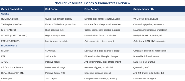
This article covers six key biomarkers — hsCRP, ESR, ANCA, complement proteins C3 and C4, IGRA, and fibrinogen — and five genetic variants — HLA alleles, TNF-alpha, IL-6, MTHFR, and PTPN22 — that are most relevant for understanding and managing nodular vasculitis. Each biomarker section explains why it matters for this specific condition, how to measure it with cost ranges, and what to do if your result is abnormal, both with and without supplements. The genetics section explains how each variant may influence your immune and vascular response, and what targeted interventions can modify the downstream effects. Following those sections, you will find a summary of Tom O'Bryan's The Autoimmune Fix — a framework for understanding how chronic inflammation accumulates silently over years — along with five complementary therapeutic modalities that have meaningful clinical evidence behind them. The overarching theme: this condition can be approached with considerably more precision than most patients are ever offered.
6 Biomarkers to Track if You Have Nodular Vasculitis
Understanding the pattern of inflammation in nodular vasculitis requires more than a single blood test. The condition involves immune complex deposition, vascular wall inflammation, fat lobule involvement, and sometimes an underlying infectious or autoimmune trigger — each of these dimensions has a corresponding biomarker signature. The six markers below collectively provide the most useful biological snapshot available outside of tissue biopsy, and together they can reveal what is actually driving the condition in a given individual.
1. High-Sensitivity C-Reactive Protein (hsCRP)
Why it matters
C-reactive protein is produced by the liver in response to inflammatory signaling, particularly interleukin-6. In nodular vasculitis, elevated hsCRP reflects the degree of systemic inflammation accompanying the localized skin and vascular lesions. It is not specific to this condition — many inflammatory states raise CRP — but tracking it over time gives a reliable signal of whether a given intervention is working or whether a flare is building before it becomes visible. The high-sensitivity version detects low-grade inflammation that standard CRP misses. Peter Attia has consistently emphasized hsCRP as one of the most actionable monitoring biomarkers precisely because it responds to lifestyle changes within weeks, making it a practical feedback tool for any anti-inflammatory protocol.
How to measure it
A simple blood draw. Most standard labs offer hsCRP as part of a cardiovascular risk panel or as a standalone test. Cost: $15–60 depending on the lab and insurance. Optimal range: below 0.5 mg/L. Concerning: above 1.0 mg/L. Above 3.0 mg/L indicates significant active inflammation and warrants investigation.
If the score is bad, the plan without supplements
Prioritize a low-glycemic, whole-food dietary pattern and eliminate refined carbohydrates and industrial seed oils completely. Time-restricted eating — a 10–12 hour eating window — has been shown in multiple trials to reduce hsCRP independently of weight loss. Consistent moderate aerobic exercise (30 minutes, four to five times per week) brings hsCRP down measurably within 8–12 weeks. Sleep optimization targeting 7.5–8.5 hours per night is often underestimated; poor sleep independently and directly raises CRP. Brief cold water exposure at 10–15°C for two to three minutes has anti-inflammatory signaling effects through norepinephrine release and reduction in prostaglandin synthesis.
If the score is bad, the plan with supplements or equipment
Omega-3 fatty acids (EPA and DHA combined): 2–4 grams per day with food. Strong human evidence for CRP reduction in inflammatory conditions. Use continuously; reassess at 12 weeks. Side effects: mild GI discomfort at higher doses, fishy aftertaste manageable with enteric-coated formulations.
Curcumin with piperine: 500–1000 mg curcumin combined with 5–10 mg piperine, once or twice daily. Meta-analysis evidence documents significant reductions in hsCRP (Sahebkar et al., 2016). Cycle in 8-week blocks with 2-week breaks. Side effects: may have mild blood-thinning effects at high doses; avoid before surgery.
Magnesium glycinate: 300–400 mg before bed. Low magnesium is independently associated with elevated CRP through NLRP3 inflammasome activation. Side effects: loose stools if dose is too high — titrate upward slowly.
2. Erythrocyte Sedimentation Rate (ESR)
Why it matters
ESR measures how quickly red blood cells settle in a test tube, which is influenced by the concentration of acute-phase proteins. It is less specific than hsCRP but slower-moving, making it useful for tracking chronic inflammation trends over months rather than weeks. In nodular vasculitis, ESR tends to remain elevated during active disease and normalizes during remission. When ESR and hsCRP diverge — one elevated and one normal — it can indicate specific inflammatory subtypes. High ESR with near-normal CRP, for example, often points toward fibrinogen-mediated viscosity changes or chronic disease patterns rather than acute-phase flare activity.
How to measure it
Blood draw; often included in a basic inflammation panel. Cost: $10–40. Optimal values: below 20 mm/hr for women under 50; below 30 mm/hr for women over 50. Note that nodular vasculitis predominantly affects women. Values of 40–60 mm/hr during active flares are common.
If the score is bad, the plan without supplements
The same foundational lifestyle changes that reduce hsCRP also bring ESR down, though more slowly. ESR responds well to elimination of food sensitivities, which can be investigated through a structured 4-week elimination protocol removing gluten, dairy, eggs, and legumes, followed by systematic reintroduction while tracking skin lesion activity and symptom severity in a daily log.
If the score is bad, the plan with supplements or equipment
Boswellia serrata standardized to AKBA (acetyl-11-keto-beta-boswellic acid): 100–200 mg AKBA daily. Human evidence for reduction of inflammatory markers in joint and bowel inflammatory conditions translates mechanistically to vascular inflammation. Cycle: 12-week blocks. Side effects: occasional mild GI discomfort.
Infrared sauna: 20–30 minutes per session, three to four times per week at 55–60°C. Human studies document reductions in inflammatory markers including ESR and improvements in peripheral circulation. Side effects: avoid if blood pressure is uncontrolled; hydrate adequately before and after. Do not use during acute flare with active skin breakdown.
3. ANCA (Antineutrophil Cytoplasmic Antibodies)
Why it matters
ANCA are autoantibodies directed against proteins within neutrophil granules. They are strongly associated with systemic vasculitides — granulomatosis with polyangiitis, microscopic polyangiitis, eosinophilic granulomatosis with polyangiitis — that can be confused with or coexist alongside nodular vasculitis. Testing for both c-ANCA targeting PR3 and p-ANCA targeting MPO helps differentiate idiopathic nodular vasculitis from more aggressive systemic disease that requires entirely different treatment decisions.
A positive ANCA in a patient presenting with cutaneous vasculitis changes the clinical picture significantly, pointing toward systemic involvement requiring nephrology and pulmonology evaluation. Even a weakly positive result warrants specialist follow-up. Thomas Dayspring and clinicians who work extensively with vascular biomarkers flag ANCA as a critical rule-out in any patient with recurring vascular skin lesions, particularly when accompanied by constitutional symptoms like fatigue, weight loss, or joint involvement.
How to measure it
Blood test — order an ANCA panel with reflex testing for PR3 and MPO specificity. Cost: $80–250 depending on whether reflex testing is included. Request both immunofluorescence (IF) and ELISA testing for maximum sensitivity and specificity.
If the score is bad, the plan without supplements
A positive ANCA result requires specialist review before any self-management. That said, dietary strategies that reduce neutrophil activation are relevant in parallel: a low-lectin, anti-inflammatory dietary pattern reduces the immune stimulus that drives ANCA-mediated vascular damage. Consistent stress management is essential, as psychological stress activates neutrophil degranulation through adrenergic pathways and directly amplifies ANCA-mediated vascular inflammation.
If the score is bad, the plan with supplements or equipment
Under specialist guidance: low-dose naltrexone (LDN) at 1.5–4.5 mg nightly has been studied in autoimmune contexts and shows a favorable anti-inflammatory profile with minimal side effects through transient opioid receptor blockade that upregulates endogenous endorphin production. Requires physician prescription and monitoring.
Vitamin D3 with K2: maintain serum 25-OH-D at 50–80 ng/mL, as low vitamin D correlates with ANCA-positive vasculitis severity in observational data. Dose: 4000–6000 IU D3 with 100–200 mcg MK-7 K2 daily. Retest at 3 months and adjust accordingly.
4. Complement Proteins (C3 and C4)
Why it matters
The complement system is a key branch of innate immunity. In vasculitis involving immune complex deposition — which nodular vasculitis can involve — complement proteins are consumed faster than they are produced, leading to low C3 and C4 levels. Low complement in the context of active skin lesions is a meaningful signal that immune complexes are actively being deposited within vessel walls, which is mechanistically important for understanding the severity of the vascular component and for guiding treatment toward immune complex clearance rather than simple anti-inflammatory suppression.
Conversely, persistently elevated complement in someone not experiencing an acute flare may indicate chronic activation without adequate resolution — a smoldering state that predisposes to repeated episodes. Allan Sniderman has emphasized complement as an underused clinical marker in vascular inflammation, noting that its directional shifts often precede symptom changes.
How to measure it
Blood test. C3 and C4 are ordered together as a complement panel. Cost: $40–120. Normal ranges: C3 at 90–180 mg/dL; C4 at 16–47 mg/dL. Values below the lower limit of normal during active skin lesion activity are diagnostically meaningful and should prompt rheumatology consultation.
If the score is bad, the plan without supplements
When complement is low, the priority is identifying and reducing the immune complex burden rather than supplementing complement directly. This means actively investigating underlying triggers — latent TB (see IGRA below), chronic bacterial infections, autoimmune coexistence — and removing the source of antigen driving complex formation. Reducing dietary inflammatory load (particularly removing sources of advanced glycation end-products and oxidized lipids) reduces immune complex formation over time. Avoid alcohol entirely during active periods, as it compounds complement consumption through multiple pathways.
If the score is bad, the plan with supplements or equipment
Quercetin: 500–1000 mg twice daily with food. Evidence supports complement pathway regulation and mast cell stabilization, reducing one source of complement activation. Cycle: 8-week blocks. Side effects: generally mild at these doses; may interact with certain medications including some antibiotics and blood thinners.
NAC (N-acetylcysteine): 600 mg twice daily. Reduces oxidative stress that amplifies complement activation through alternative pathway engagement. Side effects: GI discomfort at higher doses; sulfur odor is normal. Compatible with continuous use but periodic reassessment of need is reasonable.
5. Interferon-Gamma Release Assay (IGRA / QuantiFERON-TB Gold)
Why it matters
Erythema induratum — the subtype of nodular vasculitis most closely associated with Mycobacterium tuberculosis — accounts for a clinically significant proportion of cases, particularly in populations with higher background TB exposure. Studies have found that 30–50% of nodular vasculitis presentations may represent tuberculid reactions: delayed hypersensitivity responses to mycobacterial antigens circulating in a host with latent or prior TB infection, rather than active infection spreading directly to the skin. The skin lesions are essentially a local immune reaction, not a site of active mycobacterial replication.
The IGRA test detects T-cell sensitization to TB antigens without the false positives associated with BCG vaccination that complicated the older tuberculin skin test. A positive IGRA in a patient with nodular vasculitis changes the treatment approach entirely — antituberculous therapy becomes central to management, and the skin lesions frequently resolve with TB treatment alone, often without requiring chronic immunosuppression.
How to measure it
Blood test sent to a reference laboratory. The QuantiFERON-TB Gold Plus is the current standard. Cost: $60–200. Results in 3–7 days. Also consider a chest X-ray as a baseline, and CT chest in selected cases to evaluate for subclinical pulmonary involvement or lymphadenopathy that might not be apparent clinically.
If the score is bad, the plan without supplements
A positive IGRA requires infectious disease or pulmonology consultation to confirm latent TB status, rule out active disease, and determine the most appropriate prophylactic regimen. If latent TB is confirmed, isoniazid for 6–9 months or rifampin for 4 months is the standard approach and commonly leads to resolution of nodular vasculitis lesions within months of initiating treatment. No supplement replaces this step when TB is the underlying driver.
If the score is bad, the plan with supplements or equipment
During and after antituberculous therapy, support hepatic function: milk thistle (silymarin) at 140–420 mg three times daily, as isoniazid carries hepatotoxic potential that warrants monitoring and support. Monitor liver enzymes (ALT, AST) monthly during treatment — this is standard clinical protocol, not optional.
Vitamin B6 as pyridoxine at 25–50 mg daily prevents isoniazid-induced peripheral neuropathy; this is standard adjunct therapy recommended alongside isoniazid. Do not skip it.
6. Fibrinogen
Why it matters
Fibrinogen is a coagulation protein that also functions as an acute-phase reactant — it rises during inflammation and contributes directly to blood viscosity and clot formation in small vessels. In vasculitic conditions, elevated fibrinogen reflects both the inflammatory component and increased thrombotic risk within affected vessel segments. The combination of vascular wall inflammation and elevated fibrinogen is particularly concerning in nodular vasculitis because the condition predominates in the lower legs, where venous circulation is already gravitationally challenged and blood flow is slowest.
Peter Attia has consistently highlighted fibrinogen as an underappreciated cardiovascular and vascular risk biomarker. In the context of nodular vasculitis specifically, fibrinogen is also a useful index of treatment response: normalization of fibrinogen during therapy typically indicates that the vascular inflammatory process is resolving.
How to measure it
Blood test — part of a coagulation panel or ordered standalone as fibrinogen activity. Cost: $20–60. Optimal: below 300 mg/dL. Above 400 mg/dL in the context of active inflammatory disease warrants attention. Above 500 mg/dL represents significant hypercoagulable risk.
If the score is bad, the plan without supplements
Graduated compression therapy for the lower legs (20–30 mmHg medical-grade compression stockings) reduces venous stasis and appears to lower local fibrinogen deposition in vasculitic lesion areas. Consistent walking — even 20–30 minutes daily on flat terrain — significantly improves venous return and reduces systemic fibrinogen over 8–12 weeks. Leg elevation during rest periods reduces local vascular pressure and assists fibrinolytic clearance. Avoid prolonged standing without movement.
If the score is bad, the plan with supplements or equipment
Nattokinase: 100–200 mg (2000–4000 FU) twice daily on an empty stomach. A fibrinolytic enzyme derived from fermented soybeans with human evidence for fibrinogen reduction. Cycle: 12 weeks on, 4 weeks off. Important: avoid if you are on anticoagulants or antiplatelet medications — discuss with a physician before starting.
Lumbrokinase: an alternative fibrinolytic derived from earthworm extract with different enzyme selectivity. 20–40 mg twice daily on an empty stomach. Similar cycling protocol. As with nattokinase, require physician guidance if on any blood-thinning agent.
Omega-3 fatty acids: beyond the hsCRP effect, EPA specifically reduces plasma fibrinogen concentration. Dosing as above.
Moving from biomarkers to the underlying genetic architecture reveals a complementary angle: why some individuals are predisposed to this kind of vascular inflammation in the first place, and what can be done about inherited tendencies that laboratory tests alone cannot capture.
The Genetic Architecture Behind Nodular Vasculitis: 5 Variants Worth Understanding
Genetic testing for inflammatory and autoimmune conditions has moved from research settings into clinical and consumer-accessible testing. While no single gene causes nodular vasculitis, specific variants influence immune reactivity, inflammatory amplification, vascular tone, detoxification, and methylation — all of which bear directly on this condition's biology. Understanding these variants helps explain individual susceptibility patterns and, critically, provides specific targets for lifestyle and supplemental intervention that generic recommendations would never surface.
1. HLA Alleles (HLA-B and HLA-DR)
What the gene does
Human leukocyte antigen genes encode proteins that present antigens to T cells — determining what your immune system recognizes as foreign versus self. Specific HLA alleles are overrepresented in vasculitic and panniculitic conditions. HLA-B51 is the strongest genetic risk factor for Behçet's disease, a systemic vasculitis that overlaps diagnostically with nodular vasculitis in some patients. HLA-DR4 and HLA-DR3 alleles are associated with broader autoimmune predisposition and are overrepresented in patients with tuberculid reactions to mycobacterial antigens.
If the gene is bad, the plan without supplements
HLA alleles cannot be changed, but their downstream effect — how powerfully and inappropriately the immune response fires — is modifiable through antigen reduction. Maintaining gut microbiome diversity through a fiber-rich, diverse plant-containing diet reduces constant microbial antigen exposure that provides the chronic stimulus for over-reactive HLA-mediated immune responses. Removing confirmed dietary triggers — gluten in HLA-DQ2 and DQ8 carriers, casein in certain HLA-DR variants — is often clinically meaningful over 3–6 months of strict adherence.
If the score is bad, the plan with supplements or equipment
Vitamin D3 with K2 as above — vitamin D directly modulates HLA-related T-cell responses by supporting Treg cell differentiation and inhibiting Th17 proliferation, the cell type most associated with vascular tissue damage. Maintain serum 25-OH-D at 50–80 ng/mL year-round. Beta-glucans from medicinal mushrooms (reishi, turkey tail): 500–1000 mg daily. Evidence for immune modulation toward more balanced Th1/Th2 response rather than hyper-reactive states. Cycle: 12-week blocks.
2. TNF-alpha Promoter Polymorphism (-308G/A)
What the gene does
Tumor necrosis factor-alpha is one of the central inflammatory cytokines, and the -308G/A promoter polymorphism determines how much TNF-alpha is produced in response to an inflammatory trigger. Carriers of the A allele (minor allele) produce significantly more TNF-alpha after any stimulus — meaning greater amplification of any inflammatory response. This variant has been associated with panniculitis severity and with more aggressive vascular inflammation in multiple chronic inflammatory conditions. In nodular vasculitis, it may explain why some individuals have dramatically worse flares than others with identical clinical presentations.
If the gene is bad, the plan without supplements
Reduce upstream TNF-alpha stimulation: eliminate trans fats and high-fructose corn syrup completely, as both directly induce TNF-alpha secretion from macrophages. Prioritize sleep — sleep deprivation specifically amplifies TNF-alpha through NF-κB activation. Moderate exercise is anti-inflammatory; however, overtraining paradoxically raises TNF-alpha — keep training volume appropriate and recovery adequate.
If the score is bad, the plan with supplements or equipment
Curcumin with piperine: as above; curcumin specifically inhibits NF-κB, the primary transcription factor driving TNF-alpha production. This mechanism is particularly relevant for TNF-alpha polymorphism carriers. Resveratrol: 250–500 mg daily with food. Modulates NF-κB and specifically reduces TNF-alpha gene expression. Cycle: 8 weeks on, 2 weeks off. Side effects: mild at typical doses; caution in hormone-sensitive conditions at high doses. EGCG (green tea extract): 400–800 mg daily. Direct TNF-alpha inhibitory effects documented in human studies. Avoid on an empty stomach to prevent GI discomfort.
3. IL-6 Promoter Polymorphism (-174G/C)
What the gene does
Interleukin-6 drives the liver to produce acute-phase reactants — including CRP and fibrinogen — and plays a central role in the transition from acute to chronic inflammation. The -174G/C promoter variant influences baseline IL-6 production. High-producer genotypes maintain elevated baseline inflammation that is functionally similar to a chronically primed immune state, making flares more frequent and more severe when any additional trigger appears. This variant also influences why some individuals respond more dramatically to the same dietary or environmental trigger than others.
If the gene is bad, the plan without supplements
Caloric moderation reduces IL-6 independently of weight. Even modest caloric restriction (10–15% below maintenance for 8–12 weeks) has been shown in multiple human trials to significantly lower IL-6. Regular aerobic exercise directly suppresses IL-6 through myokine signaling from contracting skeletal muscle — IL-6 released during exercise paradoxically has anti-inflammatory downstream effects that reduce basal IL-6 secretion. Target 150–200 minutes of moderate aerobic activity per week, not all in one session.
If the score is bad, the plan with supplements or equipment
Magnesium glycinate or malate: reduces IL-6 through NLRP3 inflammasome inhibition. 300–400 mg nightly. Berberine: 500 mg twice daily with meals. Activates AMPK, which suppresses IL-6 transcription through multiple downstream pathways. Cycle: 8 weeks on, 4 weeks off. Side effects: GI adjustment in the first 1–2 weeks; monitor blood sugar carefully if diabetic. Melatonin: 0.5–3 mg nightly has documented IL-6-reducing effects independent of its sleep benefit — even this small dose activates MT1 and MT2 receptors that inhibit inflammatory cytokine production.
4. MTHFR (C677T and A1298C Variants)
What the gene does
Methylenetetrahydrofolate reductase converts folate into its biologically active form, which is essential for the methylation cycle and — critically — for the conversion of homocysteine to methionine. Carriers of one or two copies of the C677T variant have reduced MTHFR enzyme activity by 35–70% depending on zygosity. The result is elevated homocysteine and impaired methylation across multiple physiological systems.
Elevated homocysteine directly damages vascular endothelium, promotes coagulation, and amplifies vascular inflammation — all of which are directly relevant to a condition involving blood vessel wall involvement within subcutaneous tissue. MTHFR is one of the most clinically actionable genetic variants in any condition with a vascular component.
If the gene is bad, the plan without supplements
Increase dietary natural folate: leafy greens, liver, asparagus, and legumes. Critically, avoid folic acid from fortified foods — synthetic folic acid competes with natural folate for MTHFR processing and can actually worsen the functional deficiency in variant carriers. Reduce methionine load by moderating high-methionine animal protein somewhat. Avoid alcohol entirely during active disease, as alcohol depletes B vitamins required for the methionine-homocysteine cycle.
If the score is bad, the plan with supplements or equipment
This is one area where targeted supplementation has clear mechanistic evidence. Take methylfolate (5-MTHF) at 400–800 mcg daily — never folic acid. Combine with methylcobalamin (B12) at 500–1000 mcg daily. Add pyridoxal-5-phosphate (P-5-P) at 50 mg daily — the active form of B6 that bypasses conversion steps compromised by other gene variants. Riboflavin (vitamin B2) at 100 mg daily specifically supports MTHFR enzyme function even in variant carriers by acting as a cofactor that partially restores activity (McNulty et al., 2016). Retest homocysteine at 12 weeks; target below 7 µmol/L.
5. PTPN22 (R620W Variant, rs2476601)
What the gene does
Protein tyrosine phosphatase non-receptor type 22 regulates the activation threshold of T cells and B cells. The R620W variant lowers this threshold — immune cells fire more readily in response to self-antigens, increasing the probability of autoimmune tissue damage. This variant is among the most replicated genetic risk factors for multiple autoimmune conditions including rheumatoid arthritis, systemic lupus erythematosus, and type 1 diabetes.
Its relevance to nodular vasculitis lies in the autoimmune component of the condition. When the vascular inflammation has an autoimmune rather than infectious driver, PTPN22 variant status can help explain why immune activation is ongoing and why the condition recurs even after apparent triggers are removed.
If the gene is bad, the plan without supplements
Reduce the total immune activation burden: prioritize gut barrier integrity, as the pathway from intestinal permeability to systemic immune activation is well documented and PTPN22 carriers activate more intensely in response to the same antigen load. Remove industrial seed oils and refined sugars as immediate priorities. Implement consistent daily stress management — chronic psychological stress activates the same T-cell pathways that PTPN22 renders hyperreactive, creating a compounding effect.
If the score is bad, the plan with supplements or equipment
Colostrum: 500–1000 mg daily on an empty stomach. Supports gut barrier integrity and contains growth factors that modulate T-cell regulatory activity. Cycle: 12 weeks. Low-dose naltrexone (LDN) at 1.5–4.5 mg nightly: as described above, relevant particularly in PTPN22 carriers with confirmed autoimmune activity. Requires a physician prescription. Specific probiotic strains — Lactobacillus rhamnosus GG and Bifidobacterium longum — have documented T-regulatory cell-supporting properties that are directly relevant to autoimmune conditions driven by low immune activation thresholds. Use strains with confirmed human evidence rather than generic multi-strain formulations.
From the genetic landscape, it is natural to ask the broader question: how did this inflammatory state build in the first place, and what framework best explains it? A book published nearly a decade ago addresses this question with unusual precision.
A Book That Reframes Inflammatory Conditions: The Autoimmune Fix
Tom O'Bryan is a functional medicine educator and clinician who spent decades working with patients whose conventional labs looked unremarkable but who were clearly unwell. The Autoimmune Fix, published in 2016, is built around a central argument: autoimmune and chronic inflammatory disease does not begin the day you receive a diagnosis. It builds over years — in a predictable sequence — driven by intestinal barrier compromise, molecular mimicry, and chronic low-grade inflammation that standard testing cannot detect until organ or tissue damage has already accumulated.
For people with nodular vasculitis, particularly those in whom the condition comes and goes without an obvious trigger, O'Bryan's framework offers a meaningfully different clinical lens.
10 Key Takeaways from The Autoimmune Fix
1. Autoimmunity exists on a spectrum long before diagnosis. Most people are in autoimmune territory — producing autoantibodies, experiencing subclinical inflammation — for years before a clinical condition is named. Skin manifestations like nodular vasculitis may be early-stage expressions of a process that has been building silently.
2. Leaky gut is the central upstream driver. When intestinal barrier integrity is compromised, undigested proteins and microbial fragments cross into systemic circulation and persistently stimulate immune reactivity. This is the starting point of most autoimmune cascades, including those that manifest primarily in skin and vasculature.
3. Gluten is the most commonly implicated molecular mimic in autoimmunity. Gliadin fragments share structural similarity with proteins in multiple tissues, including vascular endothelium. The immune system that learned to attack gluten may cross-react with vascular proteins — a mechanism directly relevant to a condition involving vessel wall inflammation.
4. You can have intestinal permeability without any GI symptoms. The majority of people with a compromised gut barrier do not present with obvious digestive complaints. Their primary symptom is a skin condition, joint pain, fatigue, or — as in this case — recurring vascular inflammation in subcutaneous tissue.
5. Standard celiac and gluten testing misses most gluten reactivity. Standard anti-gliadin IgG and IgA testing covers only one of dozens of wheat proteins and peptides that can drive immune reactivity. More comprehensive panels that test multiple wheat fractions detect a substantially larger proportion of gluten-reactive individuals who test negative on standard panels.
6. A strict 21-day gluten elimination functions as both diagnostic test and therapy. O'Bryan recommends a minimum three-week complete elimination as a practical first-line investigation. Gluten has a tissue half-life of several weeks, meaning shorter elimination periods may not clear the system sufficiently to produce a measurable clinical response.
7. Environmental triggers compound genetic vulnerability additively. HLA variants, PTPN22, and TNF-alpha polymorphisms create susceptibility — but they do not cause disease alone. The trigger is almost always an environmental insult — infection, dietary antigen, toxin exposure — landing on top of genetic predisposition. Removing the trigger changes the equation even when the gene cannot be changed.
8. Neurological involvement accompanies most systemic inflammatory states. O'Bryan emphasizes that systemic inflammation consistently crosses the blood-brain barrier. Cognitive fog, mood disruption, and neurological sensitivity in people with vascular skin conditions often share the same upstream driver as the skin condition itself.
9. Gut healing is measured in months, not weeks. Intestinal cell turnover is rapid, but rebuilding mucosal integrity, restoring microbial diversity, and calming systemic immune reactivity is a process that requires 6–12 months of consistent, non-reactive dietary and lifestyle practice. Short-term elimination diets create temporary windows of improvement that collapse when old patterns return.
10. The body consistently signals early warning before clinical disease. Recurring skin lesions, unexplained fatigue, unpredictable food reactions, and cognitive fog are not random. O'Bryan argues that investigating these signals upstream — rather than suppressing each individually with targeted medications — is the fundamental shift required for meaningful long-term improvement.
The evidence-based lifestyle approaches discussed in this book connect naturally to a set of complementary therapeutic modalities that have documented clinical evidence in inflammatory conditions with overlapping biology.
Complementary Approaches with Documented Relevance
The following modalities have meaningful human clinical evidence for inflammatory and autoimmune conditions. None replace medical diagnosis and specialist care. Each represents a legitimate additional lever with a defined mechanism and practical application.
The Autoimmune Protocol from Sarah Ballantyne
The Autoimmune Protocol (AIP), developed by Sarah Ballantyne (Ph.D., medical biophysics), is a comprehensive elimination-based dietary and lifestyle intervention designed specifically for autoimmune and inflammatory conditions. It removes all foods with documented potential to stimulate immune reactivity or compromise gut barrier function — grains, legumes, nightshades, eggs, dairy, nuts, seeds, and refined foods — while emphasizing micronutrient-dense animal and plant foods, sleep optimization, stress management, and appropriate movement. It goes substantially further than standard elimination diets and is more systematically evidence-informed than most marketed autoimmune dietary approaches.
A 2017 pilot study published in Inflammatory Bowel Diseases demonstrated significant reductions in clinical disease activity and endoscopic inflammation in Crohn's disease and ulcerative colitis patients following AIP over 6 weeks (Konijeti et al., 2017). While nodular vasculitis-specific AIP trials do not exist, the mechanistic pathway — gut barrier integrity, immune reactivity reduction, inflammatory cytokine normalization — is directly applicable given the condition's immune-mediated component.
Implement for nodular vasculitis with a minimum 60-day strict elimination phase before drawing conclusions. Reintroduce foods systematically, one every 5–7 days, while tracking skin lesion activity, inflammatory symptom severity, and energy levels in a daily log. Work with a registered dietitian who understands the protocol to ensure nutritional completeness during elimination.
Mindfulness-Based Stress Reduction (MBSR)
Psychological stress is a well-documented trigger and amplifier of vascular inflammatory skin conditions through a clearly defined biological pathway. Stress activates the HPA axis and sympathetic nervous system, increasing cortisol and catecholamines that directly upregulate TNF-alpha, IL-6, and NF-κB — the same inflammatory mediators addressed in the genetic and biomarker sections above. MBSR, the structured 8-week mindfulness program developed by Jon Kabat-Zinn, has the strongest evidence base of any mindfulness intervention for chronic inflammatory conditions.
A 2013 randomized controlled trial published in Brain, Behavior, and Immunity showed that MBSR training led to significantly lower inflammatory markers following a stress challenge compared to active controls (Rosenkranz et al., 2013). In dermatological conditions specifically, MBSR has been shown to accelerate skin healing. Evidence is limited but mechanistically coherent — stress reduction reduces the inflammatory signaling that maintains and amplifies vasculitic lesions.
For practical implementation: enroll in an 8-week MBSR course, available through certified programs online and in most major cities. Commit to the full protocol — 45 minutes of daily formal practice during the 8 weeks is what generates measurable biological effects; casual 10-minute sessions produce minimal physiological impact. After completion, a maintenance practice of 20–30 minutes daily sustains the autonomic and inflammatory benefits.
Low-Level Laser Therapy (Photobiomodulation)
Low-level laser therapy uses specific wavelengths of red and near-infrared light (630–850 nm) to stimulate mitochondrial function and reduce local inflammation in targeted tissue. The mechanism — stimulation of cytochrome c oxidase in mitochondria, leading to reduced reactive oxygen species and decreased pro-inflammatory cytokine production locally — is relevant to the vascular inflammation seen in nodular vasculitis lesions on the lower legs. It is most established in wound healing and musculoskeletal pain, but the anti-inflammatory tissue-level effects translate logically to inflammatory panniculitis.
A 2014 meta-analysis in Lasers in Medical Science confirmed anti-inflammatory effects of photobiomodulation across multiple tissue types and clinical conditions. Nodular vasculitis-specific trials are not available, but the safety profile and tissue-level mechanism make it a reasonable adjunct for reducing local inflammation in accessible lesion sites.
Apply a red or near-infrared device (commercially available panels or handheld devices at 30–100 mW/cm², 630–850 nm) to affected lower leg areas for 10–20 minutes per session, four to five times per week. Allow 48 hours between sessions on the same treatment area. Expect to see potential benefit within 4–8 weeks of consistent use. No significant side effects at standard parameters; do not apply over areas of active infection or open wounds.
Microbiome-Directed Therapies
The gut-immune axis is increasingly recognized as central to systemic inflammatory and autoimmune conditions, including those with cutaneous vascular manifestations. Research in related conditions shows that gut dysbiosis amplifies systemic inflammatory tone through multiple convergent pathways: increased intestinal permeability, reduced short-chain fatty acid production that normally supports Treg cell activity, and altered Th17/Treg balance that promotes autoimmune tissue targeting. Targeted microbiome support modifies the upstream inflammatory environment in a way that no single anti-inflammatory supplement can replicate.
A 2019 systematic review in Autoimmunity Reviews documented associations between gut dysbiosis patterns and cutaneous inflammatory and autoimmune conditions, supporting microbiome modification as a therapeutic target (Salem et al., 2019). Specifically reduced Faecalibacterium prausnitzii and Roseburia alongside elevated Proteobacteria are consistently associated with heightened inflammatory burden.
For practical application in nodular vasculitis: pursue comprehensive stool analysis (GI-MAP or Genova GI Effects) to identify specific dysbiosis patterns before selecting probiotic strains. This avoids the mistake of supplementing generic multi-strain probiotics without knowing the actual microbiome state. Once the pattern is characterized, use targeted probiotic strains alongside prebiotic fiber (partially hydrolyzed guar gum, psyllium husk) to support deficient populations. Reassess at 3–4 months with repeat testing.
Breathing-Based Therapies
Diaphragmatic breathing, box breathing, and related practices shift the autonomic nervous system from sympathetic dominance toward parasympathetic tone, reducing adrenergic-driven inflammatory signaling and enhancing vagal activity. Vagal tone has a direct anti-inflammatory effect through the cholinergic anti-inflammatory pathway: increased vagal activity suppresses TNF-alpha, IL-1β, and IL-6 secretion from macrophages through nicotinic acetylcholine receptor signaling. This pathway was characterized extensively by Kevin Tracey's group at the Feinstein Institutes and represents one of the most direct mechanistic links between stress reduction practices and measurable inflammatory change.
A 2017 paper in Frontiers in Immunology reviewed vagal nerve stimulation and breathing-based approaches as anti-inflammatory interventions, confirming the mechanistic and clinical validity of this approach (Pavlov et al., 2017). In conditions where flares track with high-stress periods — which nodular vasculitis frequently does — this is among the most accessible and cost-free tools available.
Protocol: 5 minutes of box breathing (4 seconds inhale through nose, 4-second hold, 4-second exhale, 4-second hold) twice daily, morning and evening. Extend to 10 minutes per session over 2–3 weeks. The physiological sigh (two short nasal inhales followed by a long oral exhale) is effective for acute stress moments between sessions. No equipment needed; benefit is measurable in heart rate variability within 2–4 weeks of consistent daily practice.
Conclusion
Nodular vasculitis sits at the intersection of vascular inflammation, immune dysregulation, and — in a meaningful proportion of cases — an identifiable underlying driver that can be targeted rather than suppressed. The six biomarkers covered here — hsCRP, ESR, ANCA, complement proteins, IGRA, and fibrinogen — collectively map the inflammatory landscape of this condition in a way that symptom observation alone cannot. The five genetic variants — HLA alleles, TNF-alpha, IL-6, MTHFR, and PTPN22 — add a layer of understanding about why your immune system may be predisposed to this kind of vascular reactivity and which nutritional and lifestyle adjustments are most likely to be meaningful for your specific biology.
None of this replaces specialist care, particularly a dermatologist experienced with panniculitis and a rheumatologist for systemic vasculitis rule-out. But the best specialist conversations happen when the patient arrives with meaningful data and well-formed questions. The next smart step is to request the key biomarkers from your physician, understand where your numbers sit, and begin adjusting the most modifiable variables — diet, sleep, stress, targeted supplementation — in a systematic and measurable way. Better information leads to better decisions, and in a condition as variable as nodular vasculitis, knowing your specific biological picture is where genuine progress begins.
Cardiovascular: Vascular Conditions
Skin: Inflammatory Skin Conditions
Autoimmune: Inflammatory Conditions
Infectious: Bacterial Infections